


Introduction
Anterior cervical spine surgery (ACSS) is a common procedure that offers excellent quality of life improvements in terms of pain and functionality.1 Cervical hematoma is a rare but significant complication post ACSS, with acute airway obstruction being the most common presentation.2 Postoperative hematomas result in a relatively high rate of surgical re-intervention, occurring in an estimated 46.1% of cases.3 Retropharyngeal hematomas typically develop within a critical 6 to 24-hour window following ACSS, underscoring the importance of perioperative monitoring.4 Here, we discuss the challenges encountered in managing the airway of a patient with an enlarging hematoma in the retropharyngeal space after ACSS. Difficult airway management is integral to emergency situations, intensive care settings, and clinical anesthesiology practice. Rapid identification and decision-making are essential to mitigating severe patient outcomes, including cerebral hypoxia, cardiopulmonary compromise, and death.
Case Description
A 41-year-old male presented to the emergency department with aphonia, dyspnea, and worsening dysphagia, saturating 90% on room air. The patient exhibited neck extension and anterior swelling, restricted mouth opening (<3cm), and drooling that required continuous self-suctioning with a Yankauer suction tip. Dexamethasone 10 mg IV and acetaminophen 1000 mg IV were administered for inflammation and pain control.
CT scan was deferred due to the exacerbation of airway obstruction in the supine position. X-ray of the neck showed marked prevertebral soft tissue swelling (>7cm) and mass effect indicative of a large hematoma in the retropharyngeal space (Figure 1). An anesthesiology consult led to a discussion of airway management strategies, comparing awake fiberoptic intubation with video laryngoscopy. Given the edematous and narrowed airway, glidescope video laryngoscopy (GVL) was chosen for maneuverability of the mandible. Ketamine 250 mg IV was administered to maintain spontaneous ventilation, with preoxygenation to 100% SpO2 prior to intubation. A laryngeal mask airway and cricothyrotomy kit remained available at bedside.

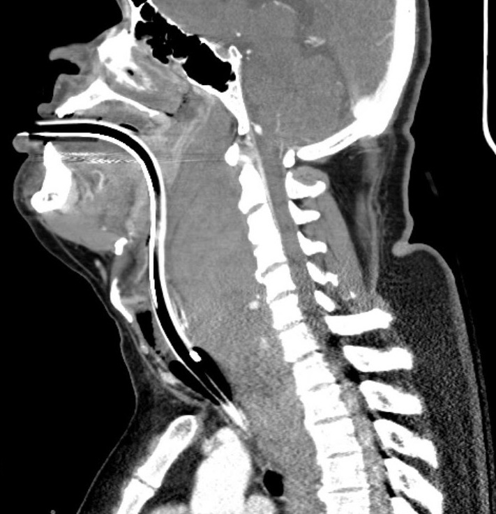
Upon insertion of the glidescope, vocal cord visualization was hindered by oropharyngeal excessive bleeding. Despite extensive suctioning, only the epiglottis was visible due to hematoma compression and soft tissue swelling obscuring distal anatomical structures. The patient’s SpO2 declined to the 80s despite spontaneous breathing, prompting two-person bag-mask ventilation with an oral airway insertion. A Grade III Cormack-Lehane classification was given, and subsequent successful glidescope intubation was made with a gum-elastic bougie (GEB) after SpO2 recovered to 100%. During this intubation, the GEB was blindly advanced below the epiglottis in the anterior direction, followed by the endotracheal (ET) tube passing over the GEB. Successful placement of the ET tube was confirmed by a CO2 color changer, auscultation, and chest x-ray. Propofol infusion at 50 mcgs/kg/min was started for sedation. A post-intubation CT scan showed evidence of compression from the nasopharynx to subglottic airways with the ET tube in good position above the carina (Figure 2). Midazolam 4 mg IV and fentanyl infusion at 50 mcg/hr was initiated for comfort. Once the airway was secured and the patient was hemodynamically stabilized, he was transferred to a higher-level care facility.
Discussion
The American Society of Anesthesiologists (ASA) recommends awake intubation in cases with airways with suspected difficult intubation, ventilation, or desaturation risk5 (Figure 3). While awake fiber-optic intubation is often preferred, it was deferred in this case given its limited capacity for suctioning large volumes through its small aspiration port.6 Studies suggest that sedation can diminish voluntary efforts to maintain airway patency, potentially increasing the risk of progression from partial to complete obstruction.7 To mitigate this risk in our case, we utilized IV ketamine alone for induction, allowing the patient to maintain spontaneous breathing before intubation.
Hematoma formation post ACSS is associated with increased risk for ventilator requirement, wound infection, pneumonia, and reintubation, with up to 12% of patients requiring emergent cricothyrotomy.8,9 This case highlights the importance for providers to be skilled in next-level treatment plans, including incisional decompressions and cricothyrotomy.7 While there are no established standardized algorithms for airway management post ACSS, some proposed protocols have demonstrated effectiveness in reducing postoperative complications. Recommendations include patients remaining intubated overnight if they had specific risk factors, such as exposure of more than three cervical levels, operative site on C3-4 or above, operative time exceeding 5 hours, blood loss >300mL, or significant medical comorbidities like obesity and obstructive sleep apnea.4 Additionally, implementing preventative strategies like fiber-optic assessments, cuff leak tests, and serial radiographs at intervals before extubation has been proven to significantly reduce the incidence of airway complications and hospital readmission following ACSS.4
An ideal outcome in our case would be a successful first-pass intubation attempt, as failed attempts can result in posterior pharynx trauma, increased airway blood and sections, and subglottic edema – all of which can complicate subsequent intubations.6 To further enhance intubation success rate in emergent cases, the adoption of recent technological innovations can be advantageous. Real-time ultrasonography for structural guidance and artificial intelligence for precise facial contour mapping represent progressive approaches that can support physicians in managing difficult airways and significantly improve patient outcomes.10
_difficult_airway_algorithm.png)
Conclusion
Anterior cervical spine surgery is performed widely for its benefits in enhancing quality of life and carries a relatively low incidence of associated risks. However, it is critical to acknowledge that airway compromise can manifest within minutes to hours after surgery, even when initial outcomes appear favorable. Postoperative airway complications, such as pharyngeal edema and hematomas, can compromise perfusion and escalate into multisystem organ failure if airway management is delayed. It is therefore essential for anesthesiologists to be able to rapidly identify and address these complications, and be open to adopting preventative strategies and technical advancements.