

Introduction
Elbow dislocation is the second most common major joint dislocation, following the shoulder joint.1 Most elbow dislocations, particularly posterior or posterolateral types, are treated non-operatively with closed reduction, splinting and physical therapy. Dislocations are most commonly due to falls, direct blows, rapid stops or missteps during a jump, and hyperextension injury where the elbow is forced beyond its normal range of motion.2
Open dislocations involve a breach in the skin and the underlying joint structures or bones are exposed, which increases the risk of infection and other complications. When an elbow dislocation involves a vascular structure—usually the brachial artery—this injury becomes a limb-threatening emergency. It can result in rapid blood loss, limb ischemia, or even amputation if not identified and treated promptly.3 The brachial artery is located near the elbow and is prone to injury during high energy trauma. Open dislocations are associated with higher complication rates, including compartment syndrome nerve injury, and infection.4 Prompt recognition and treatment are paramount.
Traumatic vascular injuries of the upper extremity are rare and are often underdiagnosed. This is especially true when distal pulses are present, which can be misleading because of collateral circulation. Collateral circulation is an alternate blood flow around the injury site that can maintain distal pulses, despite a major arterial injury.5 Such misdiagnoses can delay the diagnosis and treatment of the injury, putting the limb at increased risk of ischemia.
In this case, the authors describe an open posterolateral elbow dislocation with complete brachial artery transection. The novel aspect of this case report is the description of an uncommon injury pattern and the rare use of a vein graft interposition for vascular repair in this injury pattern. The patient’s limb was saved by timely multidisciplinary surgical intervention of vascular repair and joint reduction. This report describes an effective treatment strategy for an injury that is rarely encountered and emphasizes the need for careful vascular assessment in traumatic elbow dislocations.6
Case presentation
The patient is a 38-year-old female with an injury presenting on the right upper extremity following a fall. The patient presented to the hospital with an open wound and deformity to the right upper limb. The patient reports she had been wearing new shoes, and had lost balance, slipped and fallen down the stairs while exiting her home. She landed on her right arm with significant injury, after which she was promptly brought to the emergency department.
The patient’s vital signs were temperature 97.5°F, pulse 75, respiratory rate 20, blood pressure 178/95 mmHg and oxygen saturation 97% on room air. Patient was morbidly obese, with a BMI of 42. A physical examination revealed swelling and tenderness of the right elbow, forearm, and wrist. The patient had been put in a posterior splint and wrapped with ace bandages, and by report of EMS, had an exposed distal humerus. Unwrapping the extremity revealed an open fracture (Figure1). Mild discoloration of the digits of the right hand was identified; however, the oxygen saturation was 97% by pulse oximetry on all digits. Doppler pulse was intact to the radial artery. Her neurological examination including cranial nerves, motor and sensory system, and reflexes was normal, including good sensation in the radial, median, and ulnar nerve distribution of the right arm.
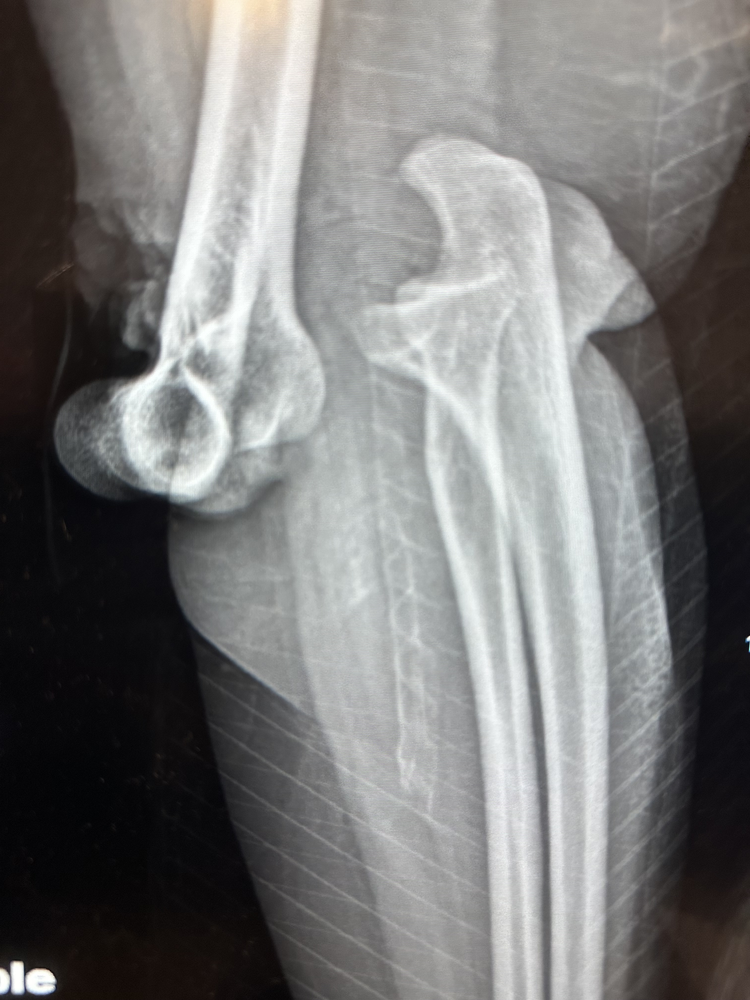
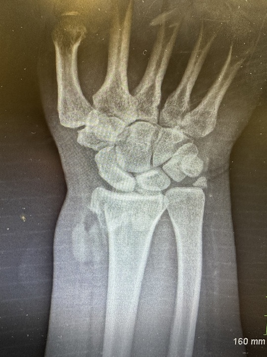
X-rays of the right elbow and wrist were obtained. The x-ray of the right elbow confirmed complete posterior dislocation of the elbow with the distal humerus anteriorly and inferiorly subluxed in relation to the olecranon fossa, with significant soft tissue swelling (Figure 2). Radiograph of the right wrist demonstrated a distal radial diaphyseal fracture, and ulnar styloid process fracture (Figure 3). Orthopedics was consulted for further evaluation and management of the extremity injury, and the patient was admitted for urgent intervention of open right posterolateral dislocation and traumatic brachial artery disruption of the right arm.
Open reduction of the elbow dislocation was performed by the orthopedic surgical team. During the procedure, traumatic brachial artery disruption from posterior elbow with clot formation at the area of proximal injury of the vessel was determined and was repaired with interposition basilic vein graft placement. The comminuted intra-articular radial diaphyseal and ulnar styloid process fractures were managed conservatively. Patient was started on medication for pain management and prophylactic antibiotics.
The following day the patient reported symptoms of paraesthesia of the 5th digit of the right hand, and a weakened ability to flex the interphalangeal joints of the thumb and index finger, suggestive of anterior interosseous nerve palsy. The patient was observed for possible progression of the new onset symptoms, however they resolved spontaneously over the next day.


Discussion
The second most common cause of large joint dislocations is the elbow, with an incident of 5.21 per 100,000 person.7 The number one most common dislocation is typically posterior or posterolateral and is caused by a fall on an outstretched hand /Fall On Outstretched Straight Hand (FOOSH) injury, deceleration, or hyperextension injury that drives the ulna and radius back relative to the humerus.8 Most can be treated using nonoperative methods of management.9
In the presence of a brachial artery injury, however, the picture is different. The brachial artery is the main vessel to the upper extremity distal to the shoulder and runs in the antecubital fossa.10 It crosses the elbow on its way to the forearm, and is prone to injury in high energy trauma, as seen in this case. It is possible for a patient to sustain a complete brachial artery transection during an open or grossly displaced dislocation.11 In this case, the patient sustained a posterolateral dislocation of the elbow, with transection of the brachial artery at the level of dislocation.
The danger of brachial artery injury is that it can often appear normal, even in the setting of massive damage. The collateral circulation around the elbow, including, from the profunda brachii and other smaller vessels, allow for enough blood flood that even pulse oximetry, capillary refill, and doppler assessments may be normal in the presence of severe injury.12 Physical examination of the patient in this case revealed a normal oxygen saturation at all digits of the affected arm, as well as Doppler study finding normal movement of blood at the radial artery level. It is important to maintain a high index of suspicion for arterial injury in any case with gross deformity, soft tissue injury, open wounds, or signs of ischemia, such as pallor, coolness, paresthesias, or motor weakness.13
In the case of missed or delayed treatment, arterial injuries in the upper extremity can lead to catastrophic consequences. Ischemia can quickly lead to irreversible tissue damage, particularly in the forearm muscles, which are particularly susceptible to compartment syndrome.14 As the edema accumulates, it causes pressure within the muscle compartments, which in turn causes increased ischemia and further swelling, leading to necrosis. In cases of neglect, the limb can become non-functional, or in certain cases, may require amputation. Certain nerves are also often damaged in the setting of elbow dislocation, with the median, radial, and ulnar nerves in close proximity to the joint, and the anterior interosseous branch of the median nerve in particular is prone to injury.15 Injuries to this branch of the median nerve can cause difficulty flexing the distal phalanx of the thumb and index finger, leading to the classic “pinch sign” abnormality, as well as fine motor weakness.
In this case, the patient suffered an anterior interosseous nerve palsy, with a persistent weakness and paresthesia to the thumb and index finger. While function may return over time, long-term effects may require additional evaluation or intervention. Traumatic neuropathy may be due to direct stretch, swelling, or even entrapment in scar tissue during healing.16
The first priority in the setting of brachial artery injury is to restore perfusion. Vascular surgeons must explore the injury and attempt to revascularize the limb. In the case of laceration or transition, the lacerated or transected portion of the artery is excised and an interposition graft using the patient’s own vein is created.17 In this case, the vein harvested was the basilic vein. Angiography can be used to confirm the level and extent of the vascular injury, which can be performed preoperatively with CT angiogram or intraoperatively with a contrast injection during surgery.
Once revascularization is performed, the next step is to reduce and stabilize the joint.18 In cases without fractures, this can often be performed by closed reduction. In cases with fractures, such as distal radius fracture in this case, long-term orthopedic planning is needed. In some cases, the initial orthopedic focus is short-term stabilization until the vascular status is secure, followed by staged repair of the fractures at a later time.19 The initial splinting is often posterior splinting, followed by physical therapy after the swelling subsides.
Cases like this are valuable because of how infrequently they are described in literature. The combination of an open posterolateral elbow dislocation and complete brachial artery transection is not common and catastrophic if missed. Fewer than 5% of elbow dislocations have vascular injury, and open dislocations are even rarer.20
Conclusion
The major significance of this case is that it illustrates the urgent nature of diagnosis and treatment in a case of open dislocation of the elbow in the presence of a brachial artery injury. Although dislocations of the elbow are usually managed non-operatively, the occurrence of a brachial artery transection changes the condition into a limb-threatening emergency. Due to collateral circulation, presence of distal pulses can be misleading, and, in the absence of vascular imaging, diagnosis may be delayed. Rapid recognition, repair of the brachial artery with a vein graft, and a multidisciplinary approach were critical for achieving perfusion, preventing further damage, and preserving function.