Introduction
Snapping syndrome could be defined as a popping sensation during knee motion sometimes associated with pain. It presents many difficulties to be defined and diagnosed due to instrumental limitations and a variety of intra and extra-articular structures that could be involved in lateral snaps. Among the extra-articular causes of the snapping phenomenon, the subluxation of the distal biceps femoris tendon over the fibula head is a rare etiology often undiagnosed.1,2 Usually, conservative treatment is unsuccessful making necessary a surgical approach. Several surgical treatments have been described to manage the dynamic snapping of the biceps femoris tendon over the fibular head. These include relocation of the long head biceps femoris insertion or resection of the fibular head.3,4 We report a case of symptomatic snapping knee due to a biceps femoris tendon trauma in a young soccer player with a 12-month follow-up.
The case
A 21-year-old football player was admitted to the outpatient department due to increasing left knee pain. In his medical history, there was a varus knee injury about 2 years before. The patient referred that he experienced pain in the lateral compartment of the knee at first but gradually he started to feel a snap during active flexion and deep squat with worsening symptoms over the past 10 months and inability to practice. The previous conservative treatments were unsuccessful. On clinical examination, there was tenderness on palpation of the lateral aspect of the knee along the distal biceps femoris tendon, especially on the fibular head. Full active and passive ROM was maintained but on the passive motion at 100-110° flexion with intra-rotation of the tibia, the subluxation of the distal biceps femoris tendon was palpated and visualized at the fibular head. The varus stress test was performed to evaluate the fibular collateral ligament, the lateral capsule, and the posterolateral corner (PCL) but no evidence of lateral instability was found. The rest of the examination was normal. The X-ray and the MRI of the knee did not reveal any alteration of the fibular head morphology or other intra-extra articular remarkable findings. Considering the clinical situation and failure of previous conservative management the surgical treatment was decided.
Surgical Approach
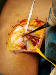
The patient was supine with a tourniquet at the root of the operative limb. The leg was placed in a leg holder. Diagnostic arthroscopy was used to evaluate knee conditions and especially to check the PCL: no intra-articular disorders were found and the drive-through sign was negative. A lateral hockey-stick incision was made over the iliotibial band extended to the fibular head. After plans dissection, the common peroneal nerve was visualized and isolated. We also visualized and isolated the fibular collateral ligament. The posterior arm of the long head of the biceps femoris tendon was partially detached from the posterolateral edge of the fibular head and a prominence on the lateral aspect of the fibular head was found with a neo-bursa over it [Fig. A].
Intraoperatively dynamic subluxation of the distal biceps femoris tendon over the fibular head was directly visualized by flexing the knee at 100° and intra-rotating the tibia. Debridement of the neo- bursa and osteotomy of the prominent lateral portion of the fibular head were performed. The cancellous bone was exposed and the distal arm of the long head of the biceps femoris tendon reinserted on the posterolateral aspect of the fibular head with three anchors Y-Knot Conmed [Fig. B]. Sutures were tied with the knee in full extension. The repair was reinforced with a No. 0 Vicryl suture [Fig. C]. There were no perioperative complications. Postoperative management consisted of partial weight-bearing for 10 days and progressive load-bearing on the operated knee for 20 days. The patient wore a hinged knee brace locked in extension at 0° for 10 days, then locked at 0°-90° for 2 weeks and later on unlocked for the last 2 weeks. Immediate passive motion with a Kinetec device was allowed for 30 days 3 times per day for 1 hour (gradually increasing the ROM from 0°-90° the first 2 weeks to 0°-120° the last 2 weeks). Flexor thigh isolated exercises and exercises against resistance were not allowed for the first 3 months postintervention. He had post-surgical rehabilitation and physiotherapy for 4 months and returned to the sport with no further problems. Outcomes were assessed postoperatively at 1 month, 3-month and one year.
Discussion
The remarkable finding in this case of dynamic snapping knee was the combined etiology of a varus trauma with abnormal morphology of the fibular head, formerly not revealed by imaging. The surgical approach allowed us to better understand the pathogenetic mechanism of the subluxation of the biceps femoral tendon and to achieve the resolution of symptoms. In literature numerous articles report the difficulties to reveal any alteration on X-ray and MRI5–9 therefore, clinical diagnosis has a key role in the management of the biceps femoris tendon snapping knee. Some authors suggested the useful role of the dynamic US to identify the structures involved in the snap: involvement of the bicep tendon can be easily confirmed using dynamic sonography by applying the probe against the fibular head during active motion of the patient knee,10–12 but this diagnostic method is not yet being used widely. The etiology of the snapping phenomenon includes intra-articular and extra-articular causes: among intra-articular structures such as fabellas,13 lateral meniscus, ganglion cysts,14 intra-articular tumours15 can lead to a lateral knee snap. Extra-articular causes can be acquired and congenitally abnormal movements of tendons or ligaments and impingements between tendons and bony structures16: for instance, extrusion on the popliteus tendon along the edge of the popliteal groove, or friction of the iliotibial band against the lateral condyle12 or, as discussed before in our report, impingement of the distal biceps femoris tendon over the fibular head. In the latter case, the most common predisposing factor is an anomalous tibial insertion of the long head of the biceps femoris tendon5–7 other causes described in the literature are an abnormal fibular morphology or a true exostosis8,17 or, even rarer, a direct injury18 to the tendon insertion.
Our patient had a prominent fibular head and a history of trauma. Published surgical options include reinsertion of the anomalous arm of the long biceps femoris tendons through a tunnel into the fibular head or directly onto the periosteum with anchors,1,3,19 partial osteotomy of the fibular head4 or simple partial release of the anomalous tendon.19 In our patient, the prominence of the fibular head and the previous trauma resulted in painful subluxation of the biceps tendon over the later aspect of the fibula. Probably the trauma led to local inflammation of the tendon insertion with the partial detachment of his direct arm resulting in a neobursa and progressive fibrosis over a fibular head that was yet slightly prominent. Therefore, we decided to perform both fibular osteotomy and anatomic repair of the long biceps femoris to the posterolateral aspect of the fibular styloid as recommended by LaPrade technique.1 Neurolysis of the common peroneal nerve was necessary to avoid any iatrogenic injury to the nerve during the surgical. The reconstruction of the insertion of the distal biceps tendon was also necessary since we partially detached it from the posterolateral edge of the fibular in order to perform the debridement of the neo-bursa and osteotomy of the prominent lateral portion of the fibular head. The surgical technique as described by LaPrade requires the restoration of the anatomic position of the biceps femoris tendon.
Conclusion
Snapping knee femoris tendon is a syndrome that is rarely encountered in everyday clinical practice and with limited review in literature. The symptoms are often not recognized and the patient with this condition may visit several specialists before a diagnosis is made. Therefore, the snapping knee femoris biceps tendon represents a challenging for doctors. It would be beneficial to increase the body of literature about this pathology and to investigate further the pathogenetic mechanisms, the diagnostic tools to use, and the treatment options. It must be considered that snapping the knee is not a disabling disease, but operative repair is indicated for patients with high functional demands, such as athletes. The clinical examination and imaging is essential and given the area of interest, the lateral compartment and the posterolateral corner must be examined carefully. We recommend weight-bearing X-rays and MRI both to exclude the involvement of other structures that could be interested in varus trauma such as popliteus tendon, fibular collateral ligaments, both to reveal other such disorders as exostosis, oedemas, tendon lesions. We also suggest performing the arthroscopy which could be useful to diagnose intra-articular associated disorders.
AUTHOR CONTRIBUTION
Dr. Massimo Mariconda and Dr. Claudio Zorzi participate in the conception and design of the study. Venanzio Iacono, Simone Natali and Amedeo Guarino worked on acquisition, analysis and interpretation of data. Luca Padovani and Ludovica Auregli draft the article. All Authors revise it critically for the important intellectual continent. All authors agree to be accountable for all aspects of the work if questions arise related to its accuracy or integrity, especially the corresponding author.
CONFLICT OF INTEREST
All authors declare that there is not any conflict of interest.


