1. INTRODUCTION
Back pain in adolescent athletes is common with a reported incidence of 21% and a prevalence ranging between 37% - 66%.1–3 Most commonly pain is reported in the lumbar region.2,4 Prior wisdom indicated that girls are more likely than boys to have lower back pain and typically have a longer duration of symptoms than boys,3 though recent evidence suggests that the prevalence of back pain is equal among gender.2
Adolescents are at an increased risk for back pain related to several factors including the rapid growth of the vertebral column during puberty.5 Back pain in the adolescent population preferentially affects athletes. Many sporting activities have been thought to potentially play a role in adolescent back pain with certain sports, however, only until recently have sport-specific risk factors been identified. Further, a large portion of the conversation around back pain in the adolescent athlete is centered around diagnosis and treatment.
The purpose of the present investigation, therefore, is to describe sport-specific risk factors that were not well known until recently, to provide an update on the pathoanatomic through recent cadaveric and kinematic studies, to describe the growing emphasis on prevention/screening, and to report recent successes in minimally invasive non-surgical options and minimally invasive surgical options.
2. MATERIALS AND METHODS
Search Strategy
The literature search was performed using Medical Search Headings (MeSH) in Mendeley version 1.19.8. Articles published between January 1975 to December 2021 were Search fields were varied until no new articles were collected at which point the search was considered exhaustive.
Study Screening and Selection
All articles were screened by title and abstract. An initial decision to include a given article was made based on the relevance of the information within the abstract as determined by our inclusion/exclusion criteria (Table 1). These articles then underwent a full-text screening process. This included reading the article in its entirety. Any question regarding the inclusion of an article was discussed by all authors until an agreement was reached. The bibliographies of these articles were also hand-searched to identify any missing articles.
3. RESULTS
3.1. Risk Factors
Adolescent athletes have a higher risk of developing spondylolysis and spondylolisthesis than their non-athletic counterparts. Participation in athletic activity alone has demonstrated a higher risk of developing spondylolysis and spondylolisthesis differences regardless of pelvic incidence.2 Varsity and nationally/internationally competitive athletes have been identified as having a higher yearly incidence of back pain as well.2 Increased time participating in a sport has been shown to be a risk factor for the development of back pain.5 Activities such as all-day sports camps and tournaments are risk factors for the development of back pain.5
Increased lumbar lordosis, abdominal muscle weakness, hip flexor tightness, hamstring tightness, thoracolumbar fascia tightness, femoral anteversion, genu recurvatum, and thoracic kyphosis can predispose adolescent athletes to develop back pain.6 Female adolescent athletes have been shown to have increased lordosis, and pelvic rotation than their male counterparts, putting them at increased risk for back pain,7 though this was not seen in the most recent cross-sectional study in the United States.2 Other risk factors identified include BMI, previous back pain, use of rolling backpacks, and use of backpacks with one strap have also been identified as risk factors.2
Specific sports have been shown to have an increased risk of back pain in adolescent athletes. 27% of college football players, 50% of an artistic gymnasts, and 86% of rhythmic gymnasts frequently have back pain.5 The sport of weightlifting was looked at specifically and found to have an increased rate of back pain.8 The back pain associated with weightlifting can be attributed to performing fixed movements, lifting heavy weights, and poor form.8 Pitchers can often present with non-dominant arm facet pain.9 In soccer, playing surface, previous back injuries, previous groin injuries, and playing the position of goalkeeper all had statistically significant increases in back pain.10,11 Female gender and previous back or groin injury caused the most significant increase in the development of back pain.11
Combat sports, such as boxing and lower back pain in adolescents have a strong association with back pain.12 Sports that require hyperextension including gymnastics and volleyball are also associated with back pain. Football is itself a risk factor for back pain with offensive linemen having a higher rate of back pain.13
3.2. Anatomy and Pathoanatomy
Adolescent athletes are at an increased risk of back pain.14 A postulated hypothesis is that spine rotation restriction caused by rapid growth that occurs during puberty plays a role in the development of back pain.14 The vertebral ring apophysis is the most vulnerable area as it undergoes rapid expansion during the growing phases. Injury to this site is seen in adolescent athletes but not in their inactive age-matched counterparts.14
There are important relationships in the pathoanatomy of back pain outside of the vertebral column. These include tightness in the hip flexors and hamstrings, weakness of the abdominal muscles and gluteals, and an excessive lordotic posture.6 Individuals with decreased flexibility at the hips have been shown to be at an increased risk for lower back pain.14 Strong abdominal core muscles have been considered protective against lower back pain.14 Early adolescent athletes have been shown to have decreased trunk muscle strength putting them at increased risk for back pain.7
Interestingly, in the past five years, there have been a few kinematic studies that have further elucidated biomechanical factors that may play a role in back pain in adolescent athletes. Arampatzis et al. 2019 demonstrated that there are greater muscle strength imbalances in early adolescence, which combined with spinal restriction could explain the incidence in the adolescent range. The mean sacral slope has been shown to be significantly lower in adolescents with spondylolysis. Variations in this parameter may load the pars intercalates and thus predispose them to spondylolysis.
3.3. Etiology
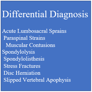
The differential diagnosis of back pain in adolescent athletes is broad. The key elements of the differential diagnosis for back pain in adolescent athletes are summarized below for the reader.

Acute soft tissue sprains, strains, and muscle contusions
Soft tissue injury in the lumbar region is the most common cause of lumbar pain in adolescent athletes. Causes can include a blow to the twisted spine, direct contact, and forced hyperextension.15,16
Spondylolysis
Spondylolysis is a non-displaced fracture of the pars interarticularis. Repetitive flexion, extension, and torsion of the lumbar spine may predispose to spondylolysis. Spondylolysis can present as unilateral or bilateral. Spondylolysis is a common finding among adolescent athletes presenting with back pain and involves the L5 vertebrate over 90% of the time.17
Spondylolisthesis
Spondylolisthesis refers to anterior/posterior vertebral slippage17 and can result from unaddressed spondylolysis. Spondylolisthesis commonly occurs at the L5/S1 vertebral level and can be classified as disruption and stress fracture of the par interarticularis, elongation of the pars without disruption due to microfracture, or acute fracture of the pars.16
Stress Fractures
Stress fractures are those caused by repetitive loading and unloading of bone. They can be thought of as either fatigue fractures or insufficiency fractures. Fatigue fractures are caused by repetitive loading of normal bone and can be seen in long-distance athletes. Insufficiency fractures are caused by repetitive loading of abnormal bone and can be seen in patients with osteopenia, osteomalacia, rickets, hypovitaminosis D, or the female athlete triad. Classic injury mechanisms leading to stress fractures in adolescent athletes include repetitive back hyperextension and rotation.5
Herniated Nucleus Pulposus
Disk herniation refers to the extrusion of the nucleus pulposus from the annulus fibrosis. The symptomology can include neurogenic pain. Though uncommon in young patients, disk herniation occasionally presents in elite adolescent athletes, especially those participating in high-impact sports (wrestling, gymnastics, and football). Body mass index, repetitive strenuous motion, and repeated lumbar flexion/extension are all risk factors for disk herniation.17
Slipped Vertebral Apophysis
Fracture of the osseocartilaginous junction between the vertebral body and apophysis results in the disc and spinal segment displacement into the vertebral canal. This most often occurs in wrestling and gymnastics and may result from acute or repetitive trauma.16
3.4. Clinical Presentation
Clinical presentation of lower back pain in adolescent athletes depends on the mechanism and extent of the injury. Common presentations according to etiology are as follows:
Spondylolysis and Spondylolisthesis
When symptomatic, spondylolysis will often present as low back pain in extension. Pain is typically described as achy and non-radiating. Spondylolisthesis presents similarly, though patients experiencing spondylolisthesis will also show characteristic posturing known as the Phalen-Dickson sign.16
The diagnostic utility of history and physical exam features in assessing spondylolysis and spondylolisthesis is controversial. Certain historical elements, including male gender, age <20, and acute onset of symptoms can heighten clinical suspicion. Hamstring tightness and palpable step-off are physical exam findings that have been proven to have both high sensitivity and specificity for spondylolisthesis.18,19
Disk Herniation and Slipped Vertebral Apophysis
Common signs of disk herniation include lower back pain, progressive radicular pain, and occasionally neurologic deficits.20 Pain while sitting, limited mobility of the lumbosacral region, and excessive hamstring tightness are also typical findings. In general, the clinical presentation of slipped vertebral apophysis closely resembles that of disc herniation.
In addition to the physical exam findings noted above, certain maneuvers, including the straight leg and reverse straight leg test, can be done to support the diagnosis of disc herniation. Though the specificity and sensitivity of these tests are variably limited, they provide a high index of suspicion of disc pathology. Imaging of the lumbar spine and visualization of the nucleus pulposus is confirmatory.9
3.5. Diagnosis
General
The lack of clinical criteria to diagnose lower back pathologies in adolescent athletes means that clinicians must integrate history, physical exams, and image studies. Distinguishing the cause of lower back pain is often complicated by unreliable special tests, inaccurate imaging techniques, and diverse presentations of each condition.21 Recent studies have shown that common clinical features, including pain with extension, the difference between action and resting pain, and male sex may be used to guide practitioners in the diagnosis of back pain in adolescent athletes. Further, there exists some discrepancy in diagnosis between the general orthopedic community and spine surgeons.
Imaging
Adolescent athletes with LBP who are suspected of having lumbar spine injuries are typically assessed using imaging studies. Physicians often begin with x-rays to evaluate the integrity of the pars interarticularis using a lateral oblique view.22 MRI remains to be the primary imaging modality to evaluate spine pathologies in athletes.23 CT is not required as often in the workup of an adolescent athlete with back pain but may provide insight into a mechanical cause of back pain if vertebral column injury is suspected. It can also be used to evaluate bony anomalies, as a surgical planning tool, and in the case of suspected congenital osseous abnormalities.6,24,25
Static and Dynamic Control
Assessing truncal stability may be useful in the evaluation of patients presenting with lower back pain. Though a lack of central static and dynamic control has been shown to correlate with lower back pain in the general population, no such correlation has been found in adolescent athletes.26,27 Nevertheless, there is evidence that high physical workload correlates with a back injury in this population. Thus, isokinetic testing is useful to gauge muscle strength of the trunk and gain a better idea of risk for each individual patient.
3.6. Screening and Prevention
Screening
Various exercises to assess trunk strength have been used to screen adolescent athletes with lower back pain. It has been shown that isokinetic testing can be done to assess trunk peak torque and flexion/extension ratios, with the expectation that strength in extension and flexion is decreased with symptomatic lower back pain. Trunk muscle activity can be evaluated using electromyography (EMG), with recent evidence demonstrating that EMG amplitudes are increased for dorsal muscles in rotation and extension but not for ventral muscles in flexion in adolescent athletes with lower back pain.28 These results provide the framework for evaluating biomechanical data in the context of screening for the development of back pain in adolescent athletes.
Prevention
As imbalances in trunk muscle strength and alterations in lumbopelvic alignment are suggested risk factors of lower back pain, it has been postulated that strength development of trunk muscles and pelvis stabilizers is one preventative measure that can reduce the incidence in the adolescent athlete population.29 Further, utilizing improved neuromuscular control to enhance spinal stability may prevent lower back pain. Arampatzis et al. showed that the prevalence of lower back pain in adolescent athletes significantly decreased with perturbation-based exercise intervention designed to strengthen trunk flexors/extensors and improve neural synchronization.29 Though several studies have found that exposure to perturbations leads to an improvement of motor control and a higher level of coordinated activation of muscle groups, more work must be done to explore the exact role of central nervous system modulation in preventing adolescent athletic back pain.
3.7. Conservative Treatment
First-line treatment for adolescent low back pain remains conservative treatment prior to any surgical or otherwise invasive intervention; most cases resolve with only supportive care.30 Conservative management involves activity modification, bracing, and physical therapy. Lumbar bracing may be used, however, should be saved for patients who fail to improve without bracing, as there is controversy regarding the impact of bracing on clinical outcomes and concern that the restriction may increase movement at the lumbosacral junction.31–33 Several primary articles support early initiation of physical therapy as opposed to long periods of immobilization, which allows earlier return to sport, possibly due to minimizing muscle atrophy and deconditioning.32–37 Physical therapy should be guided towards strengthening deep abdominals and multifidus muscles in conjunction with sport-specific exercises. Dynamic weight-bearing exercises can also be used in later stages of therapy.31–33 Hamstring tightness has been correlated to low back pain in certain sports disciplines, however, research regarding addressing hamstring tightness in treating low back pain and the integration of this into physical therapy protocols is required.38 The results of conservative treatment are generally excellent. Estimated rates of return to sport are above 90% within six months with the majority of patients reporting resolution of pain symptoms.31,32,35 The high likelihood of return to sport should be outlined in order to minimize hesitation of activity and fear of re-injury.32,36
3.8. Minimally Invasive Treatment
In the event, that conservative treatment fails to resolve symptoms, minimally invasive treatment may be pursued. Minimally invasive treatment should be guided based on the underlying diagnosis. External bone growth stimulators have shown some limited support for healing pars fractures in spondylolysis.39–41 Intradiscal steroid injection or transforaminal steroid injection may offer therapeutic benefits to adolescents with inflammatory low back pain; however, it has only been extensively studied in adults and requires further study in the pediatric population.9,42,43 Patients diagnosed with herniated disc alongside a chief complaint of leg pain (rather than back pain), positive straight leg test, and a soft protruded disc may seek chemonucleolysis over discectomy due to its minimally invasive nature, as well as reports of shorter hospital stay, early rehabilitation, and no back muscle trauma during the procedure.44 However, it is important to note that chemonucleolysis can result in early disc degeneration compared to operative discectomy.44
3.9. Surgical Treatment
Surgical treatment for adolescent low back pain varies based on the underlying cause. In general, there has been a significant trend toward minimally invasive surgical techniques including percutaneous approaches, endoscopic surgeries, and shorter incisions. These are associated with decreased operative time, decreased operative cost, increased healing, and improved patient outcomes.
Herniated discs are often repaired through percutaneous endoscopic discectomy, or open lumbar discectomy.9,45,46 Discectomy has been done endoscopically with great success, where an 8 mm cannula is placed approximately 10 cm lateral to the midline. Once the cannula can visualize the epidural space and annulus fibrosus, the herniated annulus fibrosus could be removed. Recent results demonstrate a 90% likelihood of return to their usual intensity of sport 6-8 weeks after surgery, as well as less than 10% likelihood of recurrence.9
Direct repair is preferred with either Buck’s technique, Scott’s technique, or modified Scott’s technique and the screw-rod-hook technique are acceptable.47–50 The literature favors Buck’s technique due to decreased time operating, blood loss, and postoperative hospital stay.50,51 Furthermore, Buck’s technique stabilizes and better restores physiological lumbar biomechanics.51 In recent years, direct pars repair has been shown to be capable through irrigation endoscopic technique which is favorable to patients as it involves only two 0.5 cm access points and no muscle dilatation, minimizing scarring and patient discomfort.51 This procedure is well-tolerated and patients can typically be discharged on a postoperative day one with oral antibiotics, non-steroidal anti-inflammatory agents, and about 8 weeks of a lumbosacral support brace. Pedicle screw-rod-hook technique was notably associated with an earlier return to sport compared to Buck’s technique, though increased surgical time and blood loss were also reported.
There is concern regarding the treatment of spondylolysis with pedicle screw fixation and fusion. This technique often results in a loss in the range of motion and may result in increased loading on adjacent spinal segments.50,52,53 The additional loading raises concern for symptomatic degeneration later in life.50,52
4. CONCLUSIONS
Back pain in adolescent athletes is common, maybe under reported and may not lead to appropriate alterations in athletes’ level of participation. Tightness of hip flexors, hamstrings, excessive lordosis, and decreased trunk muscle strength, in combination with the rapid growth of the vertebral column during puberty, may place patients at higher risk. Athletes with a higher body mass index should be counseled regarding the benefits of losing pain with regard to addressing back pain. Adolescent athletes should be counseled to use backpacks with both straps and avoid excessive tightening of waist straps. Weight lifters, gymnasts, football players, and combat athletes may be at higher risks. There is a paucity of literature regarding isokinetic testing and electromyographic data use as a screening tool, though there is some evidence to suggest that strength deficits and postural control could play a role in the screening of patients at risk for worsening of existing back pain. A single perspective control trial supplies high-level evidence that perturbation-based exercise interventions can reduce the incidence of adolescent athletic back pain. However, more literature is required in this effort. There is a large body of evidence to support the efficacy of physical therapy. There is some data to support minimally invasive treatments including external bone growth simulators, steroid injections, and chemonucleolysis for specific pathologies. Many causes of low back pain in adolescents can be treated endoscopically, which has the potential to decrease operative time, decrease cost, promote healing.
Author Roles
Study concept and design: N. V.; I. U.; O.V.; Acquisition of data: N. V.; A.R.; I. U.; O.V.; Analysis and Interpretation of data: N. V., I.F; C.H.; A.K.; I.U.; O.V.; Drafting of the Manuscript: N. V., I.F; C.H.; A.R.; A.K.; I.U.; O.V.; critical revision of the manuscript for important intellectual content: N. V., I.F; C.H.; A.R; A.K.; I.U.; O.V.; Statistical Analysis: none; Administrative, technical, and material support N. V.; I. U.; O.V.; Study supervision: N. V.; A.K; I. U.; O.V..
Disclosures Statement
We have no disclosures or potential conflicts of interest.
Further Information
This manuscript did not receive any funding and has not been presented at many conferences.