INTRODUCTION
Total Knee Arthroplasty (TKA), is one of the most frequent surgical procedures and an effective treatment option for knee advanced osteoarthritis, by decreasing pain and improving function; the development of new knee prosthetic models over the last decade has involved, in addition to the search for reproduction of the ‘physiological’ kinematics, a number of sizes and shapes sufficient and necessary to fit the prosthetic components to the morphological differences related to individual, gender and race variability.1
Nevertheless, some patients achieve implant fails or periprosthetic fractures and a revision surgery is required. The aging of the worldwide population, due to the enlarging of life expectancy, and the age-related increasing of both osteoporosis and sarcopenia are affecting a constant rise of incidence of knee periprosthetic fractures.2
It has been estimated that the incidence of femoral periprosthetic fractures after TKA ranged between 0,3 to 2,5%,3–6 but increases to 38% when considering revision TKA.6 The majority of these fractures occurs between 2 to 4 years post-operatively, affecting principally the distal femur (2%) and less frequently the proximal tibia (0,3-0,5%).6 An important factor that acts in this field is the implant design.7 The intraoperative rate of periprosthetic TKA fracture is 4%, but is probably underreported.8 Patient-related risk factors for T.K.A. periprosthetic fracture (T.K.A.P.F.) include osteoporosis, age, female sex, revision arthroplasty and peri-implant osteolysis.9 On the other hand, surgical-related risk factors are use of long tibial stems, cementless press-fit fixation, malalignment of the tibial component, tibial tuberosity osteotomy and bony defects in revision arthroplasty.10,11 The aim of treatment of TKAPF should be similar to other fragility fractures, in other words early mobilization and weight bearing in order to reduce the patient’s morbidity and mortality.
However, the complication rate of TKAPF surgical treatments is relevant; a review of 415 cases reported a nonunion rate of 9%, fixation failure in 4%, infection rate of 3% and revision surgery in 13%.12,13 A grate orthopedic debate concerns the choice of the most appropriate fixation device for T.K.A.P.F.: closed or open reduction with internal fixation with either locked plate or intramedullary nail is the most commonly used for treating these fractures, proving good outcomes in terms of fracture union and joint function.13
In order to produce a therapeutically flow-chart in the treatment decision making of TKAPF, this article reports a comprehensive review of the last 10 years literature on TKAPF.
ETIOLOGY AND RISK FACTORS
The age group most affected by aseptic TKAPF is that between 65 and 75 years old, in which the risk of mechanical loosening is higher depending on the bone quality.
Femoral supracondylar periprosthetic fractures are the most common with an incidence rate of 0,3 – 2,5% in post-primary Total Knee Replacement (TKR), followed by tibial fractures with an incidence rate approximately 0,9% in revision TKR and patella with an incidence of 0,2 – 21% of cases, depending on the eventual patellar resurfacing, which can increase the incidence.
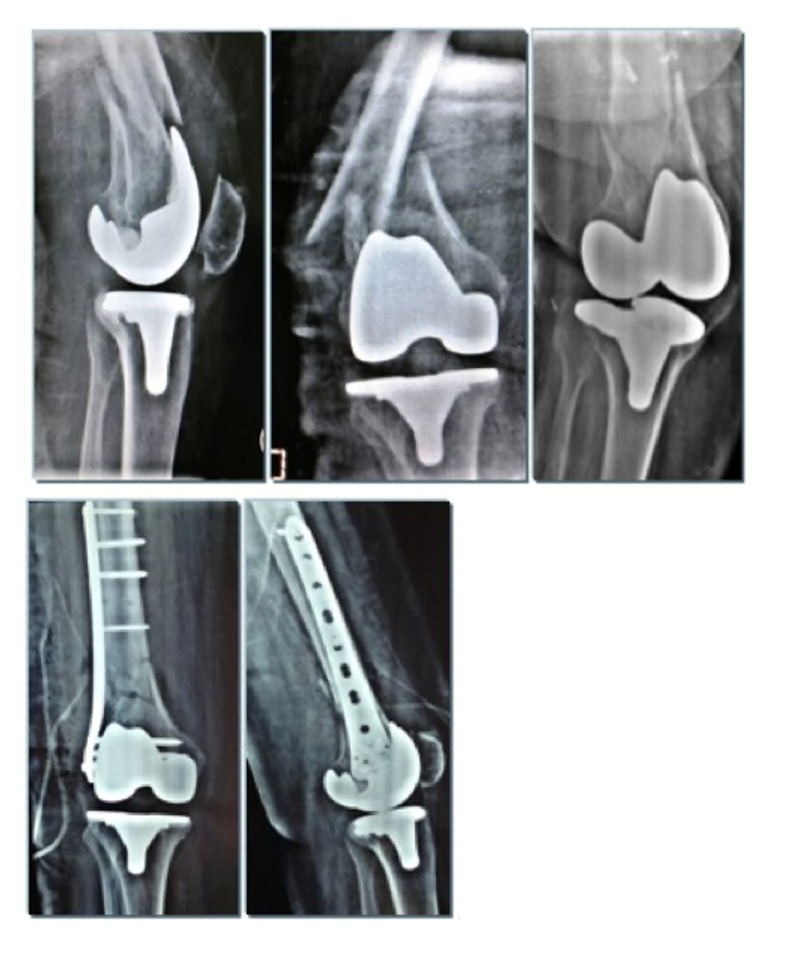
_treated_with_liss_plate_(fig._1b).png)
Advanced age is a major risk factor regarded as an individual risk factor within itself, as well as a risk factor for osteoporosis and recurrent falls.
Additional risk factors include the chronic use of steroid therapy, inflammatory arthroplasty such as rheumatoid arthritis, and patients suffering from neurological diseases including epilepsy, Parkinson’s disease, poliomyelitis, and myasthenia gravis which all appear to increase the risk of periprosthetic fractures. Diabetes mellitus is an additional risk factor that has important effects on post-surgical healing due to microvascular and neural damage, as well as increasing the patient recurrent falls rate while an increase in mortality is reported due to the association of the fracture with Covid 19 infection in recent years due to the recent Covid 19 pandemic14 Revision TKA was in itself described as another major risk factor for the development of periprosthetic fractures (facture risk after primary TKA 0,6% vs TKR 1,7%).15
DIAGNOSIS OF TKAPF
The correct approach to a patient afferent to the emergency room should start from acquiring the complete history of the patient about comorbidities, chronic taking of medicines, recent trauma or if not, investigating on chronic pain or joint instability, which could be indirect signs of pre-existing aseptic loosening of the implant, possibly due to component loosening, polyethylene wear with osteolysis, ligamentous laxity, arthrofibrosis or patello-femoral complications.
An accurate X-rays study, followed by a CT-scan, may both describe the fracture pattern and evaluate typical signs of prosthetic loosening, such as a radiolucent line of 2 mm or more around the prosthesis at the bone-cement interface in case of femoral stem displacement.16
CLASSIFICATION
There are different Classification Systems for TKAPF, selective for the femur, the tibia and the patella, but nowadays the AO/OTA “Association for the study of Internal Fixation” has chosen the Unified Classification System (UCS) as the primary classification for periprosthetic fractures; in this classification, there are six different classes of fractures, categorized from A-F. Each category describes a specific anatomical description of a periprosthetic fracture; all of the classes can be utilized in different anatomical locations, as long as one follows the classification principles.17
FEMURAL PERIPROSTHETIC FRACTURE
The femoral supracondylar periprosthetic fractures are considered all solution of continuity within 15 centimeters from the joint line. Femoral supracondylar periprosthetic fractures are the most common in the periprosthetic fractures with an incidence rate of 0.3–2.5%18 post primary TKA, that can rise up to 38%19 in case of revision surgeries. The female sex, dementia, motor alteration or Parkinson disease and femoral overcut previous were factors associated with the increased risk of this type of fractures.20
Several classification systems are used for peri-TKA femoral fractures, but the most commonly used is Lewis and Rorabeck Classification (1997), that divides the fractures in three types: Type I (Nondisplaced; component intact), Type II (Displaced; component intact), Type III (Displaced; component loose or failing). Another recent classification system is Su and Associates’ Classification of Supracondylar Fractures of the Distal Femur, that divides in three types: Type I (Fracture is proximal to the femoral component), Type II (Fracture originates at the proximal aspect of the femoral component and extends proximally), Type III (Any part of the fracture line is distal to the upper edge of anterior flange of the femoral component).
For a stable and non-displaced fracture (Rorabeck and Su Type I) the conservative treatment is a valid option and involves cast application with or without a traction period. Agarwal et al.21 evaluated the outcome in patients treated for peri-prosthetic knee fractures. Out of 15 patients with supracondylar femoral fractures, 2 were Rorabeck Type I (non-displaced, with intact prosthesis–bone interface). They were treated with immobilization in a long leg cast. Follow-up was of 24 and 34 months and both patients showed excellent results in the range of motion, knee score, and functional score.
Merkell and Johnson evaluated 36 supracondylar fractures of the femur. Of those, 26 fractures had been treated using non-operative treatment. Seventeen of those fractures (65.4%) healed without surgical treatment. Fourteen of the 17 were followed for over 2 years and did not present with significant differences in the knee score over this time. The remaining patients required knee revision surgery due to nonunion, malunion, loosening of the component, and extension lag. Nonetheless, they concluded that traction or application of a cast, or both, should be the primary treatment options, and usually one will result in healing of the fracture and a satisfactory outcome.22
The operative treatment is considered the best option for unstable, displaced fractures, because it allows early range of motion and ambulation.23 The implant stability, the fracture pattern, an active infection process, the bone’s quality, and bone stock are the parameters to considering. Common surgical techniques include both external fixation and internal fixation (i.e. blade plates, condylar screws, retrograde intramedullary nails or locking plates) and revision TKA.
Finally, some authors, to stimulate the healing of the fracture, recommend starting an integrative therapy immediately and in recent literature an important role of pulsed magnetic fields in the healing process has also emerged24
PLATE FIXATION
Open reduction and internal fixation (ORIF) allow the surgeon to perform anatomical reconstruction, enabling the patient to perform early rehabilitation. It’s indicated for intact/stable prosthesis (Lewis-Rorabeck II or Su Types I or II) with fracture unable to accommodate intramedullary device and for fracture distal to flange of anterior femoral component (Su Type III).
Several implants are used to treat this fracture’s: a) condylar buttress plate (non-locking/conventional plate), but does not resist varus collapse, b) locking supracondylar / periarticular plate and polyaxial screws allow screws to be directed into best bone before locking into plate, and can avoid femoral component, c) blade plate / dynamic condylar screw difficult to get adequate fixation around PS implant
Conventional plates have some complications such as loss of reduction and varus deformity, often in the fractures with medial wall multi- fragmentary. In these cases, dual plating might be considered to prevent the secondary varus deformity of the femur associated with single lateral conventional plate.
The locking plates assure a more rigid fixation, than a conventional plates, in the periarticular, comminuted, and osteoporotic fractures. Locking plates are fixed-angle devices designed to engage with the screw threads, and so they have highest union rate, yet complete healing took 6 months and early full weight-bearing.
The ORIF needs a large surgical exposure with extensive soft tissue damage that could lead to an increased risk of nonunion due to damage to the periosteum and blood vessels. Introducing the minimally invasive percutaneous plate osteosynthesis (MIPO) injury to the adjacent soft tissue and periosteum are reduced, thus promoting rapid bone union with a low risk of complications. Closed reduction can be obtained with use of percutaneous screw insertion or a retractor based on the principle of ligamentotaxis.25
Kregor et al. performed LISS fixation (fid.1a-b) for the treatment of supracondylar fractures and obtained bone union in 36 of 38 knees without any complications.26 Hofmann et al. retrospectively reviewed 111 fractures in 106 patients who underwent locked plate fixation due to periprosthetic fractures around the knee. Thirty-six fractures were treated with the open reduction method, and 75 fractures were treated with minimally invasive submuscular plate application. Of the total number of fractures, 91% healed completely. There was a decreased frequency of nonunion among those whose fractures were treated by the minimally invasive submuscular technique compared to those treated with the open technique.
However, the use of locking plates for distal femoral fractures is still debated, because of the reported healing problems and complications. In particular, a nonunion rate ranged from 0 to 19%, a delayed union ranged from 0 to 15%, and an implant failure ranged from 0 to 20% were reported.
We can conclude that the “mini open” technique with cerclage wiring and the use of a polyaxial locking plate is the preferable technique today with regard to soft tissue preservation, as it also emerged for the treatment of hip periprosthetic fractures,27 but requires an experienced surgeon and a careful preoperative planning is needed when choosing a locking plate, to identify the correct plate length, fixation working length, and screw distribution.
RETROGRADE INTRAMEDULLARY NAILING
Retrograde intramedullary nailing (RIN), compared to conventional plate fixation, is associated with less soft tissue damage, less operative time and less intraoperative blood loss with a relative risk reduction for developing a nonunion and for requiring revision surgery.
Currently used interlocking intramedullary nailing takes advantage of interlocking screws that allow higher resistance to axial compression and torsional forces. RIN yields high union rates and excellent functional outcomes in Rorabeck type II supracondylar femoral fractures, especially if it should be long enough to reach the level of the lesser trochanter, considering that the engagement of the isthmus prevents a windshield wiper effect and improves stability. It is also indicated in intact/stable prosthesis with open-box design to accommodate nail, fracture proximal to femoral component (Su Type I) and fracture that originates at the proximal femoral component and extends proximally (Su Type II).
Before performing a RIN osteosynthesis, the surgeon must be aware of the shape of the femoral component of the implanted TKA, to be sure that the distal entry point between the condyles is ‘opened’. In fact, TKA with a box (e.g. posteriorly stabilized) or with a stem cover the RIN entry point. In this case, a plate might be used. Hyperextension of the femoral component may occur in the sagittal plane because reaming and insertion are performed with the knee in flexion position and can be more evident if the insertion site is posterior to the intercondylar notch. According to Pelfort et al., hyperextension of the femoral component does not significantly affect stability of the prosthesis, bone union, or knee joint function. Valgus malalignment of the distal fragment in the coronal plane is often encountered. To avoid this, it is recommended to use a blocking screw as a guide for proper insertion of the intramedullary nail.28–32
In addition, the application of poller screws or pins is a useful technique utilized to improve the reduction and the final alignment of the femur. Taken together with this technique, it was found that an increased number of distal interlocking screws were found to have reduced the risk of nonunion and reoperation rates.
Contraindications to intramedullary nailing include patellar baja, joint ankylosis precluding intramedullary nail insertion, < 11 mm intercondylar distance or narrow medullary cavity, preexisting intramedullary stem in the proximal femur from previous total hip arthroplasty, severe comminution or extremely distal fracture precluding stable internal fixation and unstable TKA prosthesis.
REVISION TOTAL KNEE ARTHROPLASTY AND ENDOPROSTHESIS
Previous literature has already shown satisfactory results in the treatment of of periprosthetic hip fractures with Modular tapered conical revision stem33 and megaprosthesis in more difficult cases34
Revision TKA (RTKA) should be considered in case of femoral component instability (Rorabeck type III and Su type III), severe comminution or fracture periarticular extension that precluding internal fixation, failure of previous treatments, and severe malalignment of the TKA.
Revision TKA with a long-stemmed cemented femoral component is used in femoral fracture with a good bone stock; if fracture stability cannot be obtained using only the long stem, strut allograft or cerclage wiring should be used to improve stability of both the fracture and the femoral component. The long stem is inserted through the fracture site into the proximal femur overcoming the fracture site.
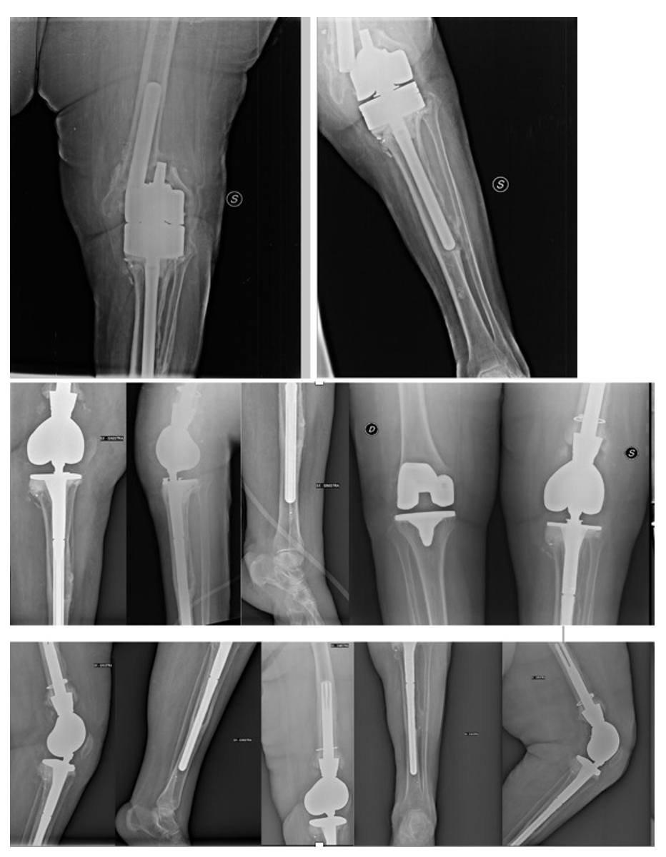
If the fracture is characterized by severe comminution or the bone is so weak that we have to deal with three components simultaneously: bone fracture, unstable prosthesis, and loss of bone, we used distal femoral replacement with a megaprosthesis (Endoprosthesis) as a limb salvage procedure to preserve minimum knee function and maintain the length of the leg (fig 2a-b). Distal femur endoprosthesis should be considered in patients with advanced age and poor bone quality who require early mobilization who have no other treatment option. However, this procedure requires highly experienced orthopedic surgeons who are familiar with this procedure and special arthroplasty equipment and implants.35,36
_and_implant_loosening__treated_with_a.png)
TIBIAL PERIPROSTHETIC FRACTURE
Periprosthetic fractures of the tibia associated with TKA have an incidence of 0.4-1.7% on first implant; among all the specific fracture risk factors are listed: prior tibial tubercle osteotomy, component loosening or malposition and insertion of long-stemmed tibial components. The treatment of severely displaced, unstable periprosthetic tibial fracture can be performed using open reduction and internal fixation. The best option is locking plates with several locking screws inserted around the tibial stem to assure a valid fixation.37 The major classification is the Felix and associates’ one, that combines the site of fractures (Type 1: At the level of tibial plateau, Type 2: Inferiorly and adjacent to the prosthetic stem, Type 3: Distal to the tibial stem, Type 4: Fracture of tibial tubercle) and the stability of the prosthetic implant (Type A: Stable prosthesis, Type B: Component loosening, Type C: Intraoperative fracture). The surgical approach should be based on the status and site of the fracture. Generally, Felix Type 1A nondisplaced fractures with stable prosthesis are managed conservatively with knee extension cast or brace, while unstable fractures with stable prosthesis could be managed with lateral plate fixation. Even Type 1C “intraoperative” fractures could be treated with ORIF. For unstable Type 1B fractures, screw fixation of fragments is followed by insertion of a long-stemmed tibial revision prosthesis through the fracture site into the tibial medullary cavity; bone allograft could be a valid option in order to increase the stability of the revision prosthesis. Type 2B and 2C fractures should be treated by a long-stemmed tibial component and bone graft at the fracture site, while Type 2A fractures, is to say post-operative fractures with stable prosthesis, could be managed with rigid immobilization and loading abstention for six weeks. Type 3C fractures are managed both with 4.5mm plates internal fixation and conservative treatment, depending on fracture site and pattern. Postoperative fractures can be treated as isolated tibial fractures by plaster immobilization followed by a late revision, once the fracture is consolidated, in case of prosthetic component loosening. Type 4 fractures should be treated by screw fixation or wire cerclage of tibial tubercle, keeping attention to avoid extensor mechanism disruption; a possibility of both stabilize the fracture and reinforce the extensor apparatus is the polypropylene mesh tape or semitendinosus rerouting.38
Because of the weak thickness of the skin overlying the tibial plateau, MIPO technique should be preferred in order to preserve the local blood supply and minimize the soft-tissue complications.
Haller et al. suggested the use of intramedullary nail for management of Felix Type 3A fractures choosing a more distal transpatellar entry point, hand reaming the proximal tibia next to the implant, using a suction tip to navigate the guidewire past the posterior cortex, inserting a 9 mm diameter nail while bending it with a bending press. Good results in terms of fracture union and patients walking ability have been showed at 14 months follow-up.39
Subtype B periprosthetic tibial fractures should be managed with a revision TKA with a long-stem tibial component, in order to overpass the fracture site40,41; in case of remaining unstable bone fragments, additional internal fixation should be required and metal augmentation with thick polyethylene insertion used for managing any tibial shaft defects inferior to 5 cm. More severe bone defects or comminuted fractures should be managed using either bone allograft or tumor megaprosthesis.38 The main indication for reconstruction with megaproshesis is Felix Type 1B fractures with severe comminution or major cortical destruction due to osteolysis,37 while in all the other cases is preferred to restore the insertion of the extensor apparatus. It is important to remember that, according to literature review, the revision rate associated with megaprosthesis ranged up to 55%.42
In case of unicompartimental prostheses, the treatment is usually accomplished with open reduction and internal fixation if tibial component is stable and fracture reducible, while in case of prosthesis loosening or fracture irreducible, a revision prosthesis will be better.
PATELLA PERIPROSTHETIC FRACTURE
Periprosthetic fractures of the patella associated with TKA have an incidence of 0.2-21% in resurfaced patella and of 0.05% in unresurfaced one. The principal fracture specific risk factors are: patellar osteonecrosis, asymmetric resection of patella, inappropriate thickness of patella, and implant related factors like central single peg implant, uncemented fixation, metal backing on patella and inset patellar component. The mail patellar periprosthetic fractures Classification is the Ortiguera and Berry one, that analyze the stability of the implants and the integrity of extensor mechanism: Type 1) Extensor Mechanism intact and Patellar Component stable, Type 2) Extensor Mechanism disrupted with stable of loose Component, Type 3) Extensor Mechanism intact and Patellar Component loose, subclassified in 3A) if the bone stock is reasonable (patellar thickness ≥ 10mm) and 3B) if bone stock is poor (patellar thickness < 10mm or marked comminution). Another valid classification is the Goldberg one, that classifies patellar fractures in: Type I) Fracture not involving implant/cement interface or quadriceps mechanism, Type II) Fracture involving implant/cement interface and/or quadriceps mechanism, Type IIIA) Inferior pole fracture with patellar ligament rupture, Type IIIB) Inferior pole fracture without patellar ligament rupture, Type IV) All types with fracture dislocations.
The main indications to casting or bracing in extension are stable implants with intact extensor mechanism and non-displaced fractures, followed by a hinged orthosis in order to increase progressively the range of flexion in a protected way. In case of loose patellar component and/or extensor mechanism disruption a surgical strategy should be planned, ranging on ORIF with or without component revision, partial patellectomy with tendon repair, patellar resection arthroplasty and fixation and total patellectomy.
In case of traumatic fracture, the skin may be bruised or macerated and this may increase the risk of infection, so it should be indicated to delay the surgery until ensuring good skin quality. Technically, a middle incision is preferred in order to clean the hematoma carefully, paying attention not to damage the surrounding soft tissues, very important to avoid patellar devascularization. The ideal would be to use two partially threaded cannulated screws to perform a tension band cerclage, since biomechanically it has proven to be the strongest and most resistant fixation method. If the fracture has small, non-repairable fragments, a partial patellectomy would be preferred. In Type 3 fractures, when the patellar component is loose and the extensor mechanism intact, we must think that a patellar osteonecrosis has occurred. Even with a loosened component, if the patient maintains the leg in extension against gravity, a period of conservative treatment is recommended, especially if there is a history of trauma. If symptoms persist beyond three weeks, either a revision of the patellar component should be performed (if the remaining bone is adequate), or the component should be removed and a patellar remodeling performed in order to obtain optimal tracking.
When direct repair attempts fail, it is not recommended to continue because of the high rates of associated periprosthetic infection and the low success rate of extensor reconstruction; in these cases, it is recommended to repair using a complete allograft of the extensor mechanism.43,44
CONCLUSION
In conclusion, in patients with non-displaced fractures and stable prosthesis or those who are not eligible for surgery due to medical comorbidities, conservative non-surgical methods can be used that yield acceptable results. In patients who suffer from stable or unstable fractures, but possess good bone stock and stable prosthesis, the choices include both locking plate and intramedullary nailing. In case of component loosening, if the bone stock is adequate, fracture reduction and a stemmed revision arthroplasty are a functional option. We should consider this option when the ligamentous structures provide adequate stability and there is an adequate bone stock after primary prosthesis removal. When we encounter fractures that involve bone stock deficiency, the choices that we have are allograft prosthesis composite or distal femur replacement endoprosthesis.
Net of the foregoing, the primary goal of surgical treatment in TKAPF is to achieve a satisfactory fixation and to restore proper alignment, in order to allow the most immediate recovery of the patient’s walking and quality of life.
ACKNOWLEDGEMENTS
None
AUTHORS’ CONTRIBUTION
A.P; A.V.C; A.S; G.P and M.P contributed to the design and implementation of the research, to the analysis of the results and to the writing of the manuscript.
FUNDING
None
CONFLICT OF INTEREST STATEMENT
All authors disclose any financial and personal relationships with other people or organizations that could inappropriately influence (bias) their work. Examples of potential conflicts of interest include employment, consultancies, stock ownership, honoraria, paid expert testimony, patent applications/registrations and grants or other funding.
HUMAN AND ANIMAL RIGHT
For this type of study is not required any statement relating to studies on humans and animals.