Loading [Contrib]/a11y/accessibility-menu.js
1.
Al-Jabri T, Wood MJ, Faddul F, et al. Periprosthetic Distal Femoral Fractures Around a Total Knee Arthroplasty: a Meta-analysis Comparing Locking compression Plating and Retrograde Intramedullary Nailing. Orthopedic Reviews. 2024;16. doi:10.52965/001c.91507
Download all (9)
  • Figure 1. Preferred Reporting Items for Systematic Reviews and Meta-Analysis (PRISMA) flow diagram outlining study inclusion.
  • Table 1. MINORS criteria scoring for included studies.
  • Table 2. Overview of study characteristics.
  • Table 3. Fracture healing, complications and patient reported outcome measures (PROMS).
  • Figure 2. Forest Plot showing comparison of non-union rate between RIMN and LCP. No statistical significance found. M-H= Mantel–Haenszel, CI= Confidence Interval
  • Figure 3. Forest plot showing comparison in reoperation rate between RIMN and LCP. No statistical significance found. M-H = Mantel–Haenszel, CI= Confidence Interval
  • Figure 4. Forest plot showing comparison of infection rate between RIMN and LCP. No statistical significance found. M-H = Mantel–Haenszel, CI= Confidence Interval
  • Figure 5. Forest plot showing comparison in mortality rate between RIMN and LCP. No statistical significance found. M-H = Mantel–Haenszel, CI= Confidence Interval
  • Figure 6. Recommended treatment flowchart for distal femoral peri-prosthetic fractures.

Abstract

Purpose

The number of total knee replacements (TKRs) performed per year has been increasing annually and it is estimated that by 2030 demand would reach 3.48 million procedures per year in the United States Of America. The prevalence of periprosthetic fractures (PPFs) around TKRs has followed this trend with incidences ranging from 0.3% to 3.5%. Distal femoral PPFs are associated with significant morbidity and mortality. When there is sufficient bone stock in the distal femur and a fracture pattern conducive to fixation, locking compression plating (LCP) and retrograde intramedullary nailing (RIMN) are commonly used fixation strategies. Conversely, in situations with loosening and deficient bone stock, a salvage procedure such as a distal femoral replacement is recognized as an alternative. This meta-analysis investigates the rates of non-union, re-operation, infection, and mortality for LCPs and RIMNs when performed for distal femoral PPFs fractures around TKRs.

Method

A search was conducted to identify articles relevant to the management of distal femoral PPFs around TKRs in adherence to the Preferred Reporting Items for Systematic Reviews and Meta-Analyses (PRISMA) checklist. Articles meeting the inclusion criteria were then assessed for methodological quality using the methodological items for non-randomised studies (MINORS) criteria. Articles were reviewed, and data were compiled into tables for analysis.

Results

10 articles met the inclusion criteria, reporting on 528 PPFs. The overall incidence of complications was: non-union 9.4%, re-operation 12.9%, infection 2.4%, and mortality 5.5%. This meta-analysis found no significant differences between RIMN and LCP in rates of non-union (9.2% vs 9.6%) re-operation (15.1% vs 11.3%), infection (2.1% vs 2.6%), and mortality (6.0% vs 5.2%), respectively.

Conclusion

This meta-analysis demonstrated no significant difference in rates of non-union, re-operation, infection, and mortality between RIMN and LCP and both remain valid surgical treatment options.

Introduction

There are now over 1.3 million primary knee replacements recorded in the United Kingdom (UK) National Joint Registry (NJR), the world’s largest arthroplasty registry. Approximately 90% of these are total knee replacements (TKRs) with the remaining being partial knee replacements.1 The global number of primary knee replacements has been increasing tremendously annually and in the United States of America (USA) it is estimated that by 2030 demand for primary TKRs would grow to 3.48 million procedures per year.2 In keeping with this trend, the number of knee revisions has been on a steady incline with 1,225 revisions performed in the UK in 2004 and 7,005 in 2019.1 Although Theophilus Gluck is often credited with performing the first ever TKR in 1891, reports of periprosthetic fractures (PPFs) remained scarce and only began appearing in the late 1970s.3 It wasn’t until the last two decades that PPFs became more frequently spotlighted with published incidences ranging from 0.3% to 3.5% for distal femoral PPFs, though large joint registries estimate the rates to be between 1.1% and 1.3%.4–6 Of the 87,535 revision procedures recorded in the UK NJR, 3,358 revisions (3.84%) were performed for a PPF. Data from the Scottish arthroplasty project has demonstrated that the number of PPFs recorded per year has more than doubled between 1997 and March 2008 in line with the increase in TKRs performed.7 This rise in the number of procedures performed reflects the number of at-risk implants in the UK NJR dataset and the wider population.

There is significant mortality and morbidity associated with distal femoral PPFs and despite advances in our understanding of fracture management and implant design, the subsequent reduction in quality of life and joint function remains high. Mardian et al. reported only 20% of patients were able to mobilise without assistance postoperatively.8 This perhaps may account for the findings of two studies by Shields et al. and Streubel et al. which demonstrated mortality rates of 14% at 3 months and 17.7% at 12 months postoperatively, with 80% of the deaths occurring within 3 months of surgery.9,10 In the UK, the national hip fracture database has recently been collecting data on PPFs and it has been recommended that these fractures should be considered life-threatening injuries that need to be approached with the same urgency and treatment objectives as native hip fractures (e.g. early mobilisation and weightbearing) to improve the prognosis for patients.10–12

Unfortunately, these fractures remain challenging to treat owing to a number of factors including the advanced age of patients and the difficulty in achieving stable fixation in patients with poor bone stock and fragile bone quality. The primary aim of this article is to provide a meta-analysis of specific outcomes (infection, non-union, re-operation, and mortality) of locking compression plates (LCPs) and retrograde intramedullary nailing (RIMN) for distal femoral PPFs around TKRs.

Methods

Literature search strategy

This systematic review and meta-analysis was conducted in adherence to Preferred Reporting Items for Systematic Reviews and Meta-Analyses (PRISMA) checklist.13 Searches were conducted on PubMed/Medline looking at literature published between January 2012 and December 2022. The search terms were as follows: ‘Total knee arthroplasty (All Fields) OR Total knee replacement (MeSH Terms) AND Periprosthetic Fracture (All Fields) OR Peri-prosthetic Fracture (MeSH Terms)’. Articles generated were then screened and excluded in accordance with outlined eligibility criteria.

Eligibility

Inclusion criteria: articles published in the last 10 years (January 2012 to December 2022), patients aged 55 years and older, primary procedures (revision procedures of failed fracture treatment not included), minimum post-operative follow up of 6 months, articles of evidence levels I-III and English language texts only. Procedures included were RIMNs and LCPs.

Exclusion criteria: non-traumatic indications for arthroplasty, patients under 55, native bone fractures, case series, reports, letters, conference articles and abstracts, repeated studies and data, cadaveric and animal studies and studies deemed poorly designed and of evidence levels IV-V. Procedures excluded were revision arthroplasties, distal femoral replacement (DFR), combination fixation strategies (LCP + RIMN hybrids), external fixation and conventional non-locked plating.

Data extractions and analysis

The authors independently extracted data from the selected articles, with discussions held for any disputes in extracted data. Study author, publication year, patient demographics, details of the intervention and outcomes were collated onto a spreadsheet using Microsoft Excel 16.64.

Review Manager 5.3 was used for data analysis. Mean difference was used to analyse continuous data, and odds ratio and the risk difference was used to analyse discrete data. Statistical significance was defined as P < 0.05, with confidence intervals of 95%. Chi squared was used to assess for heterogeneity in the pooled studies, with results represented through I2. I2 cut offs for defining heterogeneity were as outlined in the Cochrane Handbook for Systematic Reviews of Interventions, 0-40% = Low, 30-60% = Moderate, 50-90% = Substantial 75%-100 = Considerable.14 Results, along with all studies featured in the meta-analysis, were presented as forest plots.

Outcome Measures

The primary outcome measures of this review were postoperative rates of non-union, re-operation, infection and mortality. Data reporting complications, time to union, malunion, post-operative range of motion, refracture and patient reported outcome measures (PROMS) were also collected.

Methodological Quality

With consideration of the non-randomised nature of the selected studies, the methodological index for non-randomized studies (MINORS) criteria was used to score their methodological quality.15 Global scores were taken of each study, with a maximum score of 24 for comparative studies, and 16 for the non-comparative studies.

Results

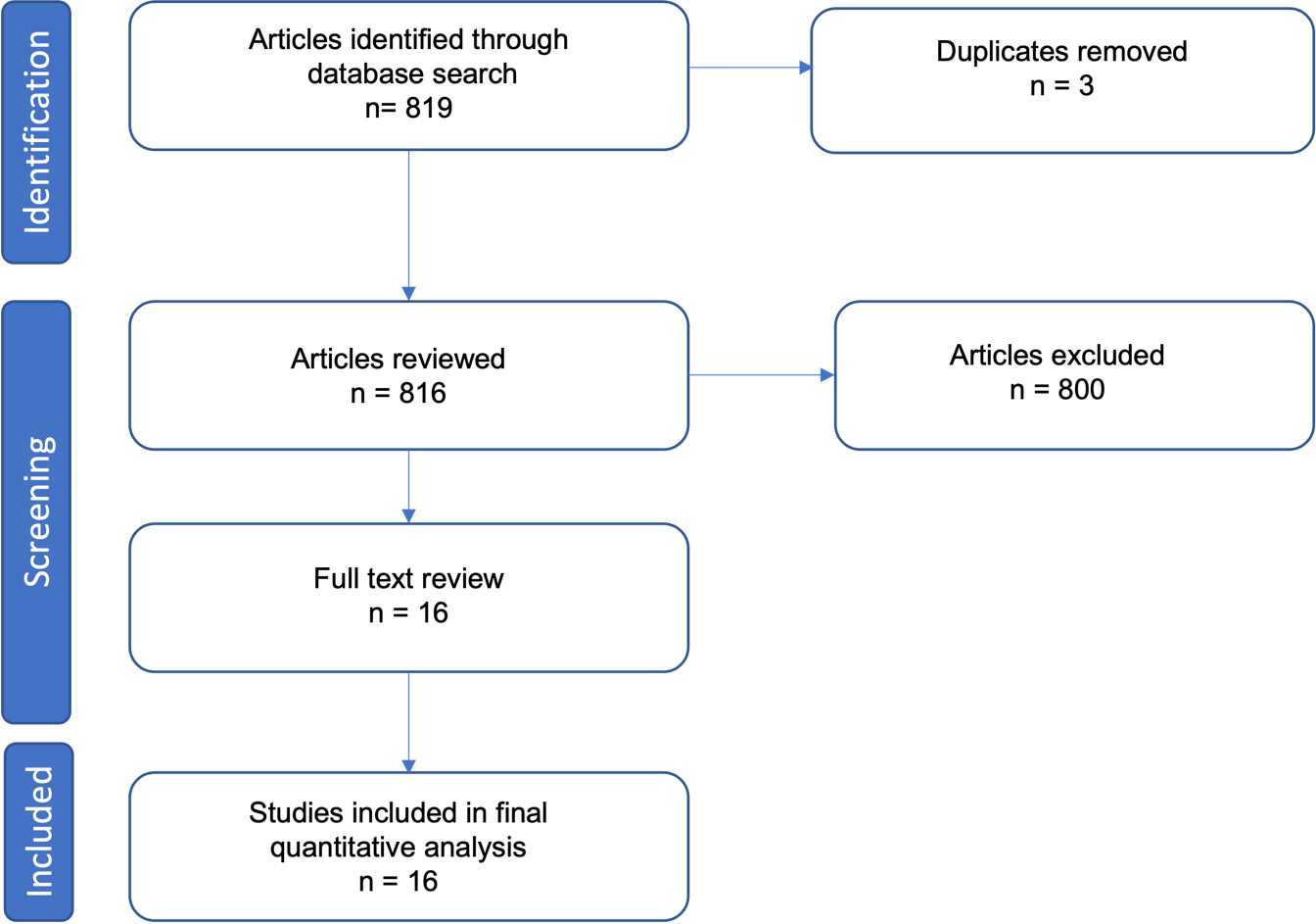
The literature search initially produced 969 articles. These 969 articles were screened by reviewing their title and abstract. Of the screened articles, a final 10 were included in this review for assessment. The PRISMA flow diagram is below (see Fig. 1). Exclusions were made in accordance with the listed eligibility criteria. Of the included studies, 9 were retrospective cohort studies and 1 was a prospective cohort study.

Figure 1
Figure 1.Preferred Reporting Items for Systematic Reviews and Meta-Analysis (PRISMA) flow diagram outlining study inclusion.

Quality Assessment

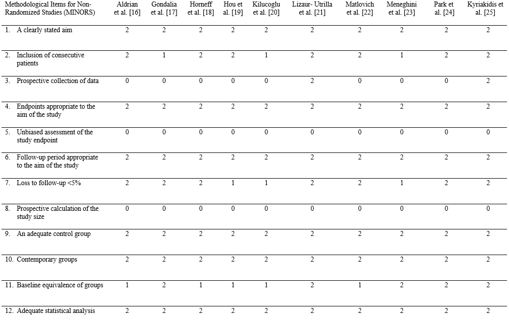
The methodological quality of the included studies was assessed using the methodological items for non-randomised studies (MINORS) criteria,15 all included studies were deemed to meet quality standards (see Table 1).16–25

Table 1
Table 1.MINORS criteria scoring for included studies.

Study Characteristics

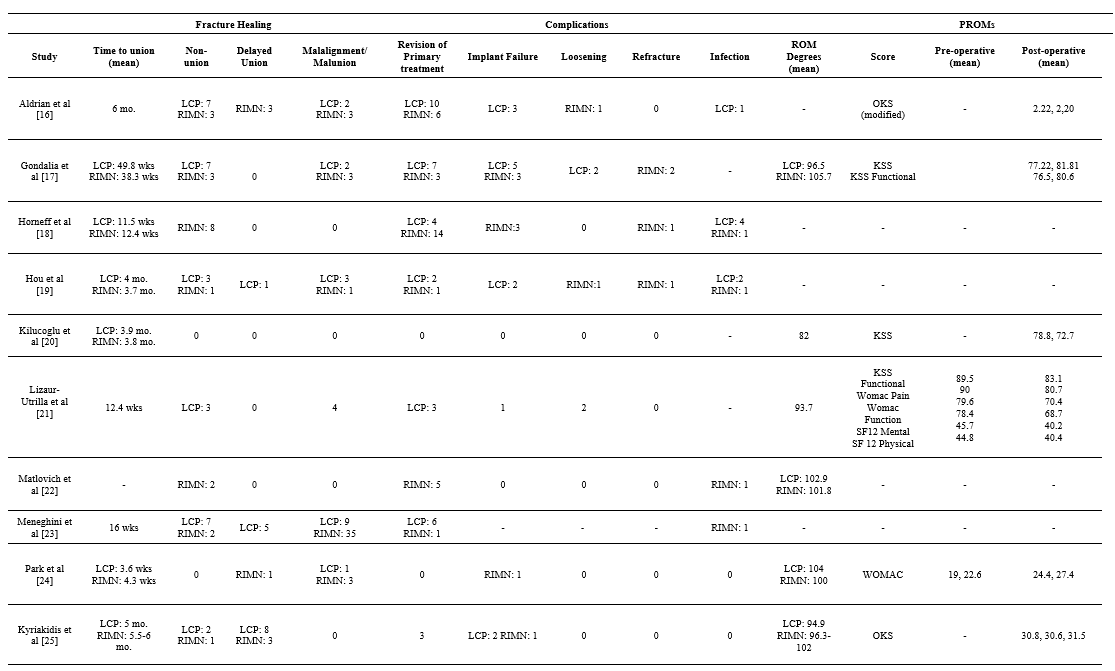
Across all included studies a total of 536 PPFs were reported. 318 LCPs and 218 RIMNs were performed. The characteristics of all 10 included studies (see Table 2),16–25 and further outcome measures (see Table 3)16–25 have been outlined. An overall summary of our results is provided in Table 4.

Table 2
Table 2.Overview of study characteristics.
Table 3
Table 3.Fracture healing, complications and patient reported outcome measures (PROMS).

ROM= range of motion, LCP= lockikng compression plate, RIMN= retrograde intramedullary nail, RA= Revision Arthroplasty, KSS= Knee Society Score, WOMAC= Western Ontario and McMaster Universities Osteoarthritis (WOMAC), OKS= Oxford Knee Score, SF12= Short form survey, HSS: Hospital for Special Surgery Score

Table 4.Incidence of complications for: RIMN, LCP, and overall cohort.
RIMN LCP Overall
Non-union 20/218 (9.2%) 29/301 (9.6%) 49/519 (9.4%)
Re-operation 33/218 (15.1%) 34/301(11.3%) 67/519 (12.9%)
Infection 4/188 (2.1%) 7/272 (2.6%) 11/460 (2.4%)
Mortality 6/100 (6.0%) 8/153 (5.2%) 14/253 (5.5%)

Non-union

49 (9.4%) cases of non-union were reported in a total of 519 patients across 10 studies comparing RIMN and LCP, with moderate heterogeneity (I2 =44%). Comparative analysis found no statistical significance (P = 0.63) (see Fig. 2).16–25

Figure 2
Figure 2.Forest Plot showing comparison of non-union rate between RIMN and LCP. No statistical significance found. M-H= Mantel–Haenszel, CI= Confidence Interval

Re-operation rates

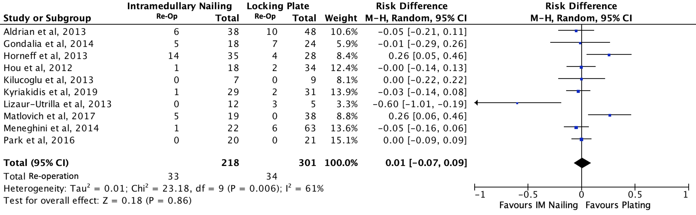
67 (12.9%) cases of re-operation were reported in a total of 519 patients across 10 studies comparing RIMN and LCP, with moderate heterogeneity (I2=61%). Comparative analysis found no statistical significance (P = 0.86) (see Fig. 3).16–25

Figure 3
Figure 3.Forest plot showing comparison in reoperation rate between RIMN and LCP. No statistical significance found. M-H = Mantel–Haenszel, CI= Confidence Interval

Infection

11 (2.4%) cases of infection were reported in a total of 460 patients across 7 studies comparing RIMN and LCP with low heterogeneity (I2=0%). Comparative analysis found no statistical significance (P = 0.80) (Fig. 4).16,18,19,22–25

Figure 4
Figure 4.Forest plot showing comparison of infection rate between RIMN and LCP. No statistical significance found. M-H = Mantel–Haenszel, CI= Confidence Interval

Mortality

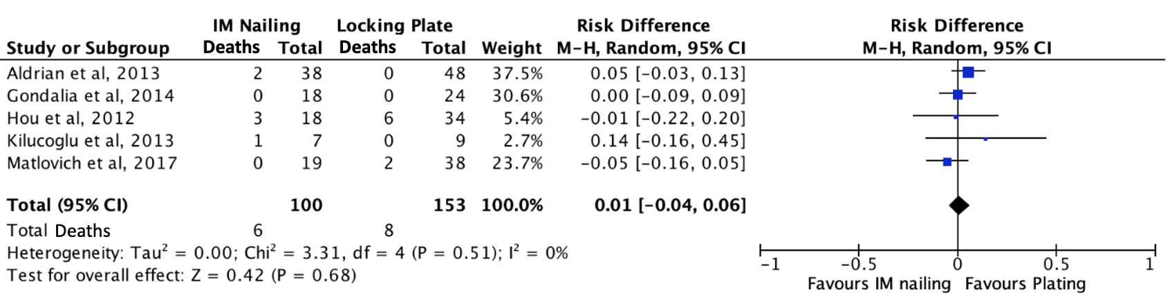
14 (5.5%) mortalities were reported in a total of 253 patients across 5 studies comparing RIMN and LCP, with low heterogeneity (I2 = 0%). Comparative analysis found no statistical significance (P = 0.68) (Fig. 5).16,17,19,20,22

Figure 5
Figure 5.Forest plot showing comparison in mortality rate between RIMN and LCP. No statistical significance found. M-H = Mantel–Haenszel, CI= Confidence Interval

Discussion

The aim of this meta-analysis is to provide an updated review of the literature for the treatment of distal femoral PPFs over the last decade, focusing on the rates of complications of RIMN and LCP. This study suggests that the rates of complications following RIMN and LCP performed for distal femoral PPF, are comparable. The overall incidence of complications reported were: non-union 9.4%, re-operation 12.9%, infection 2.4%, and mortality 5.5%. This meta-analysis found no statistically significant difference between RIMNs and LCPs in rates of non-union (9.2% vs 9.6%), re-operation (15.1% vs 11.3%), infection (2.1% vs 2.6%) and mortality (6.0% vs 5.2%) (respectively).

The results of this meta-analysis support the findings of recent meta-analyses by Magill et al. and Shin et al., both of which also reported no significant difference in rates of non-union and re-operation between the two surgical techniques.26,27 In addition, Shah et al. described no difference in rates of non-union, but did report a significantly lower re-operation rate following LCP compared to RIMN.28 Whilst this meta-analysis did find a lower rate of re-operation after LCP compared to RIMN, this comparison did not reach statistical significance. It is worth noting that two studies in this comparison did reach statistical significance for a lower re-operation rate post-LCP and that this comparative analysis showed moderate heterogeneity (I2 =61%), highlighting the need for further high-quality research into re-operation rates in particular before any substantial conclusions can be made.18,22

The aetiology of each complication was variably reported across studies. Only Hou et al. and Matlovich et al. reported on the aetiology of non-union following RIMN, with Hou et al. citing infection and distal screw loosening for their single case of non-union and Matlovich et al. citing one case of infection and one case of aseptic loosening.19,22 Hou et al. reported this case eventually requiring definitive management with above knee amputation but Matlovich et al. did not describe how the two cases of non-union were managed.19,22

Four studies reported on the aetiology of non-union following LCP.16,17,19,21 Aldrian et al. described three cases of plate breakage but did not disclose the aetiology of the other four cases of non-union following LCP. However, all seven of the cases reported by Aldrian et al. were managed by revision LCP and bone grafting.16 Lizaur-Utrilla et al. reported one case of non-union after LCP caused by loss of plate fixation which was managed with revision to a dynamic condylar screw. The other two cases of non-union described by Lizaur-Utrilla et al. were managed by dynamic condylar screw fixation for one patient and conservatively in the other, until a delayed knee arthrodesis was performed for loosening of the femoral component.21 Gondalia et al. reported all seven cases to be due to screw failure: five broken screws and two due to screw pull out.17 Hou et al. reported two cases of plate breakage and one case of infection progressing to a chronically infected non-union.19 The latter two studies did not describe the management of non-union following LCP.17,19

Neither Meneghini et al. nor Kyriakidis et al. reported the aetiology of non-union following RIMN or LCP, but both described the management.23,25 Following RIMN, Meneghini et al. reported one case being managed with revision to LCP and one with a bone stimulator. The seven cases of non-union after LCP cited in Meneghini et al. were managed as follows: four with revision LCP and bone grafting, two converted to distal femoral arthroplasty and one managed conservatively.23 All three cases of non-union reported in Kyriakidis et al. (one post-RIMN and two post-LCP) were managed with revision total knee arthroplasty.25

Infection was a rarely reported complication after both RIMN and LCP. Aldrian et al. reported one superficial wound infection post-LCP that was successfully treated with antibiotics.16 Horneff et al. reported five cases (one post-RIMN and four post-LCP) and Matlovich et al. one post-RIMN case of infection, that all required surgical management.18,22 Meneghini et al. and Hou et al. both reported a case of infection progressing to chronically infected non-unions, following RIMN and LCP respectively.19,23 As discussed above, Hou et al. also reported a surgical infection post-RIMN eventually necessitating amputation. Finally, Hou et al. described a surgical infection post-LCP that was managed successfully non-surgically.19 Although infection was a rare complication, it had significant consequences with seven of the eleven reported cases requiring surgical management and two cases progressed to chronically infected non-unions.18,19,22,23

Besides non-union and infection, hardware failure/irritation was the third common cause of re-operation. There is clearly significant overlap between cases of hardware failure and non-union, as the former often precedes the latter, as described in all seven cases of non-union post-LCP by Gondalia et al., both cases of non-union post-LCP by Hou et al. and the single case of non-union post-LCP described by Lizaur-Utrilla et al.17,19,21 Aldrian et al. also reported four cases of hardware failure preceding non-union: one case of distal screw loosening following RIMN and three cases of plate breakage after LCP.16 Following RIMN, Horneff et al. reported three cases of unspecified hardware failure and Matlovich et al. described three cases of distal screw irritation, all of which required surgical intervention.18,22 Horneff et al. also described one case of re-fracture after RIMN necessitating surgical re-intervention.18

Femoral PPFs are associated with significant mortality: reported as 17.7% at one year post-operatively by Shields et al. and comparable to the mortality rate after a hip fracture by Bhattacharyya et al.9,29 This meta-analysis found a lower mortality rate of 5.5% for distal femoral PPFs, although the follow up length was variable. Five studies reported on a total of fourteen mortalities. Aldrian et al. reported two mortalities from cardio-respiratory insufficiencies following RIMN.16 Hou et al. reported three mortalities after RIMN and six after LCP, all within six months of surgery and from non-fracture related causes.19 Kilucoglu et al. reported one mortality from pulmonary embolism, on post-operative day one following RIMN.20 Finally, Matlovich et al. reported two deaths post-LCP: one on post-operative day five due to congestive cardiac failure and the other on post-operative day one hundred and eleven following complications from a prolonged stay in the intensive care unit.22

There is limited and conflicting literature comparing native to periprosthetic distal femoral fractures. For example, Streubel et al. reported shorter survival in the PPF group (p = 0.005) whilst Kaufman et al. found no significant difference in mortality (p = 0.55).30,31 Bhattacharyya et al. reported femoral PPF to have a mortality rate comparable to that after a hip fracture, but they did not make any comparison to native distal femoral fractures.29 Further high-quality research is required to draw any substantial conclusions on this comparison.

Six studies reported some data on PROMs using a total of nine different scoring systems.16,17,20,21,24,25 There was no one scoring system used commonly across all six of these studies. Only two of these studies had pre-operative baseline scores to compare to.22,24 The heterogeneity of PROMs data collected across these six studies meant no meaningful conclusion could be made about PROMs after RIMN and LCP for distal femoral PPF.

Whilst this meta-analysis contributes to the growing literature that there is no significant difference in complication rates of RIMN and LCP for distal femoral PPFs; the management strategy is a multi-factorial decision, requiring consideration of fracture pattern, implant stability, co-morbidities of the patient, bone stock and design of the in-situ TKR.32 Non-operative management of supracondylar distal femoral PPFs yields less favourable results than operative management, particularly with regards to non-union and malunion.33–36 Culp et al. reported rates of non-union and malunion as 43% for non-operative management and 13% for operative management.37 However, non-operative management may be the only option for a patient with unacceptably high anaesthetic risk and may be considered in undisplaced epicondylar fractures, provided there is regular follow up to assess for fracture union and preservation of stability.32

For displaced fractures with stable implants, operative fixation is recommended. Historically this involved non-locking plates with dynamic condylar screws,38 which provided improved rates of union compared to non-operative management but often caused significant morbidity due to the extensive incisions and considerable soft tissue stripping.33,34,39 Minimally invasive modern surgical techniques such as RIMNs and LCPs have reduced these complications and there is increasing data to suggest that the outcomes and rates of complications between RIMN and LCP are equivocal.26,27 Deciding between RIMN and LCP often comes down to surgeon preference and experience, but one must consider a handful of contraindications to RIMN. The nail must facilitate safe insertion and recognised contraindications preventing this include: the presence of a patella baja, a closed box design of the in situ TKR and an ipsilateral hip arthroplasty with a long femoral stem which may prevent nail insertion or risk the creation of a stress riser.

Distal femoral PPFs with unstable femoral components require revision arthroplasty or distal femoral replacement (DFR), depending on the quality of bone stock, as outlined in our treatment recommendation flowchart below (Fig. 6). Two recent meta-analyses by Wadwa et al.40 and Ponugoti et al. have reported no significant difference between DFR compared to open reduction and internal fixation (ORIF: a combined cohort of patients who either underwent RIMN or LCP) in rates of complications, re-operation and mortality.40,41 Wadwa et al. also described comparable functional outcome scores between DFR and ORIF, but better post-operative range of motion in the ORIF group.40 These studies also highlight the advantage of early mobilisation and weight bearing post-DFR, reducing length of hospital stay and immediate sequela of immobility. This is supported by Tandon et al., who reported shorter length of hospital stay and lower complication rate (19% vs 40%) following DFR when compared to ORIF.42 These initial findings are encouraging but further research is necessary to illuminate the overall risks and outcomes of DFRs for distal femoral PPFs.

This meta-analysis is not without limitations. There is a scarcity of high-quality data that underpins the topic of distal femoral PPFs. Nine of the ten studies included in this meta-analysis, were retrospective cohort studies that lacked randomisation. There are limited case numbers involved in these studies and both aetiology and management of complications are variably reported across studies, further reducing quantity of data and the statistical power of the subsequent analyses. Surgeon preference greatly influences choice of fixation method and is a multi-factorial decision, influenced by experience with each technique, institutional trend, fracture pattern and bone quality. Fracture pattern was both variably reported and not standardised across the ten studies. Without this information, we were unable to elucidate what contribution fracture pattern had in deciding surgical technique and any potential influence on rates of complications.

Figure 6
Figure 6.Recommended treatment flowchart for distal femoral peri-prosthetic fractures.

Conclusion

This meta-analysis finds no significant difference in rates of non-union, re-operation, infection, or mortality between LCP and RIMN. At present, the framework provided, offers recommendations for the management of distal femoral PPFs. Further high-quality research is essential to evaluate both poorly reported outcomes, and emerging treatment modalities in the future.


Abbreviations

Arbeitsgemeinschaft für Osteosynthesefragen (AO)
Orthopaedic Trauma Association (OTA)
Distal femoral replacement (DFR)
Intramedullary (IM)
methodological items for non-randomised studies criteria (MINORS)
National Joint Registry (NJR)
NICE (National Institute for Health and Clinical Excellence)
Open reduction and internal fixation (ORIF)
Periprosthetic fracture (PPF)
Preferred Reporting Items for Systematic Reviews and Meta-Analyses (PRISMA)
Total knee replacements (TKRs)
United Kingdom (UK)
United States of America (USA)

Accepted: December 10, 2023 EDT

References

1.
National Joint Registry. 18th Annual Report of the UK NJR.; 2021. Accessed October 31, 2022. https://reports.njrcentre.org.uk/Portals/0/PDFdownloads/NJR%2018th%20Annual%20Report%202021.pdf
2.
Kurtz S, Ong K, Lau E, Mowat F, Halpern M. Projections of primary and revision hip and knee arthroplasty in the United States from 2005 to 2030. J Bone Joint Surg Am. 2007;89(4):780-785. doi:10.2106/00004623-200704000-00012
Google Scholar
3.
Short WH, Hootnick DR, Murray DG. Ipsilateral supracondylar femur fractures following knee arthroplasty. Clin Orthop Relat Res. 1981;158:111-116. doi:10.1097/00003086-198107000-00016
Google Scholar
4.
Pitta M, Esposito CI, Li Z, Lee Y yu, Wright TM, Padgett DE. Failure After Modern Total Knee Arthroplasty: A Prospective Study of 18,065 Knees. J Arthroplasty. 2018;33(2):407-414. doi:10.1016/j.arth.2017.09.041
Google ScholarPubMed CentralPubMed
5.
Yoo JD, Kim NK. Periprosthetic Fractures Following Total Knee Arthroplasty. Knee Surg Relat Res. 2015;27(1):1-9. doi:10.5792/ksrr.2015.27.1.1
Google ScholarPubMed CentralPubMed
6.
Singh JA, Jensen M, Lewallen D. Predictors of periprosthetic fracture after total knee replacement: an analysis of 21,723 cases. Acta Orthop. 2013;84(2):170-177. doi:10.3109/17453674.2013.788436
Google ScholarPubMed CentralPubMed
7.
Meek RMD, Norwood T, Smith R, Brenkel IJ, Howie CR. The risk of peri-prosthetic fracture after primary and revision total hip and knee replacement. Journal of Bone and Joint Surgery - Series B. 2011;93 B(1):96-101. doi:10.1302/0301-620x.93b1.25087
Google Scholar
8.
Märdian S, Schaser KD, Scheel F, Gruner J, Schwabe P. Quality of Life and Functional Outcome of Periprosthetic Fractures around the Knee Following Knee Arthroplasty. Acta Chir Orthop Traum Cech. 2015;82(2):113-118. doi:10.55095/achot2015/016
Google Scholar
9.
Shields E, Behrend C, Bair J, Cram P, Kates S. Mortality and Financial Burden of Periprosthetic Fractures of the Femur. Geriatr Orthop Surg Rehabil. 2014;5(4):147-153. doi:10.1177/2151458514542281
Google ScholarPubMed CentralPubMed
10.
Streubel PN, Gardner MJ, Morshed S, Collinge CA, Gallagher B, Ricci WM. Are extreme distal periprosthetic supracondylar fractures of the femur too distal to fix using a lateral locked plate? Journal of Bone and Joint Surgery - Series B. 2010;92(4):527-534. doi:10.1302/0301-620x.92b3.22996
Google Scholar
11.
Jennison T, Yarlagadda R. Mortality in patients sustaining a periprosthetic fracture following a previous extracapsular hip fracture fixation. Injury. 2018;49(3):702-704. doi:10.1016/j.injury.2018.01.001
Google Scholar
12.
Royal College of Physicians. Facing New Challenges – the NHFD Report on 2020 (January–December 2020).; 2021. Accessed October 31, 2022. https://www.rcplondon.ac.uk/file/33681/download
13.
Page MJ, McKenzie JE, Bossuyt PM, et al. The PRISMA 2020 statement: an updated guideline for reporting systematic reviews. Syst Rev. 2021;10(1):1-11. doi:10.1186/s13643-021-01626-4
Google ScholarPubMed CentralPubMed
14.
Higgins JPT, Thomas J, Chandler J, et al., eds. Cochrane Handbook for Systematic Reviews of Interventions version 6.3 (updated February 2022). In: Cochrane. ; 2022. Accessed October 31, 2022. https://training.cochrane.org/handbook/current
Google Scholar
15.
Slim K, Nini E, Forestier D, Kwiatkowski F, Panis Y, Chipponi J. Methodological index for non-randomized studies (MINORS): development and validation of a new instrument. ANZ J Surg. 2003;73(9):712-716. doi:10.1046/j.1445-2197.2003.02748.x
Google Scholar
16.
Aldrian S, Schuster R, Haas N, et al. Fixation of supracondylar femoral fractures following total knee arthroplasty: Is there any difference comparing angular stable plate fixation versus rigid interlocking nail fixation? Arch Orthop Trauma Surg. 2013;133(7):921-927. doi:10.1007/s00402-013-1730-9
Google Scholar
17.
Gondalia V, Choi DH, Lee SC, et al. Periprosthetic supracondylar femoral fractures following total knee arthroplasty: clinical comparison and related complications of the femur plate system and retrograde-inserted supracondylar nail. J Orthop Traumatol. 2014;15(3):201-207. doi:10.1007/s10195-014-0287-x
Google ScholarPubMed CentralPubMed
18.
Horneff JG III, Scolaro JA, Jafari SM, Mirza A, Parvizi J, Mehta S. Intramedullary nailing versus locked plate for treating supracondylar periprosthetic femur fractures. Orthopedics. 2013;36(5). doi:10.3928/01477447-20130426-16
Google Scholar
19.
Hou Z, Bowen TR, Irgit K, et al. Locked Plating of Periprosthetic Femur Fractures Above Total Knee Arthroplasty. J Orthop Trauma. 2012;26(7):427-432. doi:10.1097/bot.0b013e31822c050b
Google Scholar
20.
Kilucoglu OI, Akgul T, Saglam T, Yazicioglu O. Comparison of locked plating and intramedullary nailing for periprosthetic supracondylar femur fractures after knee arthroplasty. Acta Orthop Belg. 2013;79:417-421.
Google Scholar
21.
Lizaur-Utrilla A, Miralles-Muñoz FA, Sanz-Reig J. Functional outcome of total knee arthroplasty after periprosthetic distal femoral fracture. Journal of Arthroplasty. 2013;28(9):1585-1588. doi:10.1016/j.arth.2013.03.007
Google Scholar
22.
Matlovich NF, Lanting BA, Vasarhelyi EM, Naudie DD, McCalden RW, Howard JL. Outcomes of Surgical Management of Supracondylar Periprosthetic Femur Fractures. Journal of Arthroplasty. 2017;32(1):189-192. doi:10.1016/j.arth.2016.06.056
Google Scholar
23.
Meneghini RM, Keyes BJ, Reddy KK, Maar DC. Modern retrograde intramedullary nails versus periarticular locked plates for supracondylar femur fractures after total knee arthroplasty. Journal of Arthroplasty. 2014;29(7):1478-1481. doi:10.1016/j.arth.2014.01.025
Google Scholar
24.
Park J, Lee JH. Comparison of retrograde nailing and minimally invasive plating for treatment of periprosthetic supracondylar femur fractures (OTA 33-A) above total knee arthroplasty. Arch Orthop Trauma Surg. 2016;136(3):331-338. doi:10.1007/s00402-015-2374-8
Google Scholar
25.
Kyriakidis T, Kenanidis E, Akula MR, Zorman D, Tsiridis E. Locking plates versus retrograde intramedullary nails in the treatment of periprosthetic supracondylar knee fractures. A retrospective multicenter comparative study. Injury. 2019;50(10):1745-1749. doi:10.1016/j.injury.2019.04.019
Google Scholar
26.
Magill H, Ponugoti N, Selim A, Platt J. Locked compression plating versus retrograde intramedullary nailing in the treatment of periprosthetic supracondylar knee fractures: a systematic review and meta-analysis. J Orthop Surg Res. 2021;16(1):1-9. doi:10.1186/s13018-021-02222-x
Google ScholarPubMed CentralPubMed
27.
Shin YS, Kim HJ, Lee DH. Similar outcomes of locking compression plating and retrograde intramedullary nailing for periprosthetic supracondylar femoral fractures following total knee arthroplasty: a meta-analysis. Knee Surg Sports Traumatol Arthrosc. 2017;25(9):2921-2928. doi:10.1007/s00167-016-4050-0
Google Scholar
28.
Shah JK, Szukics P, Gianakos AL, Liporace FA, Yoon RS. Equivalent union rates between intramedullary nail and locked plate fixation for distal femur periprosthetic fractures – a systematic review. Injury. 2020;51(4):1062-1068. doi:10.1016/j.injury.2020.02.043
Google Scholar
29.
Bhattacharyya T, Chang D, Meigs JB, Estok DM II, Malchau H. Mortality after periprosthetic fracture of the femur. Journal of Bone and Joint Surgery. 2007;89A(12):2658-2662. doi:10.2106/jbjs.f.01538
Google Scholar
30.
Streubel PN, Ricci WM, Wong A, Gardner MJ. Mortality After Distal Femur Fractures in Elderly Patients. Clin Orthop Relat Res. 2011;469(4):1188-1196. doi:10.1007/s11999-010-1530-2
Google ScholarPubMed CentralPubMed
31.
Kaufman MW, Rascoe AS, Hii JL, et al. Comparable Outcomes Between Native and Periprosthetic Fractures of the Distal Femur. J Knee Surg. 2022;36(11):1111-1115. doi:10.1055/s-0042-1749604
Google Scholar
32.
Al-Jabri T, Ridha M, McCulloch RA, Jayadev C, Kayani B, Giannoudis PV. Periprosthetic distal femur fractures around total knee replacements: A comprehensive review. Injury. 2023;54(4):1030-1038. doi:10.1016/j.injury.2023.02.037
Google Scholar
33.
Brittain R, Howard P, Lawrence S, Stonadge J, Wilkinson M, Wilton T, et al. National Joint Registry. Natl Jt Regist. 2021;3:136-212. http://www.njrcentre.org.uk
Google Scholar
34.
Meek RMD, Norwood T, Smith R, Brenkel IJ, Howie CR. The risk of peri-prosthetic fracture after primary and revision total hip and knee replacement. J Bone Joint Surg Br. 2011;93(1):96-101. doi:10.1302/0301-620x.93b1.25087
Google Scholar
35.
Vestermark GL, Odum SM, Springer BD. Early femoral condyle insufficiency fractures after total knee arthroplasty: treatment with delayed surgery and femoral component revision. Arthroplast Today. 2018;4(2):249-253. doi:10.1016/j.artd.2018.02.013
Google ScholarPubMed CentralPubMed
36.
Alden KJ, Duncan WH, Trousdale RT, Pagnano MW, Haidukewych GJ. Intraoperative fracture during primary total knee arthroplasty. Clin Orthop Relat Res. 2010;468(1):90-95. doi:10.1007/s11999-009-0876-9
Google ScholarPubMed CentralPubMed
37.
Culp RW, Schmidt RG, Hanks G, Mak A, Esterhai JL Jr, Heppenstall RB. Supracondylar fracture of the femur following prosthetic knee arthroplasty. Clin Orthop Relat Res. 1987;222:212-222. doi:10.1097/00003086-198709000-00029
Google Scholar
38.
Wallace SS, Bechtold D, Sassoon A. Periprosthetic fractures of the distal femur after total knee arthroplasty: plate versus nail fixation. Orthop Traumatol Surg Res. 2017;103(2):257-262. doi:10.1016/j.otsr.2016.11.018
Google Scholar
39.
Singh JA, Jensen M, Lewallen D. Predictors of periprosthetic fracture after total knee replacement: an analysis of 21,723 cases. Acta Orthop. 2013;84(2):170-177. doi:10.3109/17453674.2013.788436
Google ScholarPubMed CentralPubMed
40.
Wadhwa H, Salazar BP, Goodnough LH, et al. Distal Femur Replacement Versus Open Reduction and Internal Fixation for Treatment of Periprosthetic Distal Femur Fractures: A Systematic Review and Meta-Analysis. J Orthop Trauma. 2022;36(1):1-6. doi:10.1097/bot.0000000000002141
Google Scholar
41.
Ponugoti N, Raghu A, Kosy JD, Magill H. A comparison of distal femoral replacement versus fixation in treating periprosthetic supracondylar femur fractures: a systematic review and meta-analysis. Arch Orthop Trauma Surg. 2022;143(6):3335-3345. doi:10.1007/s00402-022-04603-1
Google Scholar
42.
Tandon T, Tadros BJ, Avasthi A, Hill R, Rao M. Management of periprosthetic distal femur fractures using distal femoral arthroplasty and fixation - comparative study of outcomes and costs. J Clin Orthop Trauma. 2020;11(1):160-164. doi:10.1016/j.jcot.2019.05.015
Google ScholarPubMed CentralPubMed

Appendix

Appendix Abbreviations

Allograft Prosthesis Composite (APC)
Deep Vein Thrombosis (DVP)
Distal Femur (DF)
Distal Femur Endoprosthesis (DFR)
Full Weight-Bearing (FWB)
Global Modular Replacement System (GMRS)
Hospital for Special Surgery (HSS)
Intramedullary nail (IM nail)
Knee Society Score (KSS)
Legacy Constrained Condylar Knee (LCCK)
Legacy Knee Posterior Stabilized (LPS)
Less Invasive Stabilization System (LISS)
Minimally Invasive Plate Osteosynthesis (MIPO)
Non-Contact Bridging (NCB)
Open Reduction Internal Fixation (ORIF)
Oxford Knee Score (OKS)
Parkinson’s Disease (PD)
Pulmonary Embolism (PE)
Retrograde Inserted Supracondylar Nail (RISN)
Range of Motion (ROM)
Revision System (RSA)
Rheumatoid Arthritis (RA)
Standard Error (SE)
Standard Deviation (SD)
Supracondylar Nail (SCN)
Western Ontario and McMaster Universities Osteoarthritis (WOMAC)

Appendix Table 1.Table demonstrating raw data of studies meeting inclusion criteria, Part 1.
Study author and year of publication Study type Number of Patients (n) Number of fractures (n) Treatment Time to Surgery Implants used Patients average age per treatment Comorbidities Patient gender per treatment
Lizaur-Utrilla et al. 201321 Prospective matched cohort study comparing outcomes between 28 PPF patients with 28 age and gender matched primary TKRs for OA 56 28 1 Rorabeck type 1/AO-33B1 – compression screws 2.7 (Range 2–6) days ORIF, Synthes 75.6 (Range 56-86; SD 6.2) 26 females
2 males
5 Rorabeck type 2/AO33A1 – locking condylar plate LISS, Synthes
12 Rorabeck type 2/AO33A2 – retrograde IM nail T2, Stryker
7 Rorabeck type 2/AO33A3 – dynamic condylar screw Nolok 95, Depuy
3 Rorabeck type 3/AO33A3 – revision arthroplasty (hinged TKA) Endo-Model, Waldemar Link
11 Rorabeck type 2 –locked compression plate 67.8 (Range 45-81) 8 females
3 males
2 Rorabeck type 3 –revision arthroplasty 63 (Range 61-65) 1 female
1 male
3 Patella 3 Ortiguera and Berry type 2 – ORIF with tension band wiring 63 (Range 55-67) 3 females
2 Tibia 2 Felix type 2 – revision arthroplasty 77.5 (Range 70-85) 2 males
Kyriakidis et al. 201925 Multicentre Retrospective study between 2003 to 2014 60 60 Supracondylar fractures:
2 Rorabeck type I
48 Rorabeck Type 2
31 Fixed angle locking plates NA LISS Synthes 76.1 (SD 10.9) 8 males
23 females
15 Uncemented retrograde IM nail SCN Stryker 81.5 (SD 4.4) 3 males
12 females
14 Cemented retrograde IM nail 82.7 (SD 6.6) 4 males
10 females
Matlovich et al. 201722 Retrospective 2005 study to 2013 55 Rorabeck type 2s only 36 Locking plates NA NA 75.7 ± 11.2 9 males
27 females
19 IM nail 75.4 ± 9.2 3 males
16 females
Meneghini et al. 201423 Retrospective study 85 Rorabeck type 2s only 63 Locking plates NA NA 74 (Range 62-82) 51 females
12 males
22 IM nails 74 (Range 67-61) 14 females
8 males
Gondalia et al. 201417 Retrospective study between 2005 to 2009 42 Supracondylar fractures (17 33-A1, 8 33-A2, 17 33-A3) 24 Femur plate NA 17 LISS DF (Synthes), 7NCB plate (Zimmer) 69.9 40 females
2 males
18 Retrograde-inserted IM nails 18 RISN (4CIS)
Park et al. 201624 Retrospective between 2003 and 2014 41 Supracondylar fractures 21 Plate (MIPO) 3.5 (range 1-14) days Distal femoral locking plate (Zimmer) 75.0 (range 60-82) 1 male
20 females
20 IM nail Supracondylar IM nail (Smith and Nephews) 73.9 (Range 63-83) 17 females
3 males
Aldrian et al. 201316 Retrospective between 1996 and 2010 86 32 Su Type 1
54 Su Type 2
48 Lateral plate fixation (LISS) LISS, Synthes 75.6 (Range 59-89) 62 females
24 males
38 Rigid interlocking nail device SCN, Stryker
Kilucoglu et al. 201320 Retrospective 16 13 Neer Type 2
1 Neer Type 3
2 Neer Type 4
9 locking plates NA LISS, Synthes 76.7 (77± 4,6) 13 females
2 males
7 retrograde IM nail NA TriGen Knee Nail, Smith & Nephew 68.6 (68±5,6)
Horneff et al. 201318 Retrospective between 2001 and 2009 63 63 Rorabeck type 2 28 Locking Plate NA NA 68.3 Average BMI – 28.4 kg/m2 10 Males
18 Females
35 IM nail NA NA 69.5 Average BMI – 31.3 kg/m2 9 Males
26 Females
Hou et al. 201219 Retrospective between 2004 and 2009 52 OTA classification
(39 33A fractures
3 32B fractures
10 32A type fractures)
34 Locking plate 2.4 days NA Mean 75 23 females
11 males
18 Retrograde IM nail 1.7 days Mean 77 12 females
6 males
Appendix Table 2.Table demonstrating raw data of studies meeting inclusion criteria, Part 2.
Study author and year of publication Classification Follow-up Postoperative rehabilitation regimen Range of motion postoperatively Infection Average time to union Non-union Delayed union Malalignment/malunion
Lizaur-Utrilla et al. 201321 Rorabeck and Taylor and AO classification 6.7 (Range 5–9) years Day 1 – passive knee motion;
Day 3 postop – active motion; FWB when bridging callus seen on radiographs.
93.7 (Range 75-100; SD 11.2) degrees 1 superficial wound infection – debridement and antibiotics 12.4 (Range 8–20) weeks Plate - 3 0 3 patients with >5 degrees varus (1 retrograde nail, 2 dynamic condylar screws)
1 shortening (retrograde nail)
IM nail - 0
Kyriakidis et al. 201925 Rorabeck type 1 and 2 Plate - 22.3 (SD 12.1) months Partial and gradual weight bearing as tolerated Plate - 94.9 (SD 21.4) degrees 0 Plate - 5 (Range 4-9) months Plate - 2 (SD 6.5) Plate - 8 (SD 25.8) 0
Uncemented IM nail - 17.4 (SD 11.2) months Uncemented IM nail - 96.3 (SD 7.9) degrees Uncemented IM Nail - 6 (Range 5-6) months Uncemented IM Nail - 1 (SD 6.6) Uncemented IM Nail - 2 (SD 13.3) 0
Uncemented IM ail - 17.2 (SD 8.05) months Cemented IM nail - 102 (SD 14.6) degrees Cemented IM nail - 5.5 (Range 4-6) months Cemented IM nail - 0 Cemented IM nail - 1 (SD 7.1) 0
Matlovich et al. 201722 Rorabeck Plate - 15.6 months Fully weightbearing by 15 weeks Plate - 2.5±6.7 to 102.9±11.0 Plate - 0 ***ONLY GRAPHS PRESENT NO NUMERICAL DATA** Plate - 0 Plate - 0 0
Nail - 13.9 months IM nail - 0.3±1.2 to 101.8±16.7 IM nail - 1 infection leading to non-union IM nail - 2 (1 aseptic, 1 infected) IM nail - 0
Meneghini et al. 201423 Rorabeck and AO/OTA classification 31.8 (Range 6-176) weeks Plate - FWB 11.7 weeks NA Plate - 0 16 weeks Plate - 7 Plate - 5 Plate - 9
IM nail – FWB 9.1 weeks IM nail - 1 infection leading to non-union IM nail - 2 IM nail - 0 IM nail - 35
Gondalia et al. 201417 AO classification 34.6 (range 17.2-76.7) months Plate - Immobilisation for 6.1 weeks, FWB following clinical union Plate - 96.5 (extension to flexion 1.5-98) degrees N/a Plate - 49.8 (Range 13.3-191) weeks Plate - 5 with broken screws, 2 with medial translation (screw pull out) Plate - 0 Plate - 2 medial translation
IM nail - Immobilisation for 4.2 weeks, FWB following clinical union IM nail - 105.7 (extension to flexion 2 to 107.7) degrees IM nail - 38.3 (range 16-121.3) weeks IM nail - 3 non union with varus deformity (broken screws) IM nail – 0 IM nail - 3 varus deformity
Park et al. 201624 AO/OTA classification, Su classification, Lewis and Rorabeck classification Plate - 30.5 (Range 24-40) months ROM exercises immediately, partial weight bear 4 weeks, full weight bear 12 weeks if evidence of union Plate - 104 (range 60-140) degrees 0 Plate - 3.6 (range 3-5) months Plate - 0 Plate - 0 Plate - 1 malalignment
IM nail - 52.4 (Range 36-66) months IM nail - 100 (range 60-130) degrees IM nail - 4.3 (range 3-12) months IM nail - 0 IM nail - 1 with nail breakage managed conservatively IM nail - 3 malalignments, 1 malunion following nail breakage
Aldrian et al. 201316 Su classification At least 12 months Gradual weight bearing from day 2-3 until 6-8 weeks Plate – 34 excellent/satisfactory; 14 poor Plate – 1 superficial treated with antibiotics Average - Within 6 months in 74 patients Plate – 7 Plate - 0 Plate – 2 incorrect reduction, 3 secondary loss of reduction
IM nail – 22 excellent/satisfactory; 14 poor IM nail – 0 IM nail – 3 IM nail -3 (radiographic union and pain free by 12 months) IM nail – 3 incorrect reduction, 2 secondary loss of reduction
Kilucoglu et al. 201320 Neer classification 1.75 (2±0.7) years Range of motion exercises and toe-touch gait on day 2; FWB once union observed on radiograph 82 (Range 70-90) degrees N/a Plate - 3.9 (Range 3-6) months Plate - 0 Plate - 0 Plate - 0
5.57 (5±4.9) years IM nail – 3.8 (Range 3-5) months IM nail - 0 IM nail - 0 IM nail - 0
Horneff et al. 201318 Rorabeck Minimum 36 month Partial weight bearing immediately, FWB based on clinical data NA Plates – 4 Plate – 11.5 weeks Plate – 0 Plate – 0 Plate – 0
IM nail – 1 IM nail – 12.4 weeks IM nail – 8 IM nail – 0 IM nail – 0
Hou et al. 201219 OTA classification 29 ± 2.2 (Range 12-60) months Stability and surgeon preference NA Plate – 2 infections (1 resolved, 1 chronic infection) Plate – 4.0 ± 0.27 months Plate – 3 Plate – 1 Plate – 3 (2 inadequate reduction, 1 plate bending)
IM nail – 1 infection with distal interlock screw loosening requiring amputation IM nail – 3.7 ± 0.3 months IM nail – 1 IM nail – 0 IM nail – 2
Appendix Table 3.Table demonstrating raw data of studies meeting inclusion criteria, Part 3.
Study author and year of publication Implant failure Loosening Re-fracture Revision of primary treatment Blood loss Blood transfusion Other complications PROMs Overall complications (n)
Lizaur-Utrilla et al. 201321 1 2 0 1 AO33A1 4 months post injury (revised to a dynamic condylar screw due to loss of plate fixation)
1 AO33A2 3 years post injury (knee arthrodesis due to aseptic loosening)
1 AO33A3 7 months post injury (revision dynamic condylar screw followed by revision arthroplasty due to aseptic loosening)
N/A N/A 4 slight or occasional pain; 2 moderate or continuous pain KSS – preop 89.5 (Range 75-100; SD 6.0), postop 83.1 (Range 61–97; SD9.0)
Functional score – preop 90.0 (Range 80-100; SD 7.9), postop 80.7 (Range 50–100; SD 10.7)
WOMAC pain – preop 79.6 (Range 60-100; SD 12.7), postop 70.4 (Range 40–90; SD 11.4)
WOMAC function – preop 78.4 (Range 50-100; SD 10.6); postop 68.7 (Range 43-90; SD 9.7)
SF12 mental – preop 45.7 (Range 28-50; SD 9.9), postop 40.2 (Range 28–42; SD 9.4)
SF12 physical – preop 44.8 (Range 38-50; SD 7.7), postop 40.4 (Range 48–44;SD 7.3)
7
Kyriakidis et al. 201925 0 0 0 3 revision TKAs for non-union NA NA Plate - 2 PE (SD 6.5) OKS postop 30.8 (SD 8.1) 12
Uncemented IM nail - 1 DVT (SD 6.6) OKS postop 30.6 (SD 10.23) 4
Cemented IM nail – 0 OKS postop 31.5 (SD 8.6) 1
Matlovich et al. 201722 0 0 0 Plate - 0 NA 2 patients Plate - 22.2% Chronic postoperative pain NA 0
IM nail - 5 cases – 2 for non-union, 3 for hardware irritation 0 IM nail - 22.7% chronic postoperative pain
17.6% subjective instability
5
Meneghini et al. 201423 NA NA NA 4 revisions – locked plate + bone grafting; 2 distal femoral arthroplasty NA NA NA NA 12
1 IM nail revised to locked plate 2
Gondalia et al. 201417 Plate - 5 (broken screws) Plate - 2 (screw pull out) Plate - 0 7 NA NA Plate - 0 Plate - KSS 77.22 ± 12.7; KSS functional 76.5 ± 14.5 7
IM nail - 3 (broken distal screws) IM nail – 0 IM nail - 2 cases with fracture at level of proximal nail tip 5 IM nail - 0 IM nail - KSS 81.81 ± 8.7;
KSS functional 80.6 ± 10.9
5
Park et al. 201624 0 0 0 0 NA NA Plate - 2 skin irritations due to long distal screws Plate - WOMAC score preop 19 (range 10-42), post op 24.4 (range 11-52) 3
IM nail - 1 nail breakage IM nail - 0 IM nail - 0 IM nail - 0 IM nail - 0 IM nail - WOMAC score preop 22.6 (range 15-39), post op 27.4 (range 20-50) 4
Aldrian et al. 201316 Plate – 3 plate breakage Plate – 0 Plate - 0 Plate 10 – 7 revision surgery with bone grafting NA NA Plate – 2 respiratory failure, 2 pneumonia Plate – OKS (modified) – 2.22 Plate – 21
IM nail – 0 IM nail – 1 distal interlocking screw loosening (revision) IM nail – 0 IM nail 6 – 1 patient with screw loosening NA NA IM nail – 2 pneumonia, 2 deaths due to cardiorespiratory failure IM nail - OKS (modified) 2.20 IM nail - 13
Kilucoglu et al. 201320 Plate – 0 Plate – 0 Plate – 0 Plate – 0 NA NA Plate - 0 KSS – 78.8 (80±3.9) Plate - 0
IM nail – 0 IM nail – 0 IM nail – 0 IM nail – 0 NA NA IM nail – 1 Death (PE) KSS – 72.7 (74±4.16) IM nail - 1
Horneff et al. 201318 Plate – 0 Plate – 0 Plate – 0 Plate – 4 (all due to infection) NA Plate – 8 Plate – 0 NA Plate - 4
IM nail – 3 IM nail – 0 IM nail – 1 IM nail – 14 (1 infection, 8 non-unions, 1 refracture, 3 hardware breakage, 1 revision arthroplasty) NA IM nail - 15 IM nail – 0 IM nail - 14
Hou et al. 201219 Plate – 2 plate breakage Plate – 0 Plate – 0 Revision – 2 for broken plates Plate – 177.5 ± 23.4 mL 6 Deaths in 6 months Plate – 7
IM nail – 0 1 infection with interlock screw loosening IM nail – 1 refracture following a fall IM nail – 1 refracture requiring implant removal and anterograde IM nail fixation IM nail – 182 ± 31.6 mL 3 Deaths in 6 months IM nail – 3