


Introduction
Osteopetrosis, also known as marble stone disease, is a rare inherited disorder characterised by excessively dense bone. The term ‘osteopetrosis’ originates from the Greek words ‘osteo,’ meaning bone, and ‘petros,’ meaning stone. Albers-Schönberg first described this condition in 1904.1 The precise incidence of osteopetrosis is not well established but it is estimated to range from approximately 1 in 20,000 to 1 in 200,000 cases, depending on the type of inheritance, whether autosomal recessive or autosomal dominant.2 Osteopetrosis is caused by either a developmental or functional failure of osteoclasts, both of which result in impaired bone turnover.3 This leads to obliteration of the medullary canal, and the presence of calcified cartilage and brittle bone is one of its characteristic features.4 Also, due to obliteration of the medullary canal, haematologic abnormalities such as anaemia and thrombocytopenia may occur in these patients.5–7
While the direct relationship between osteopetrosis and osteoarthritis (OA) is still not fully understood, it has been observed that OA is common in patients with osteopetrosis.8,9 The dense subchondral bone may alter the joint biomechanics and exert pressure on the articular cartilage, potentially leading to OA.6 In patients with osteopetrosis suffering from advanced OA, total knee arthroplasty (TKA) may be considered as a treatment option.10 However, performing TKA in these cases poses significant challenges due to the high bone density, often requiring an extended surgical time and the use of multiple saw blades and drill bits for cutting.6,11 These challenges not only exhaust the surgeon but may also lead to implant malpositioning and inappropriate bony surfaces for cement interdigitation.10 The use of external alignment guides, specialised implants with shorter stems, and navigation systems by surgeons has resulted in successful short- and medium-term results, as presented in various case reports.12,13
Nowadays, advanced robotic technology has been introduced in TKA. Among the available robotic systems, robotic arm-assisted TKA (RA-TKA) includes a navigation system and a robotic arm. RA-TKA offers several advantages, such as no requirement of intramedullary guides and the use of thicker saw blades, resulting in accurate bony cutting and enhanced stability. Additionally, the robotic arm assists in relieving the physical burden of manual cutting. In this report, we present a case of a patient with osteopetrosis who underwent RA-TKA.
Case report
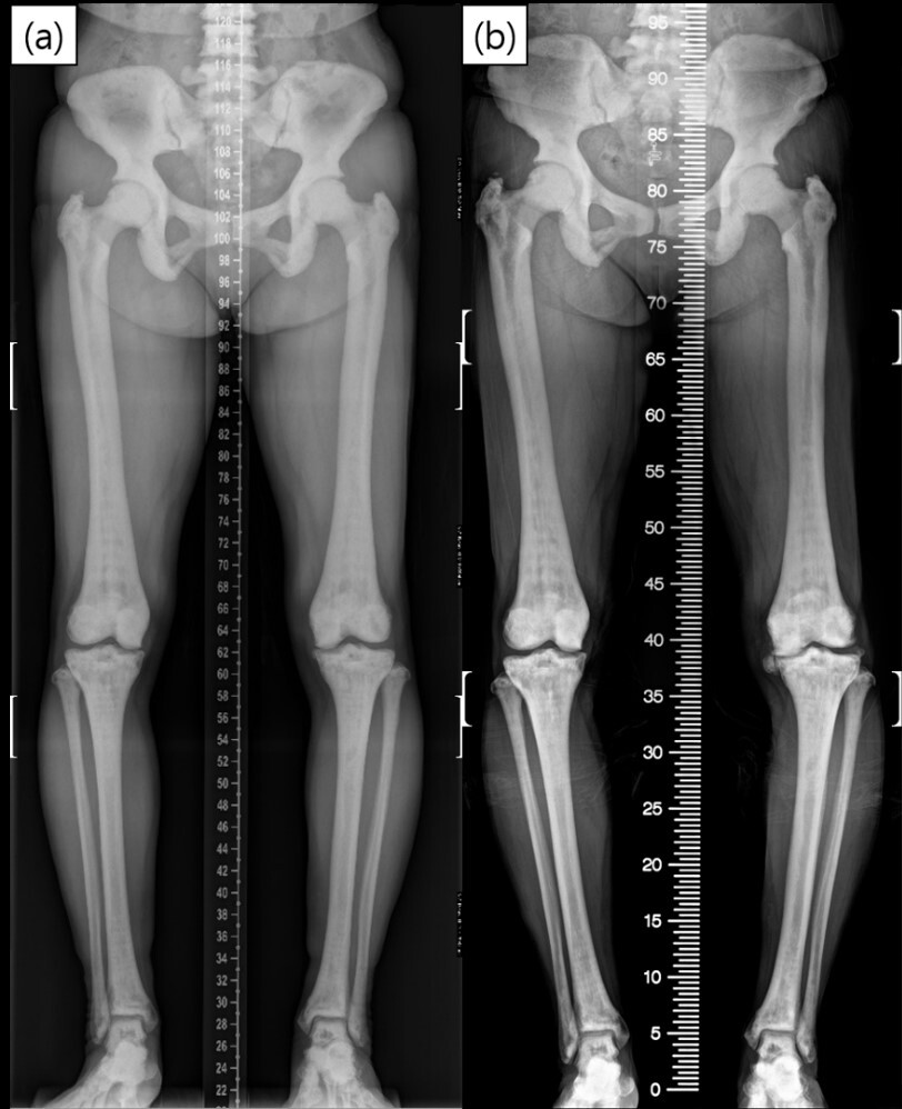
A 68-year-old woman presented with persistent medial left knee pain since the last 10 years. A decade ago, the patient had visited the outpatient clinic at the authors’ institute seeking treatment for her left knee pain. At that time, knee radiographs showed mild knee OA and osteopetrosis (Figure 1). Initially, the patient received pain medication but was subsequently lost to follow-up.
Ten years later, the patient returned with a complaint of worsening left knee pain, which she rated as a score 6 on the Numerical Rating Pain Scale, while her right knee had a pain rating of 0. The pain was particularly pronounced in the medial compartment of the knee, and it was aggravated during activities, such as walking and climbing stairs. The patient also reported the development of a bow leg deformity. Physical examination revealed tenderness along the medial joint line of the left knee. The knee flexion angles were measured at 135 degrees in both knees, without any contracture, effusion, or sensation of heat in the knee joint. Despite receiving medication and injections at local hospitals, there was no improvement in her condition, which led to her admission for RA-TKA.
Apart from knee pain, the patient did not exhibit any other clinical symptoms, including those related to haematologic abnormalities, such as dizziness or bleeding tendencies. Radiographic findings were consistent with osteopetrosis, characterised by extensive cortical thickening and increased bone density in all bones. The knee X-ray revealed severe OA of the left knee joint with subchondral bone attrition in the left medial tibial plateau. Dual-energy X-ray absorptiometry (DEXA) bone densitometry showed significantly increased bone density, with T-scores of 13.6 at the L1-4 spine, 13.3 at the femoral neck, and 14.6 at the total femur.
Laboratory tests upon admission showed a decreased platelet count of 75,000/uL. Following consultation with a haematology specialist, the patient underwent platelet transfusion, resulting in an increased platelet count of 128,000/uL. No other laboratory tests showed abnormal values.
Preoperative planning was conducted using a 0.6 mm-thick computed tomography (CT) scan to determine the size and position of the implants (Figure 2).

The RA-TKA (MAKO Surgical Corporation, Florida, USA) procedure involved the use of a tourniquet, a midpatellar vertical skin incision, and arthrotomy via a midvastus approach. Soft tissue preparation was performed without any difficulty. For fixation of femoral and tibial arrays, two bone pins with a diameter of 4 mm and length of 140 mm were used for each bone. During the bone pin insertion, one femoral pin was distorted due to the hard bone, necessitating its replacement. Although the pins were advanced only up to 5mm, the fixation remained firm. Then, patient landmark registration was performed. Subsequently, osteophytes were excised, and the navigation system was used to assess the flexion and extension gap by applying valgus and varus stress, as well as through extension and 90-degree flexion. Intraoperative adjustments were made using the robotic system based on the preplanned data, and the femur and tibia were resected using a 2 mm-thick saw blade attached to a robotic arm. The patella was preserved as the articular cartilage remained intact.
During the trial implant insertion, even a minor bony protrusion prevented a perfect fit of the femoral implant, and mallet impaction was ineffective due to hardness of the bone. To address this issue, additional bone trimming was performed using a manual saw to create a flat surface (Figure 3). After confirming the appropriate gap and range of motion, attempts were made to prepare the femoral peg and tibial keel. However, the hard bone obstructed penetration of the peg drill, reamer, and keel punch. As a solution, locations for the peg and keel were manually marked on the bone cut surface, and bone preparation was performed using a manual burr and reciprocating saw. After preparing the femoral peg and tibial keel, the final implant (Triathlon, Stryker, USA) was fixed using cement. A tourniquet was applied for 1 hour and 50 minutes, with an overall surgical time of 2 hours and 25 minutes. Only two 2 mm-thick saw blades were used.

The postoperative protocol was the same as that for the other patients. After TKA, the patient was encouraged to walk with a knee immobiliser and a walking aid while she was in the ward. Continuous passive motion exercises were initiated on the day of surgery. At the 1-month postoperative visit, no radiological abnormalities were found and laboratory tests revealed a platelet count of 80,000/uL. By the 3-month postoperative visit, her pain had resolved, except for minor discomfort in the popliteal area. X-rays showed good alignment of the implant, with no observed complications associated with surgery (Figure 4).

Discussion
To date, there are limited reports on TKA in patients with osteopetrosis, mostly consisting of case reports, which reflects the rarity of this condition.6,8,10,11,14 This study presents the first reported case of performing RA-TKA in a patient with osteopetrosis, and the clinical outcomes at the 3-month follow-up were satisfactory, with no observed complications.
Performing TKA in patients with osteopetrosis poses unique challenges due to the dense and hard bone structure, as well as obliteration of the intramedullary canal. These factors make it difficult to achieve precise bony cuts and proper positioning of intramedullary guides. Conventional manual saws, typically 1.25 mm in thickness, may bend, deviate from the intended cut line, result in blade breakage, or even cause kickback, potentially damaging surrounding soft tissues or injuring the surgeon.10 Previous studies using manual sawing have reported the use of 4 to 8 saw blades.10,11 Moreover, manually cutting a hard bone and prolonged surgical time can be physically demanding for the surgeon. In a previous case report, the required surgical time was 7.5 hours.6
Previous studies have attempted to circumvent these issues by using external femoral alignment guides, navigation systems, and patient-specific instruments in arthroplasty to overcome the challenges related to osteopetrosis.10,13,14 The use of RA-TKA can offer all these advantages and address the issues in patients with osteopetrosis. The robotic arm used in RA-TKA employs thicker saw blades (2 mm) than conventional manual saw blades, providing stability and heat endurance, particularly when cutting through harder bones, and may require fewer blade replacements. The resection boundary set by the navigation system can prevent damage caused by kickback. Additionally, indirect cutting via the robotic arm may reduce the physical burden, resulting in reduced time and effort required for the cutting procedure.
However, there were some drawbacks of using RA-TKA. One concern was the placement of femoral and tibial bone pins, which are typically fixed bicortically. In this case, the exceptionally hard bone made pin insertion challenging, resulting in a distorted femoral pin due to the convex surface of the femoral shaft. The insertion depth was limited to 5 mm for all pins, although the fixation remained stable. Additionally, there were instances where the robotic system displayed completion of bone resection but small irregular surfaces persisted, resulting from minor resection errors in RA-TKA.15 In patients without osteopetrosis, mallet impaction of the implant can effectively manage uneven bony surfaces seen in TKA, as the cut surfaces usually consist of cancellous bone. However, in this patient with osteopetrosis, even a small protruding surface did not allow for satisfactory implant seating, despite mallet impaction. To address these issues, manual trimming was performed with caution to avoid the risk of kickback. Furthermore, the RA-TKA system does not assist in femoral peg and tibial keel preparation, which requires manual preparation. However, attempts to use a peg drill, reamer, and keel punch to penetrate the bone were unsuccessful, necessitating the use of alternative instruments, such as a burr and reciprocating saw. Since the robotic arm was not utilised in this procedure, significant manual effort was required.
In this case, the patient presented with thrombocytopenia, while other haematologic test results (e.g., haemoglobin, white blood cell count, and coagulation panel) were within their normal range. Haematologic abnormalities, including thrombocytopenia, have been previously reported in patients with osteopetrosis due to bone marrow abnormalities.16 Therefore, when performing TKA in these patients, the surgeon should be aware of potential blood loss related to the haematological abnormality and the possibility of increased surgical time5,7,17
Conclusion
Although performing TKA in patients with osteopetrosis is challenging, the use of RA-TKA in this case presentation demonstrated satisfactory short-term outcomes. RA-TKA offers advantages, such as precise bony cutting, elimination of the need for intramedullary guides, reduced saw disposal, and decreased physical burden and surgical time. Surgeons should also consider haematologic abnormalities, such as thrombocytopenia, in patients with osteopetrosis during surgery.
Acknowledgements
None.
Conflict of interest
The authors have no competing interests to declare.
Funding
The authors did not receive support from any organization for the submitted work.
Ethical approval
This study was approved by the Institutional Review Board of Seoul National University Hospital (IRB No. 2308-081-1458)
Informed consent
Written informed consent for the treatment was obtained from the patient. A separate consent was taken for publishing this case.
Authors’ contributions
S.E.K. contributed to data curation, progressing of research, writing and editing of the manuscript. H-S.H performed the surgery, contributed to data analysis and editing of the manuscript.