
BACKGROUND
Osteogenesis imperfecta (OI) is the most common congenital bone disease associated with bone structure fragility and abnormal long bone shaft curvature.1 The condition results from mutations in genes that encode type I collagen, crucial for sustaining bone strength.2,3 The severity of OI differs considerably, from mild manifestations characterized by infrequent fractures to severe cases marked by frequent fractures and skeletal deformities.4 Current management employs a multidisciplinary approach, incorporating antiresorptive medications, physiotherapy, occupational therapy, and orthopedic surgery to improve bone stability and overt deformities.5
Several fixation techniques are utilized based on the patient’s age, bone quality, and the complexity of the deformity, but intramedullary fixation is the preferred method for surgical interventions in severe cases of long bone deformities and fractures.6 Nowadays, the optimal fixing method is telescopic intramedullary nailing. The advancement of telescoping intramedullary rods was motivated by the necessity to minimize the frequency of surgeries in pediatric patients with OI. Non-elongating devices often necessitate replacement as the child develops, resulting in numerous hospitalizations and heightened surgical risk.7
It was a pivotal moment when François Fassier and Pierre Duval invented a novel telescopic rod with two screw tips, since it mitigated the need for knee or ankle arthrotomy and minimized soft tissue damage.8 This system achieved significant adoption and is recognized as the standard of care in numerous centers.9
This prospective study aimed to evaluate the clinical and radiological outcomes of Fassier-Duval telescoping nails in the treatment of lower-limb deformities and fractures in children with osteogenesis imperfecta at a University hospital.
PATIENTS AND METHODS
This prospective study involved 31 children with lower-extremity deformities or fractures (40 bones) scheduled for treatment in the orthopedic department at Sohag University Hospitals, using the Fassier-Duval (FD) telescoping nail, from August 2021 to August 2023. The ethical committee in the faculty of medicine at Sohag University has approved this study. The study was registered at ClinicalTrials.gov (ID: NCT05251961). Parents of participating patients signed an informed consent. Only children with a minimum 2-year follow-up were included. Five children had a history of previous surgeries.
The inclusion criteria were children with osteogenesis imperfecta (OI), aged under 18, having surgical treatment for deformities or fractures of the femur, tibia, or both, and having at least two years of follow-up post-surgery. Rodding was conducted on children with a history of multiple extremity fractures, pain, bowing > 50 degrees, nonunion, or malunion. All patients presented with functional limitations associated with their deformities or fractures. The exclusion criteria were adult deformities, short bone deformities, osteopetrosis, and patients with very narrow medullary canals that cannot be fixed intramedullary.
Preoperative Preparation
A full workup was performed, including clinical evaluation and patient counselling, joint function evaluation, and orthogonal images for preoperative planning. OI severity and classification were assessed using the Sillence criteria.10 The Pediatric Outcome Data Collection Instrument (PODCI)11 and Brief Assessment of Mobility Function (BAMF) scores12 served as the basis for the clinical evaluation. The child’s performance during typical activities, such as sitting, walking, and other tasks, was evaluated. Parents fill out the PODCI pediatric form (ages 2 to 10). The findings of each scale are standardized to a range of 0-100 points, with 0 denoting the lowest score and 100 the highest. Coronal and sagittal CORA angles were evaluated in preoperative imaging studies.13
Operative Techniques
The original FD Rod was used on all children. In these 31 children, 8 patients had more than one bone affected and were operated on in a staged manner with an 8-week interval. Surgery was done under general anesthesia, and osteotomy was done using either the open technique or the percutaneous technique according to the degree of deformity. Operations were done either in the supine or lateral position on the radiolucent table.
Femoral Nailing
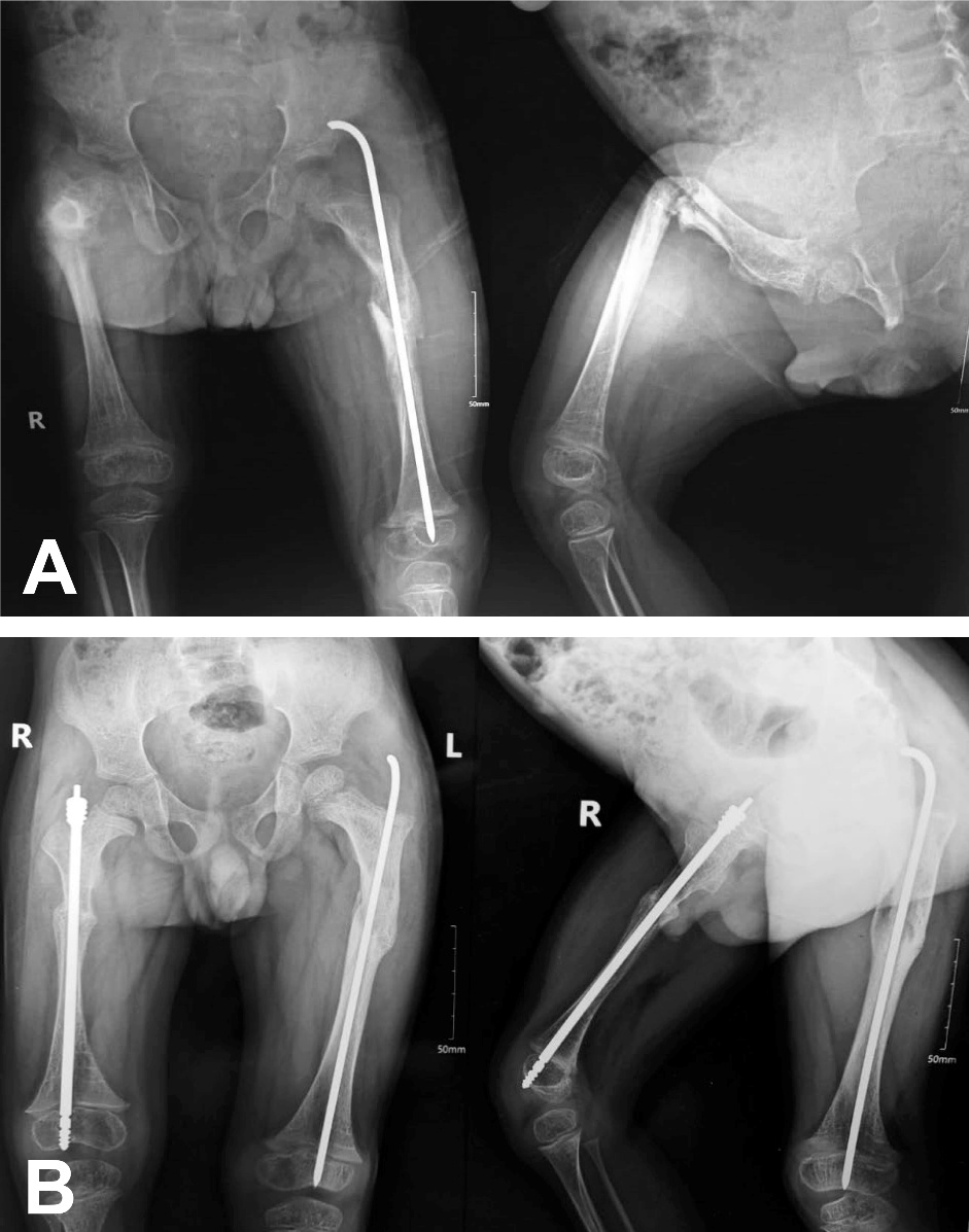
The surgical site received standard preparation using antiseptic draping. The abnormal segment was initially identified through C-arm fluoroscopy, and the number of osteotomies was established according to the severity of the deformity. The open osteotomy technique was used in most cases. The femur is exposed subperiosteally through a posterolateral approach. The proximal fragment is reamed from the first osteotomy to the greater trochanter. Insertion of a suitable-sized male rod was done till the distal physeal growth plate. The threaded part of the male rod is screwed down into the distal epiphysis. The length of the female rod was measured directly on the bone using a sterile ruler under C-arm guidance. The female rod is introduced (Figure 1).
The minimally invasive technique was used in 5 femurs. A small incision measuring 3–5 cm was made slightly lateral to the apex of the greater trochanter. A guide wire was inserted through the femoral medullary canal to the deformity site, followed by a small osteotomy incision at this location to correct the deformity. The guide wire was advanced to the center of the distal femoral epiphysis, thereby restoring the normal anatomical structure of the femur. A suitable length and diameter of the FD nail was chosen. In C-arm fluoroscopy, the male and female nails were advanced, ensuring that the male nail threads extend beyond the distal epiphyseal plate and were fixed 3–4 mm subchondrally at the center of the distal epiphysis. The female nail was secure at the greater trochanter. A hip Spica cast was applied for all patients for 6 weeks to control rotation.
_pre-operative_x-ray_showing_severe_right_femoral_deformity_in_ap_and_lateral_views__whi.jpeg)
Tibial Nailing
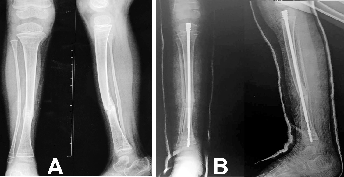
The first step involves preparing the entry point through the tibial apophysis. The patellar tendon is bisected from the lower pole of the patella to the tibial tuberosity, using a midline approach. All osteotomies were performed with an open technique. The male component of the FD telescopic nail is fitted first, followed by the female component, which is cut to size intraoperatively with dedicated instruments, all in an antegrade manner (Figure 2). Postoperatively, an above-knee cast was applied for 6 weeks. Once the cast is removed, rehabilitation begins, allowing full weight on the leg by the sixth postoperative week.
_pre-operative_x-ray_showing_mal-united_fracture_of_the_tibia_and_fibula_resulting_in_va.jpeg)
Postoperative protocol
Immediate postoperative outcomes included estimated blood loss, necessity for transfusions, operative time, and intra- and postoperative complications. The used sutures were always absorbable. X-ray follow-up was done immediately postoperatively, and at 6 weeks, 3 months, and then every 6 months. Following plaster removal, patients initiated joint rehabilitation. Rehabilitation consisted of active range of motion (AROM), strengthening exercises, and hydrotherapy in chest-high warm water. Children were re-evaluated over 2 years following rodding with progression of the home program and orthotic requirements as deemed necessary.
Outcome Evaluation
Outcomes were evaluated through clinical and radiographic examination. The clinical assessment conducted at the end of the two-year follow-up utilized PODCI and BAMF scores, which were compared to preoperative values. The range of motion in proximal and distal joints was assessed. The assessment included the child’s performance in routine activities, lifestyle improvements, and walking ability. The postoperative imaging studies evaluated the number of osteotomies, incidence of postoperative fractures, duration until continuous callus formation at osteotomy sites, instances of delayed healing or nonunion, and the frequency of revision surgery. The coronal and sagittal CORA angles were also assessed.
Statistical analysis
Statistical analysis was done using IBM© SPSS© Statistics version 26 (IBM© Corp., Armonk, NY, USA). Numerical data were expressed as mean and standard deviation or median and range as appropriate. Comparison of repeated measures was done using a paired t-test. A p-value < 0.05 was considered significant.
RESULTS
Forty-three patients were tested for eligibility, and 12 were excluded; 7 didn’t meet our inclusion criteria, and 5 declined to participate. The mean follow-up period of the studied cases was 26.5±1.5 months, ranging from 24 to 30 months.
Table 1 shows the baseline and operative characteristics of the patients involved. According to the Sillence classification, Type I was identified in 4 patients, Type III in 23, and Type IV in 13. FD nails were utilized for fixation of 28 femurs and 12 tibias in the 31 patients. Of these patients, 21 underwent osteotomy procedures for deformity correction, and 19 received FD nailing for fracture fixation. All patients received pre-operative bisphosphonate treatment. Between 1 and 3 osteotomies per long bone were carried out to obtain an acceptable alignment. A percutaneous osteotomy was accomplished in the femur in only five children, while in most children, femoral rodding was done through the open technique. In patients with tibial involvement, an open osteotomy was done for all patients. One rod was applied in 31 patients, while 8 patients had more than one rod. Four patients had two bilateral FD rods: three in the femurs and one in the tibia. Two patients had rods in the femur and tibia of one limb. One patient had bilateral femoral rods and one rod in the left tibia, and one had one rod in the left tibia and one rod in the right femur. The median hospital stay for the study population was 5 days (range, 3 to 10 days).
Clinical Outcomes
Out of the 31 patients, 16 could not walk at age-appropriate milestones before surgery. At follow-up, 25 walked unaided, 3 required aid, and 3 remained unable to walk. BAMF showed a significant improvement after operations from 5.8±1.6 to 8.2±1.3. PODCI scores showed a preoperative mean of 42.5±7.2 points (range 32-56) and a mean of 46.2±7.5 points (range 34–61) at follow-up completion (Table 2).
Radiological outcomes
Postoperative follow-up radiographs demonstrated that, in the 40 long bones, the average duration required for continuous callus formation at the surgical site was 5.6 weeks, with a range of 4 to 8 weeks. The preoperative radiographs showed a mean angular deviation of 33.5° (range 6-78°) in the coronal plane and 35.6° (range 11–89°) in the sagittal plane. Postoperative radiographs revealed significant correction of CORA angles (p<0.001). The mean postoperative coronal angle correction was 1.1° (range 0–2°) and the mean sagittal angle correction was 1.0° (range 0–2°), with a median of 2 osteotomies (range 1–3). Figure 1 displays pre- and postoperative radiographs of the femur. Figure 2 shows the preoperative and postoperative radiographs of the tibia. All patients achieved primary union within a mean of 6.5 months (4.0–7.5 months).
Of the 40 lower limb procedures carried out, the overall complication rate was 22.5% (9 procedures). There were no instances of non-union or nerve injury during follow-up. Among the procedures, we recorded three failures, one case of infection (subclinical treated conservatively), one joint intrusion (very minimal, requiring no intervention), and one bending of the telescoping nail (very minimal, requiring just follow-up) (Table 3). Revision surgery was required in three cases (7.5%), attributed to proximal migration of the female rod in two patients and nail fracture in one patient.
DISCUSSION
This prospective study demonstrates the results of FD telescopic rodding of lower limb long bones (28 femora, 12 tibiae) in children with OI with a mean age of 5.7 years and a mean follow-up of 26.5 months. Most patients had moderate to severe phenotypes (Sillence types III and IV) with recurrent fractures, progressive deformity, and high cumulative surgical burden. Two clinical features are of particular importance in the interpretation of the results. First, all patients received pre-operative bisphosphonate therapy, which is a common part of multidisciplinary OI care and may influence pain, mobility, and fracture incidence independent of surgery.14 Second, deformity correction often involved multiple osteotomies (median 2 per bone), reflecting significant pre-operative malalignment.
The current study involved mainly the femur (70%), treated mostly through the open technique due to severe bone deformities and bone angulation over 60 degrees. Open femoral osteotomy is preferred for significant deformities and non-permeable medullary canals.15 The current study applied FD rodding in 12 tibias using the open osteotomy technique, as the magnitude of deformity usually precluded percutaneous surgery. In the tibia, open osteotomy is usually used since insufficient posterior soft tissue typically prevents realignment and correction of equinus; this necessitates one or more closed wedge shortening osteotomies.9
Functional outcome
The main finding of this study was statistically significant functional improvement after surgery, with the BAMF improving from 5.8±1.6 to 8.2±1.3 (p<0.001). BAMF is a validated ordinal functional scale in children with OI and has demonstrated concurrent validity with gait parameters, muscle strength, and standardized motor development measures.12 Thus, the observed improvement is not only statistically but also clinically significant.
PODCI scores increased from 42.5±7.2 to 46.2±7.5 (p<0.001). While the magnitude of change is smaller than for BAMF, it is consistent with PODCI being a broad patient-/proxy-reported measure that captures multiple domains and could be expected to have more modest shifts over a two-year horizon, especially in more severe OI, where ceiling effects are less likely but comorbid limitations may continue.16 Importantly, PODCI physical function has demonstrated discriminant validity for the OI severity groups and correlation with BAMF, supporting the internal consistency of the current functional findings.16 A large study of 101 FD femoral rodding in children with OI presented advantages in ambulation, gross motor function, self-care, and mobility after one year.15
Also, walking ability improved substantially: 16 patients could not walk before surgery at age-appropriate milestones, whereas at the end of follow-up, 25 walked unaided, 3 walked with aid, and 3 remained unable to walk. This pattern is consistent with the anticipated goal of rodding surgery in OI, which is to restore/maintain alignment and internal support to allow for safer weight-bearing, gait training, and participation in rehabilitation.9
Radiological correction and union
The study showed almost complete radiological correction of deformity with a mean residual postoperative angulation of around one degree in coronal and sagittal planes (p<0.001 vs. preoperative angles of 33-36°). Such correction is important to minimize eccentric loading and the risk of recurrent bending or refracture around the implants.
The mean time for callus continuity at the sites of osteotomy/fracture was 5.6 weeks (range 4 to 8). All patients united with a mean time to primary union of 6.5 months (4.0-7.5). These union rates are of particular interest as delayed union and mechanical problems related to the implant are known concerns in OI rodding surgery, especially in the tibiae, and in the context of significant deformity correction.17 The overall absence of nonunion in this cohort is reassuring.
The mean intraoperative blood loss in the current study was 173±77 ml (40-300 ml), and 6 patients (15%) needed blood transfusion. A study on 23 children, 6-13 years old, tried to determine predictive risk factors for intraoperative bleeding during FD rod insertion. The mean blood loss was 237 ml, with postoperative transfusions required in 30% of cases. Patients under 10 experienced minimal blood loss. Patients not treated with bisphosphonates had a higher bleeding rate. Bisphosphonates may reduce hemorrhage by inhibiting the farnesyl pyrophosphate synthase enzyme and reducing plasma protein prenylation.18
Complications and reoperation
The overall rate of complications in the 40 procedures was 22.5%, and 7.5% of patients required revision surgery. The recorded events were failure of expansion (7.5%), proximal migration of the female rod (5%), infection (2.5%), joint intrusion (2.5%), nail bending (2.5%), and postoperative fracture (2.5%).
The complication pattern resembles well-described FD nail failure modes in the literature, particularly component migration and bending, frequently associated with purchase within the epiphysis, implant position (center-center), and residual bowing/axis deviation.17 Component migration is also one of the most frequently reported FD-related complications in larger cohorts, and its risk may be modulated by patient age, bone segment, and technical factors.19
The revision and complication rates observed are favorable compared to published series but should be interpreted with caution. For example, Azzam et al.20 reported mid-term outcomes after FD nailing with significantly higher revision requirements at longer follow-up (mean revision time ~52 months, with > 50% of patients requiring revision), and emphasized that many FD “failures” are time- and growth-dependent and may not be evident within a ~2-year time frame.20 In a double-center study, Musielak et al.19 also reported a total complication rate of 44.8%. Their study had a longer follow-up and a detailed capture of implant-related events.
Therefore, the mid-term follow-up in the present study is likely to capture early technical and early biological complications (infection, intrusion, early migration, failure of expansion), while later growth-related issues (progressive deformity recurrence, telescoping exhaustion, late migration) may be underrepresented.
Our results are comparable to those of previous studies utilizing the FD rods. In a chart review of 24 FD-rodding, the authors reported a complication rate of 40%, encompassing rod migration, restricted telescoping, and intraoperative joint intrusion, and a reoperation rate due to proximal rod migration of 13%.8 A retrospective review of FD nailing of femur and tibia evaluated motor function by the BAMF score, like the current study. Treatment resulted in enhanced mobility status; however, after 52 months, revision surgery was done in 53% of patients due to patient development and resultant fracture, while rod migration was less common.20 Rosemberg et al.21 followed children with femoral rodding for an average of 10 years. The revision rate was 46% due to fracture or rod migration and infection. Good results with enhanced mobility potential were also reported in another study.22 Probably, the low rate of revision surgery in the current study is attributed to the short follow-up period (13 months).
Clinical Significance
Our results support FD rodding with simultaneous osteotomy as an effective approach to correcting lower-limb deformity and improving functional mobility in predominantly moderate-to-severe pediatric OI. The dramatic increase in unaided walking and strong improvement in BAMF underscore that mechanical stabilization and restoration of alignment can translate into real-world functional improvements when combined with multidisciplinary management.9
The complication profile underscores the importance of meticulous technique and longitudinal surveillance at the same time. There is repeated emphasis on strategies to minimize the risk of revision, such as epiphyseal purchase, center-center positioning, and adequate correction of bowing at the CORA.17 Families should also be counseled that telescopic rods decrease, but do not eliminate, the likelihood of future revision surgery as the child grows.9
This study had some limitations. It is a single-center study with a relatively small sample size, reflecting the complexity and specificity of the procedure. Nevertheless, all children presenting to our institution over two years old eligible for the surgical procedure, as determined by expert evaluation and multidisciplinary consultation within a specified timeframe, were included. Randomization was not possible in this study, which may introduce potential bias. Most procedures employed the open technique for FD rod application, limiting the opportunity to compare outcomes between open and percutaneous approaches in this series. A mean follow-up of 26.5 months may underestimate longer-term revision needs that develop with growth and continued ambulation. PODCI is reported as a single summary score. Domain-specific reporting (Transfer/Basic Mobility, Sports/Physical Functioning, Pain/Comfort) may help to localize which aspects of function improved better.
Conclusion
This study implies that osteotomy with intramedullary fixation utilizing FD rods appears effective in the short-term management of long bone deformity or fractures in children with OI. The FD approach resulted in good clinical and radiological outcomes for the treatment of the femur and tibia, with a reasonable complication rate, adaptable blood loss, and a low reoperation rate within two years of follow-up. This method of treatment can enhance patients’ mobility and reduce their morbidity owing to the accommodation with bone growth in developing children.
List of Abbreviations
AROM: Active range of motion
BAMF: Brief Assessment of Mobility Function
FD: Fassier-Duval
OI: Osteogenesis imperfecta
PODCI: Pediatric Outcome Data Collection Instrument
Acknowledgements
Not applicable
Author Contributions
-
YS put the concept and design of the study, performed critical revision, and final approval of the manuscript
-
AW performed surgical procedures, collected data, analyzed and interpreted the results, and drafted the Article
-
MM performed surgical procedures, collected data, analyzed, and interpreted the results
-
AA shared in the concept and design of the study, performed critical revision, and final approval of the manuscript
-
AM performed surgical procedures, collected data, analyzed and interpreted the results, and revised the article
Conflicts of Interest
The authors declare that they have no competing interests
Data Availability
The datasets used and analyzed during the current study are available from the corresponding author on reasonable request
Funding
The study did not receive any funding from any source
Ethics Approval and Consent to Participate
The ethical committee in the faculty of medicine at Sohag University has approved this study. Parents of participating patients signed an informed consent. The study procedures were in accordance with the standards of the Helsinki Declaration of 1975, as revised in 2013.