
Introduction
Bicondylar tibial plateau fractures are complex, high-energy injuries that endanger both the joint surface and the mechanical axis of the knee. Even with anatomical reconstruction, they carry a substantial risk of post-traumatic osteoarthritis (PTOA), reported in approximately one-third to one-half of patients at long-term follow-up.1,2 Conversion to total knee arthroplasty (TKA) remains a clinically relevant long-term endpoint, with large series and systematic reviews indicating TKA rates of roughly 5–10% overall and higher in bicondylar fractures.3
The core treatment objective is reconstruction of the articular surface, restoration of limb alignment, and stable fixation that permits early mobilization to minimize stiffness and delay degenerative change.4,5 In this context, dual locked plating through limited anterolateral and posteromedial approaches has gained popularity for bicondylar injuries because it improves control of both columns and helps maintain reduction while potentially limiting soft-tissue compromise.2,6,7 However, prospective long-term data with regular serial clinical and radiographic assessment are scarce. The aim of this prospective study was to evaluate longitudinal clinical and imaging outcomes of bicondylar fractures managed with anterolateral and posteromedial locking plates via two mini-incisions.
Materials and Methods
Study design and setting
This prospective study was conducted at the Hospital for Trauma and Orthopaedics, Ho Chi Minh City, a tertiary orthopaedic referral center serving Southern Vietnam. Consecutive patients meeting the inclusion criteria between January 2018 and December 2020 were enrolled and followed prospectively until December 2025, yielding a minimum potential follow-up of 60 months. The study protocol was approved by the Biomedical Research Ethics Committee of Pham Ngoc Thach University of Medicine (No. 238/HĐĐĐ-ĐHYKPNT), and all participants provided written informed consent before enrolment.
Study population
Inclusion criteria were: (i) age ≥ 18 years; and (ii) closed tibial plateau fracture classified as bicondylar on plain radiographs and computed tomography (CT). Exclusion criteria were open fractures, pathological fractures, concomitant popliteal artery injury, previous ipsilateral knee surgery or fracture, and inability to provide informed consent.
Preoperative management
Upon admission, patients underwent systematic clinical assessment for neurovascular status, soft-tissue condition (Tscherne classification), and impending compartment syndrome. Standard anteroposterior and lateral radiographs of the knee and CT scans were obtained in all cases. Definitive surgery was performed once the soft-tissue envelope was judged safe (“wrinkle sign”).
Surgical technique
All operations were performed under spinal or general anesthesia with the patient supine on a standard radiolucent table. A thigh tourniquet was applied, and image intensification was available throughout. A sequential two-stage fixation was followed in every case.
Stage 1 - Posteromedial mini-incision: A 6–8 cm longitudinal skin incision was made along the posteromedial border of the proximal tibia, beginning at the medial joint line. The pes anserinus was identified and preserved. The medial collateral ligament (MCL) was inspected, when ruptured, was repaired with high-strength non-absorbable suture and reattached to bone after definitive fixation. The posteromedial fragment was directly visualized, reduced and provisionally stabilized with Kirschner wires. A pre-contoured 3.5-mm locking reconstruction or small-fragment T-shaped plate was applied as an antiglide along the posteromedial cortex.
Stage 2 - Anterolateral mini-incision: A 7–10 cm anterolateral incision was made. The joint capsule was opened and the lateral meniscus was tagged with a single retention suture for retraction. Lateral plateau depression was elevated, and bone graft was performed if needed. The lateral fragment was reduced onto the medial column and compressed with a large pointed reduction clamp. A pre-contoured proximal-tibia locking compression plate (LCP) was then tunneled submuscularly in a minimally invasive plate osteosynthesis fashion and fixed first with a distal cortical screw, followed by subchondral raft locking screws and diaphyseal locking screws. Reduction and implant position were confirmed in two planes under fluoroscopy. Meniscal tears were repaired with all-inside or outside-in sutures; bony avulsions of the anterior cruciate ligament were re-anchored with suture fixation before lateral compression. The tourniquet was deflated, distal pulses were confirmed, and a knee splint was applied.
Postoperative protocol
Intravenous cephalosporin antibiotic prophylaxis was continued for 72 hours, followed by oral antibiotics for one week. Passive and active-assisted knee range of motion (ROM) exercises were initiated on the second postoperative day. Partial weight-bearing began at 6 to 8 weeks, and full weight-bearing was permitted once radiographic union was evident, typically at 12 weeks.
Outcome assessment and follow-up
Patients were assessed preoperatively (T0), on the first postoperative day (T1), and at 6 weeks (T2), 3 months (T3), 6 months (T4), 12 months (T5), 24 months (T6), and 60 months (T7). Clinical assessments were performed by an independent orthopaedic surgeon who was not involved in the operation.
Radiographic parameters measured on standard anteroposterior and lateral views included lateral and medial plateau depression (mm), antero-posterior (AP) and lateral plateau widening (mm), posterior slope angle (PSA, °) and tibiofemoral angle deviation (°) versus the contralateral limb. Radiographic PTOA of the injured knee was graded with the Kellgren–Lawrence (KL) classification from T5 onward; the contralateral uninjured knee was graded at T5 as an internal reference. Antero-posterior and varus–valgus knee stability were evaluated clinically from T4 onward and graded on a three-level scale. Functional outcomes comprised the Visual Analogue Scale (VAS) for pain, knee flexion ROM, the Knee Society Score with separate Knee and Functional sub-scores, and the Rasmussen clinical and radiographic scores (RCS and RRS).
Statistical analysis
Continuous variables are reported as mean ± standard deviation (with range where applicable) and categorical variables as frequency and percentage. Within-patient changes between baseline and follow-up time points were assessed graphically and with paired t-tests when indicated. To explore baseline factors associated with the five predefined improvement outcomes at 60 months, separate univariable ordinary least-squares linear regression models were fitted. Results are reported as mean differences (MDs) with 95% confidence intervals (CIs) and two-sided p-values. As these were exploratory analyses involving approximately 100 tests without adjustment for multiplicity, statistically significant findings are reported as hypothesis-generating. P-values < 0.05 were considered statistically significant. All analyses were performed using R statistical software version 4.4.3.
Results
A total of 55 patients were enrolled, and the baseline characteristics are presented in Table 1. The mean age was 45.4 ± 12.4 years (range 21–68), and 33 patients (60.0%) were male. Motorcycle vehicle accidents accounted for the vast majority of cases (90.9%). Regarding fracture classification, 36 patients (65.5%) had Schatzker V fractures and 19 (34.5%) had Schatzker VI fractures. Associated soft-tissue injuries were frequent (52.7%), the most common being lateral meniscus tears (29.1%), MCL ruptures (12.7%), and ACL injuries (9.1%).
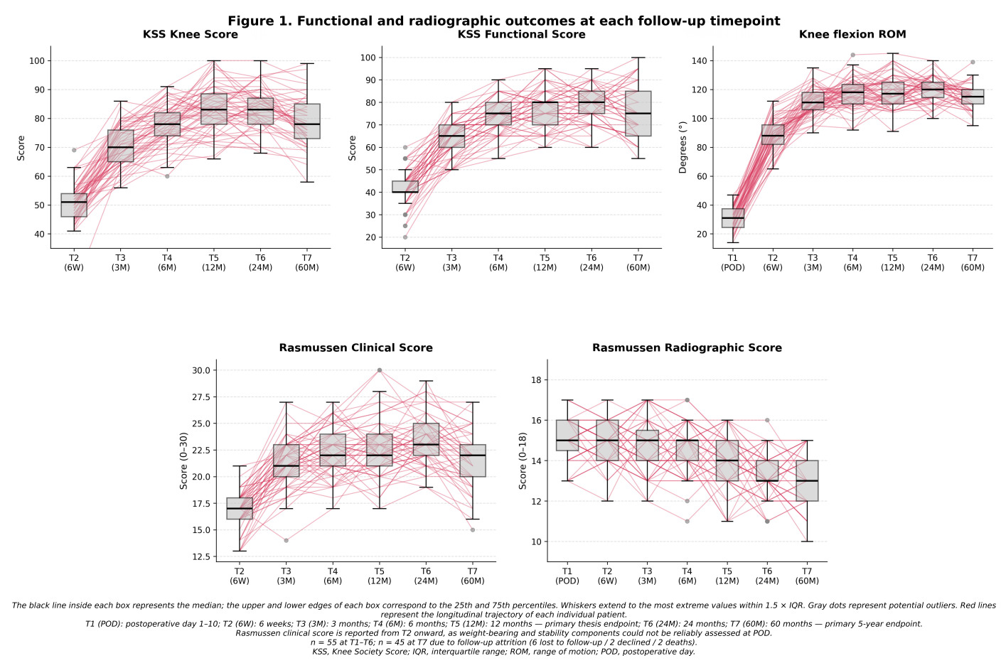
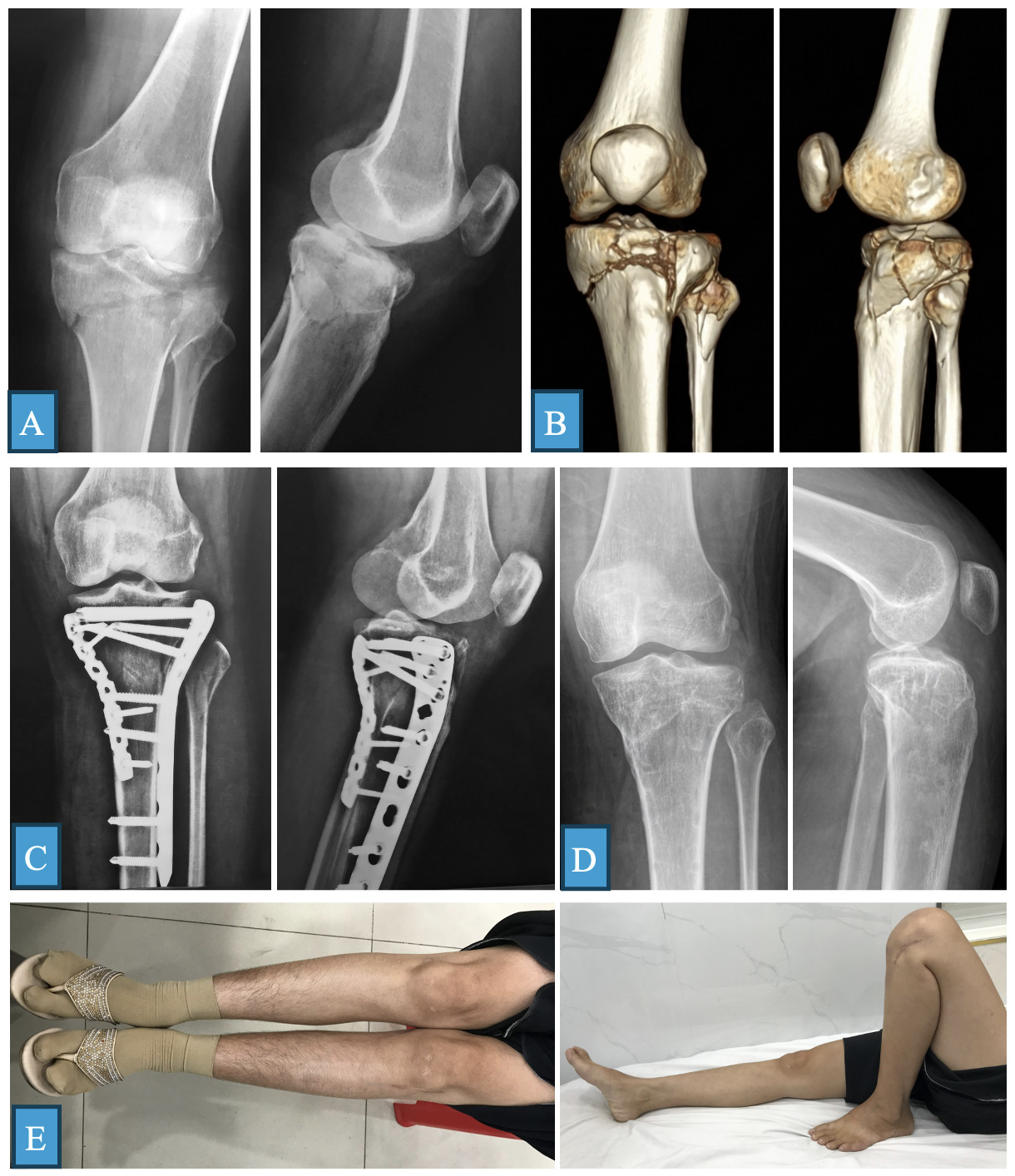
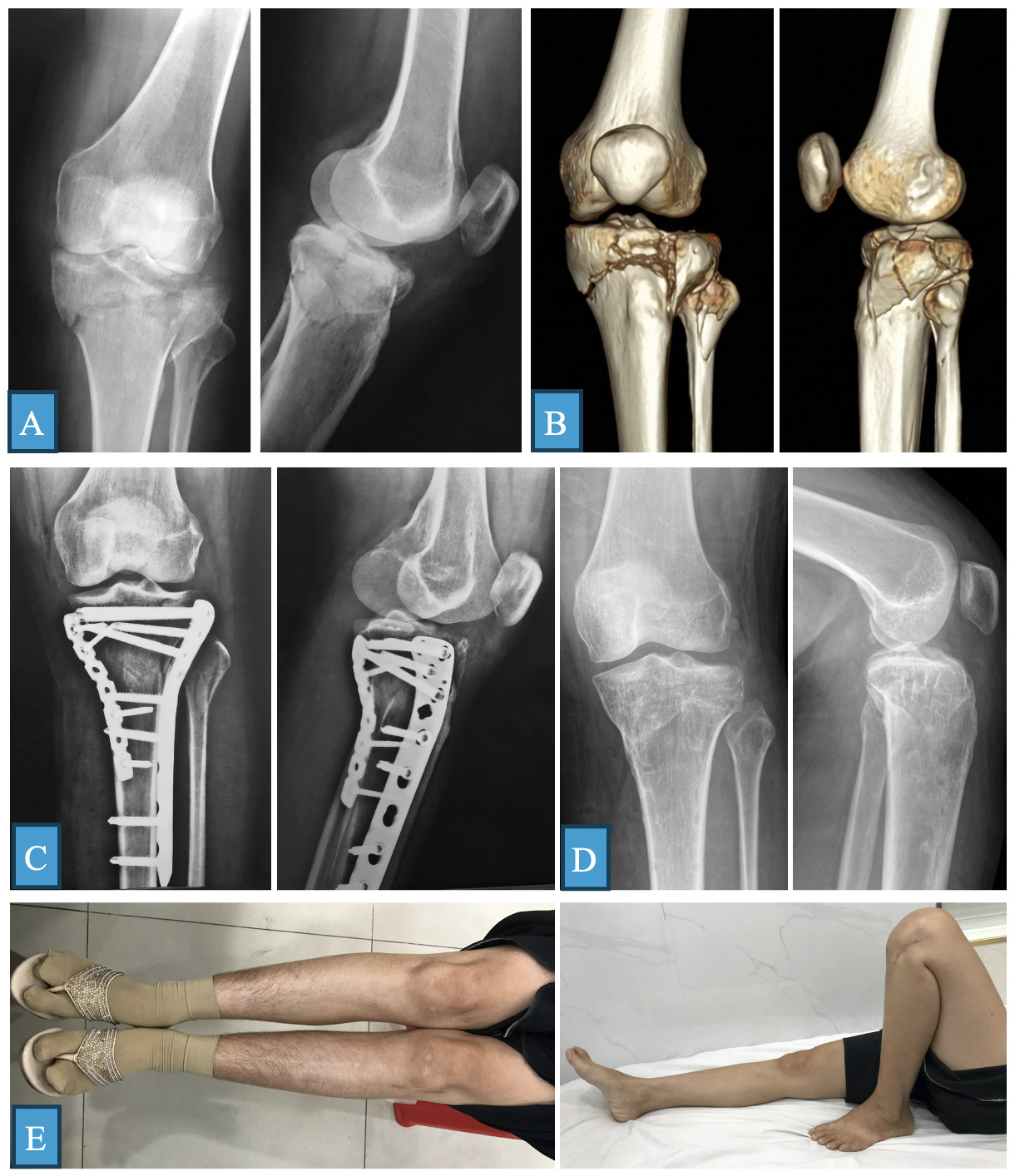
Forty-five patients (81.8%) completed the 60-month assessment (T7), while 10 patients were lost to follow-up after T6. No patient developed nonunion or required revision osteosynthesis. The mean time to radiographic union was 4.2 ± 0.7 months (range, 3–6). Metaphyseal bone grafting was performed in 23 patients (41.8%). Anatomical, clinical, and functional outcomes at each follow-up time point are detailed in Table 2 and Figure 1.
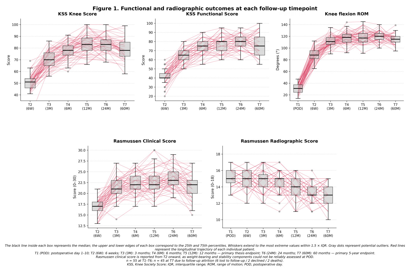
Open reduction markedly improved articular congruity and alignment. Preoperatively, the mean lateral plateau depression and AP plateau widening were 5.25 ± 4.29 mm and 10.93 ± 5.88 mm, respectively, which decreased to 1.00 ± 1.17 mm and 1.52 ± 1.65 mm at T1. The mean PSA was 7.80 ± 2.48° postoperatively and was 9.07 ± 2.54° at T7. Over time, a small but progressive loss of reduction was observed: at 60 months, lateral plateau depression and AP widening increased to 2.17 ± 1.50 mm and 3.11 ± 1.75 mm, respectively. Accordingly, the mean RRS decreased from 15.05 ± 1.03 at T1 to 13.18 ± 1.40 at T7 (Table 2; Figure 1).
Pain and knee function improved markedly during the first postoperative year and remained stable thereafter (Table 2, Figure 1). The mean VAS score decreased from 6.67 ± 1.32 at T1 to 1.13 ± 0.94 at the prespecified 12-month endpoint (T5) and was 1.67 ± 1.35 at 60 months. Knee flexion recovered from 30.25 ± 7.72° at T1 to 118.67 ± 11.39° at T5 and 114.84 ± 8.91° at T7. The KSS Knee score peaked at 12 months (83.4 ± 8.2), whereas the KSS Function score reached its highest mean value at 24 months (T6); both showed only a modest decline at 60 months (78.9 ± 8.8 and 76.6 ± 11.9, respectively). The RCS followed the same trajectory, rising from 16.78 ± 1.77 at 6 weeks to a peak of 23.25 ± 2.34 at T6 before settling at 21.47 ± 2.87 at T7. Clinical antero-posterior and varus–valgus instability greater than 5 mm or 5° was observed in 15.6% and 11.1% of knees, respectively, at the 5-year assessment.
Thirteen patients (23.6%) experienced a postoperative complication: superficial wound infection (n=4, 7.3%), malunion (n=4, 7.3%), transient peroneal nerve neuropraxia (n=3, 5.5%), deep infection (n=1, 1.8%), and deep vein thrombosis (n=1, 1.8%). The deep infection resolved with debridement and intravenous antibiotics without implant removal.
Radiographic PTOA progressed steadily over time. The prevalence of KL grade ≥ 2 in the injured knees was 14.5% (8/55) at 12 months and rose to 53.3% (24/45) at 60 months, compared with only 7.3% (4/55) in the contralateral uninjured knees at the 12-month reference assessment. Severe PTOA (grade 4) developed in 3 injured knees (6.7%); although TKA was indicated, all three patients declined the procedure.
In univariable analyses, Schatzker VI was associated with smaller RCS improvement compared with Schatzker V (MD −2.09, 95% CI −3.88 to −0.31, p = 0.023). MCL injury was also associated with greater RCS improvement (MD 2.58, 95% CI 0.02 to 5.13, p = 0.048), although this finding should be interpreted cautiously given the small subgroup size and multiple univariable comparisons. Diabetes mellitus was associated with poorer KSS Knee improvement (MD −13.42, p = 0.044) and greater VAS improvement (MD 2.66, p = 0.035). None of the remaining variables showed a statistically significant association with the five long-term outcomes at 60 months.
Discussion
The present study reports the outcomes of open reduction and internal fixation (ORIF) in 55 patients with bicondylar tibial plateau fractures prospectively followed up to 60 months. The mean age of 45.4 ± 12.4 years and the male predominance (60%) are consistent with recent reports.1,8,9 A distinctive feature of our cohort is that motorcycle accidents accounted for 90.9% of cases - markedly higher than reported by Worku (67%) and Babu (60%) - indicating a high-energy injury profile in our population.8,10 The proportion of posteromedial fragments was 50.9% - within the range reported (28–74%) - supporting the contemporary view that three-column assessment with CT and an extension of the traditional Schatzker classification are warranted.2,4,11–13 Associated soft-tissue injuries were present in 52.7% of cases (lateral meniscus tears in 29.1%, MCL ruptures in 12.7%, and ACL tibial-eminence avulsions in 7.3%), comparable to the report of Huang and colleagues, who described ACL avulsions combined with anterior meniscal root tears in up to 31% of posterior condyle fractures,14 underscoring the role of preoperative MRI assessment.
Immediate postoperative reduction was good, lateral plateau depression decreased from 5.25 mm to 1 mm, AP plateau widening from 10.93 mm to 1.52 mm. However, we observed a mild progressive loss of reduction over time (lateral plateau depression 2.17 mm and AP widening 3.11 mm at 60 months), accompanied by a decline in the RRS from 15.05 to 13.18. This pattern is consistent with the “mismatch” between favorable clinical outcomes and deteriorating radiographic findings described by Bormann,9 and suggests that long-term radiographic surveillance remains necessary even when patients are functionally satisfied. The late subsidence may reflect irreversible subchondral cartilage damage, mild residual axial malalignment, and secondary degenerative changes.
The mean VAS score decreased from 6.67 in the early postoperative period to 1.13 at 12 months and remained at 1.67 at 60 months. Knee flexion recovered to 118.67° at 12 months and 114.84° at 5 years - exceeding the 110° functional threshold required for activities of daily living. The KSS Knee score peaked at 83.4 ± 8.2 at 12 months and the KSS Function score peaked at 78.3 ± 7.7 at 24 months, comparable to the dual-plating groups of Saodekar (KSS 85.9/80.5),6 Sinha (KSS 85.4/81.0),7 Prasad,15 and clearly superior to the single-plate groups in Saodekar and Sinha (KSS Knee 73.4 and 70.9, respectively). Our findings are also consistent with other posteromedial-plating cohorts reporting similar mid-term KSS scores.16–18 The RCS peaked at 23.25 at 24 months and then declined modestly to 21.47 at 60 months, remaining within the “Good” category. The early peak followed by a modest late decline in the KSS Knee score reflects the natural history of these injuries: after early functional recovery, progressive osteoarthritic changes gradually affect knee function — in line with the long-term surveillance recommendations of Schatzker and Kfuri.4
A key procedural feature that may have contributed to these outcomes is the use of two mini-incisions rather than a single extensile midline or extended anterolateral exposure. The historical single-midline approach has been associated with deep infection rates as high as 23–87% because it requires wide soft-tissue stripping over a region already compromised by high-energy trauma; in our cohort, the two-window strategy limited the cumulative skin incision and produced only a single deep infection (1.8%), comparable to recent dual-incision series and well below the historical benchmark.3,6,7,19 The supine “floating” position adapted from Chang and colleagues allowed both the posteromedial approach and the anterolateral approach to be performed in a single setup without intra-operative repositioning, shortening operative time and avoiding the airway and pressure-injury hazards of a prone-to-supine transition.20 Direct visualization of the posteromedial fragment through the Lobenhoffer interval,17 providing adequate exposure for the near-coronal posteromedial fracture plane as confirmed by the cadaveric mapping of Krause and colleagues,21 enabled accurate reduction and buttress fixation with a pre-contoured 3.5-mm LCP, the construct with the highest load-to-failure among posteromedial fixation options in recent biomechanical testing and endorsed by current expert review.2,22,23 The lateral plateau was subsequently addressed through a separate anterolateral mini-incision using minimally invasive plate osteosynthesis of a pre-contoured LCP, with subchondral raft locking screws supporting the elevated articular surface and diaphyseal locking screws restoring the mechanical axis.6,7,24
The overall complication rate of 23.6% lies within the range reported in the literature - higher than Bhangale (16.67%) and Arya (10%),19,25 but comparable to Manikandan and Jati,1,26 and consistent with the complication spectrum summarized by Gálvez-Sirvent.3 No patient developed nonunion or required revision osteosynthesis during the 5-year follow-up. By contrast, post-traumatic osteoarthritis was observed in 14.5% of injured knees at 12 months and increased to 53.3% at 60 months, compared with only 7.3% of contralateral uninjured knees at the 12-month reference assessment. This high rate is consistent with the 32.5% osteoarthritis rate reported by Jati over a shorter follow-up and with the warning issued by Van den Berg regarding the long-term consequences of high-energy intra-articular knee fractures.1,2
Univariable analysis showed that Schatzker VI fractures were associated with a smaller RCS improvement than Schatzker V fractures (MD −2.09; p = 0.023), supporting the hypothesis that fracture complexity directly influences long-term functional recovery. Diabetes mellitus was associated with poorer KSS Knee improvement (MD −13.42; p = 0.044), but the sample size was very small (only 2 diabetic patients reached T7) and the finding should be interpreted with caution. Conversely, concomitant MCL injury was associated with greater RCS improvement (MD +2.58; p = 0.048) - a paradoxical finding that may reflect early recognition and treatment of ligamentous injury, or simply chance, given that approximately one hundred tests were performed without correction for multiplicity.
The principal strengths of the study are the 5-year prospective follow-up with eight comprehensive radiographic, clinical, and functional assessment time points, together with a contralateral-knee reference group that allowed the isolated effect of injury on subsequent PTOA to be quantified. Limitations include the single-center design, the modest sample size, the 18% loss to follow-up at 5 years, and the use of univariable analyses without correction for multiple testing.
Conclusion
ORIF using a dual-plating strategy through combined anterolateral and posteromedial mini-incisions, with bone grafting when indicated and a controlled rehabilitation program, achieves favorable and durable clinical and functional outcomes up to 5 years for bicondylar tibial plateau fractures. Nevertheless, progressive PTOA remains a clinically meaningful challenge that warrants long-term surveillance and should be a target for future preventive strategies.
Acknowledgements
None
Corresponding author:
Ngoc Quyen Le, Orthopedics, Trauma, and Rehabilitation Department, School of Medicine, University of Medicine and Pharmacy at Ho Chi Minh City, Vietnam
Email: ngocquyenbs76@ump.edu.vn
Tel: +84 903818438
Authors’ contributions
-
Ngoc Quyen Le: Conceptualization, Investigation, Data curation, Writing – review & editing, project administration.
-
Khac Xuan Hoang: Conceptualization, Investigation, Data curation, Writing-original Draft.
-
Trong Xuan Cuong Tran: Formal analysis, Visualization, Writing – review & editing.
Conflicts of interest
The authors declare no conflicts of interest.
Funding
This research did not receive any specific grant from funding agencies in the public, commercial, or not-for-profit sectors.