
1. Introduction
Orthopedic implants are widely used to restore stability, maintain alignment, and promote healing after fractures and reconstructive procedures. In many patients, retained implants remain asymptomatic and do not require additional intervention. However, a considerable subgroup of patients later undergoes elective implant removal because of persistent pain, local irritation, implant prominence, restricted motion, or dissatisfaction with retained hardware. Studies evaluating symptomatic implant removal have shown that secondary procedures are common in orthopedic practice and may produce functional improvement in selected patients, although the magnitude of benefit varies across indications and anatomical sites.1,2
Recent literature has also emphasized that elective implant removal is not a trivial event. Although symptom relief is frequently reported after removal, complication rates, patient expectations, and the balance between benefit and surgical risk remain important clinical considerations. Prospective and contemporary observational studies have demonstrated that improvement after elective implant removal is influenced by preoperative symptoms, the anatomical location of the implant, and the reason for removal, especially when pain or mechanical irritation is the principal indication.3,4 This means that identifying predictors of pain-related implant removal before the initial surgery, or at least during postoperative follow-up, may have real practical value.
Pain and irritation related to retained orthopedic hardware have been described in a variety of anatomical regions. In patellar fracture fixation, symptomatic implant removal has been linked to surgical construct characteristics and hardware prominence.5 Similar concerns have been reported after tibial plateau fracture fixation, where implant-related symptoms may persist despite otherwise successful fracture healing and alignment restoration.6 In ankle trauma, especially in syndesmotic screw fixation, hardware removal remains a recurring clinical issue, often driven by discomfort, local irritation, or surgeon preference in selected cases.7 Midshaft clavicle fixation studies have likewise shown that plate design, plate position, and implant prominence can affect the likelihood of later hardware removal.8 Together, these studies indicate that symptomatic hardware removal is not confined to a single procedure type, but rather represents a broader orthopedic phenomenon with both patient-related and implant-related determinants.9,10
Within this broader field, tibial tubercle osteotomy literature has recently provided some of the most direct procedure-specific evidence regarding pain-related hardware removal. Outpatient cohort data have shown that symptomatic hardware removal remains one of the notable reasons for reoperation after tibial tubercle osteotomy, and patient characteristics appear to influence this pattern. More specifically, recent work examining pain and hardware removal after tibial tubercle osteotomy has highlighted incidence, associated factors, and patient outcomes after secondary removal procedures. In addition, technical modifications such as headless screw fixation have been associated with lower rates of subsequent hardware removal, further supporting the idea that pain and irritation are influenced by both patient anatomy and implant characteristics.10,11
Although the orthopedic literature clearly documents pain-related implant removal across multiple procedures, evidence directly focused on BMI as a primary predictor remains limited. In many studies, BMI is included only as a secondary demographic or clinical variable rather than as the central exposure of interest. This creates an important gap. From a biomechanical perspective, BMI could plausibly influence postoperative discomfort and implant-related irritation in several ways. Higher body mass may increase mechanical loading on fixation constructs, especially in weight-bearing regions. Increased soft tissue volume may alter the interface between implant prominence and surrounding tissues. In some anatomical areas, local pressure, friction, or altered gait mechanics may increase pain perception around otherwise stable hardware. At the same time, the relationship may not be linear or uniform across all procedures. Some procedure-specific studies have suggested that body habitus, including BMI, may relate to reoperation patterns, but findings have not been consistent enough to establish a unified conclusion.10,11
Another reason this question matters is that the clinical decision to remove symptomatic hardware often occurs long after fracture healing or index fixation. When surgeons counsel patients before surgery, they routinely discuss infection, nonunion, malunion, and hardware failure, yet less attention may be paid to the possibility that the implant itself may later become a source of discomfort. If BMI is meaningfully associated with pain-related implant removal, it could affect preoperative counseling, implant selection, incision planning, soft tissue handling, postoperative surveillance, and perhaps even threshold selection for elective removal. This may be particularly relevant in lower-extremity and periarticular procedures, where both mechanical demand and local prominence can shape patient symptoms.12
Patient-centered outcome literature also supports the importance of this issue. In lower-extremity cohorts undergoing elective implant removal, patient satisfaction and symptom relief have been associated with the degree of preoperative symptoms and the appropriateness of case selection.11,12 This suggests that pain-related implant removal should not be viewed solely as a technical event, but as a clinically meaningful outcome with implications for quality of life, function, healthcare utilization, and shared decision-making. Therefore, understanding which patients are more likely to experience painful retained hardware may improve both prevention and management.12
The present study was designed to address this gap by examining BMI in relation to orthopedic implant removal due to pain or irritation in a retrospective clinical dataset. Based on the provided records, the analysis incorporated a full cohort for the primary removal outcome and a descriptive anthropometric subgroup for more detailed BMI-related characterization. In addition to the main association between BMI and implant removal, the study also evaluated age differences across BMI categories and described sex distribution patterns. We hypothesized that higher BMI categories would be associated with a greater prevalence of implant removal due to pain or irritation.
2. Materials and Methods
2.1. Study Design
This study was designed as a retrospective observational analysis based on clinical records of orthopedic implant removal procedures. The study used existing data and did not involve prospective intervention, randomization, or alteration of patient care. The main analytic approach was descriptive and inferential within the implant-removal cohort.
Because all included cases had undergone implant removal, the study was not designed to determine whether BMI predicts the occurrence of implant removal compared with patients whose implants remained in place. Instead, the analysis examined BMI distribution and internal associations within the removal cohort. This distinction is central to the interpretation of the findings. The study can describe BMI patterns among removal cases and test associations between BMI and available clinical variables, but it cannot establish causation or estimate the relative risk of implant removal in patients with high BMI compared with patients with normal BMI in the absence of a non-removal control group.
2.2. Study Population
The dataset included 88 orthopedic implant-removal procedures performed among 78 unique patients. The unit of analysis for most statistical tests was the implant-removal procedure. This approach was selected because anatomical site, indication, and implant-removal characteristics are procedure-level variables. However, the presence of 78 unique patients indicates that some patients may have contributed more than one procedure. This limitation is acknowledged because repeated observations from the same patient may introduce partial dependence between records.
Eligible records included cases with available BMI data and documentation of implant-removal procedure characteristics. BMI was calculated from height and weight, when available in the dataset. Cases with sufficient data were included in descriptive and inferential analyses. The dataset included variables for age, sex, BMI, anatomical location, and clinical notes or procedure comments.
2.3. Variables
The primary variable of interest was BMI. BMI was analyzed both as a continuous variable and as a categorical variable. For categorical analysis, high BMI was defined as BMI ≥ 25 kg/m2, representing overweight or obesity. Obesity was defined separately as BMI ≥ 30 kg/m2.
Age was analyzed as a continuous variable and also categorized into three age groups: younger than 40 years, 40 to 59 years, and 60 years or older. Sex was categorized as male or female. Anatomical location was coded from the procedure descriptions and grouped into clinically meaningful regions, including tibia/tibial plateau, ankle/foot, knee/patella, elbow/olecranon/ulna, wrist/radius, hip/femur, clavicle, and humerus.
A focused anatomical grouping was also created to examine high BMI in weight-bearing or load-related regions. This group included knee, patella, hip, and femur. These regions were compared with all other anatomical regions. The rationale for this grouping was biomechanical: knee, patella, hip, and femur are strongly involved in lower-limb loading, weight transmission, and functional mobility. Therefore, elevated BMI may be especially relevant in these locations.
A broad symptomatic-removal variable was also examined. Symptomatic removal was defined broadly when the free-text notes documented pain, irritation, prominent screw, prominent hardware, prominent metalwork, blade prominence, local pressure, or similar symptom-related language. Because this variable was derived from clinical notes rather than from a structured field, it should be interpreted as exploratory. In the expanded symptomatic example used for demonstration and presentation, 39 of 88 cases were classified as symptomatic. This coding should be clearly described as broad and dependent on the coding rules used.
2.4. Statistical Analysis
Descriptive statistics were calculated for continuous variables, including mean, standard deviation, median, and range where appropriate. Categorical variables were summarized using frequencies and percentages.
A one-sample t-test was used to determine whether the mean BMI of the implant-removal cohort was significantly higher than 25 kg/m2, the clinical threshold for overweight. Pearson correlation was used to assess the linear association between age and BMI. Spearman correlation was also used to confirm whether the association was present using a non-parametric rank-based method.
BMI differences across age groups were examined using one-way ANOVA and Kruskal-Wallis tests. The use of both tests was intended to evaluate the robustness of group differences, especially given potential non-normality and unequal group sizes. BMI differences across anatomical regions were also examined using ANOVA and Kruskal-Wallis tests.
Associations between categorical variables were tested using Fisher’s exact test or chi-square tests, depending on the structure of the table and expected cell counts. Fisher’s exact test was used for the focused comparison of high BMI in weight-bearing regions versus all other regions. Odds ratios were calculated to estimate the strength of association.
Statistical significance was defined as p < 0.05. All analyses were interpreted cautiously because the study was retrospective and did not include a non-removal comparison group.
2.5. Ethical Considerations
This retrospective study was approved by the Institutional Helsinki Committee of Nazareth Hospital EMMS . All data were anonymized prior to analysis. Due to the retrospective nature of the study, the requirement for informed consent was waived.
3. Results
3.1. Sample Characteristics
The final analytic dataset included 88 orthopedic implant-removal procedures performed among 78 unique patients. The mean age of the sample was 44.9 years with a standard deviation of 16.4 years. The mean BMI was 27.33 kg/m2 with a standard deviation of 5.06 kg/m2. These values indicate that, on average, the cohort fell within the overweight range.
Table 1 presents the general characteristics of the study cohort. The dataset included 88 implant-removal procedures among 78 unique patients. The mean BMI was above the clinical threshold for overweight, suggesting that excess body weight was common among patients undergoing implant removal.
3.2. BMI Distribution
BMI was categorized into three mutually exclusive categories: BMI < 25, BMI 25 to 29.9, and BMI ≥ 30. A total of 28 cases (31.8%) had BMI < 25, 39 cases (44.3%) were overweight, and 21 cases (23.9%) were obese. Overall, 60 of 88 cases (68.2%) had BMI ≥ 25.
Table 2 shows that most implant-removal cases were above the normal BMI range. Specifically, 60 of 88 cases (68.2%) had BMI ≥ 25, meaning that more than two-thirds of cases were classified as overweight or obese. This finding supports the relevance of examining BMI in this clinical context.
3.3. Mean BMI Compared With the Overweight Threshold
A one-sample t-test was performed to determine whether mean BMI was significantly higher than 25 kg/m2. The mean BMI was 27.33 kg/m2. The result was statistically significant, t(87) = 4.31, p < 0.001. This indicates that mean BMI in the implant-removal cohort was significantly above the overweight threshold.
Table 3 demonstrates that mean BMI was significantly higher than the threshold for overweight. This is one of the strongest BMI-related findings in the study. However, the finding describes the BMI profile within the implant-removal cohort and does not prove that BMI causes implant removal.
3.4. Sex Distribution and BMI by Sex
The sample included 52 male cases and 36 female cases. Mean BMI was 26.73 among males and 28.19 among females. Although female cases had a slightly higher mean BMI, the difference was not statistically significant. Welch’s t-test showed p = 0.203, and Mann-Whitney testing showed p = 0.423.
Table 4 shows that BMI was slightly higher among female cases than male cases, but the difference was not statistically significant. Therefore, sex was not significantly associated with BMI level in this sample.
3.5. Obesity According to Sex
Obesity, defined as BMI ≥ 30, was also compared by sex. Among male cases, 21.2% were obese. Among female cases, 27.8% were obese. Fisher’s exact test showed p = 0.612, indicating no statistically significant association between sex and obesity status.
Table 5 indicates that obesity was somewhat more frequent among female cases, but this difference was not statistically significant. The result does not support a meaningful sex-based difference in obesity prevalence within this cohort.
3.6. Association Between Age and BMI
A significant positive association was found between age and BMI. Pearson correlation showed r = 0.279, p = 0.008. Spearman correlation showed ρ = 0.286, p = 0.007. The consistency between the parametric and non-parametric tests strengthens the finding.
Table 6 shows a statistically significant positive relationship between age and BMI. In practical terms, older patients in the implant-removal cohort tended to have higher BMI values. This association was confirmed by two different correlation methods.
3.7. BMI Across Age Groups
The sample was divided into three age groups: younger than 40 years, 40 to 59 years, and 60 years or older. Mean BMI was lowest in the younger-than-40 group and higher in the two older groups. The mean BMI was 24.90 among patients younger than 40, 28.42 among patients aged 40 to 59, and 28.71 among patients aged 60 or older. Differences between age groups were statistically significant using both ANOVA and Kruskal-Wallis tests.
Tables 7 and 8 show that BMI differed significantly across age groups. The younger group had the lowest mean BMI, whereas both older groups had mean BMI values in the overweight range. Because both ANOVA and Kruskal-Wallis tests were significant, this finding can be considered relatively stable.
3.8. BMI According to Anatomical Location
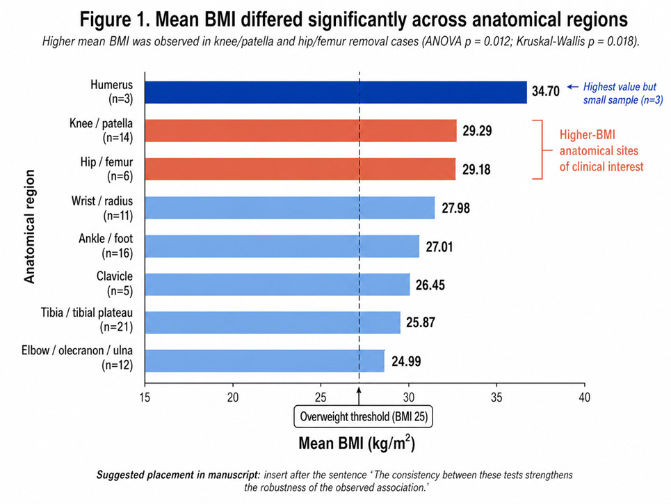
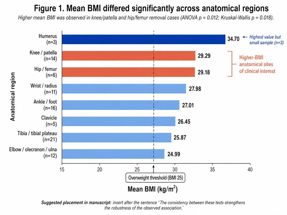
BMI was also examined according to anatomical location. Mean BMI varied across regions. The highest mean BMI was observed in the humerus group, but this group included only three cases. Knee/patella and hip/femur regions also showed relatively high BMI values.
Table 9 presents mean BMI by anatomical location. BMI appeared higher in several regions, especially knee/patella, hip/femur, and humerus. However, some anatomical groups were small, particularly humerus and clavicle, so these descriptive differences should be interpreted cautiously.
3.9. Statistical Testing of BMI Differences Across Anatomical Regions
Table 10 demonstrates that both the parametric one-way ANOVA and the non-parametric Kruskal-Wallis test identified statistically significant differences in BMI across anatomical regions. The ANOVA result (p = 0.012) indicates that mean BMI varies between at least two anatomical groups under the assumption of normal distribution, while the Kruskal-Wallis result (p = 0.018) confirms this finding without relying on distributional assumptions. The consistency between these tests strengthens the robustness of the observed association. Therefore, the variation in BMI across anatomical locations appears reliable and not driven by distributional bias or unequal group sizes, supporting the presence of true differences between anatomical regions.
3.10 High BMI and Weight-Bearing Anatomical Regions
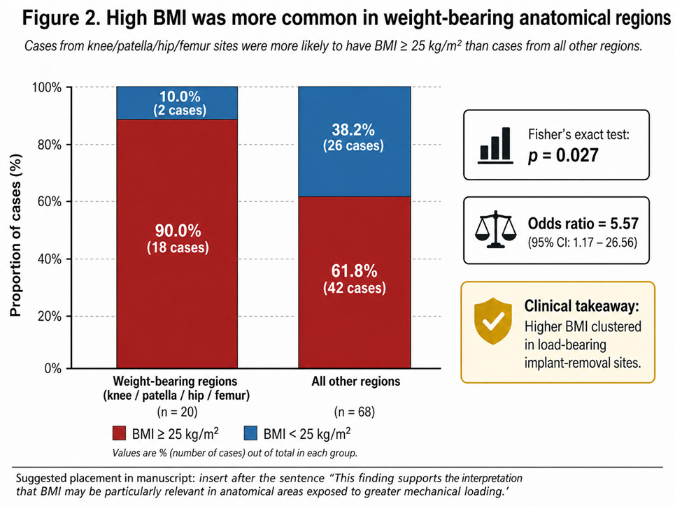
A focused analysis compared weight-bearing regions, defined as knee, patella, hip, and femur, with all other anatomical regions. High BMI was defined as BMI ≥ 25 kg/m2. Among cases from knee/patella/hip/femur regions, 18 of 20 cases (90.0%) had high BMI. In all other regions, 42 of 68 cases (61.8%) had high BMI. Fisher’s exact test showed a statistically significant association, p = 0.027, with an odds ratio of 5.57.
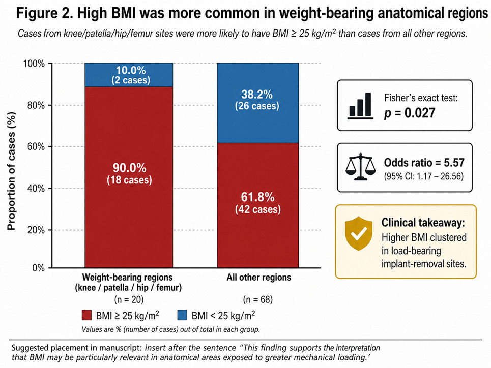
Tables 11 and 12 show a significant association between high BMI and weight-bearing anatomical location. High BMI was substantially more frequent in knee/patella/hip/femur cases than in all other anatomical regions. This finding supports the interpretation that BMI may be particularly relevant in anatomical areas exposed to greater mechanical loading.
3.11 Broad Symptomatic Removal
A broad symptomatic-removal variable was examined using clinical note language indicating pain, irritation, prominent screw, prominent hardware, prominent metalwork, or pressure-related symptoms. In the expanded coding example, 39 of 88 cases (44.3%) were classified as symptomatic, and 49 cases (55.7%) were not classified as symptomatic.
Table 13 presents the broad symptomatic-removal classification. This variable was based on free-text clinical notes and should therefore be considered exploratory. The classification captures symptom-related indications such as pain, irritation, prominent screw, prominent metalwork, or local pressure.
3.12 Broad Symptomatic Removal According to Weight-Bearing Anatomical Regions
The broad symptomatic-removal variable was then examined according to weight-bearing anatomical region. Among knee/patella/hip/femur cases, 18 of 20 cases (90.0%) were classified as symptomatic. Among all other anatomical regions, 21 of 68 cases (30.9%) were classified as symptomatic. Fisher’s exact test showed p < 0.001, with an odds ratio of approximately 20.1.
Tables 14 and 15 show a strong association between weight-bearing anatomical region and broad symptomatic-removal classification. Symptomatic removal was much more frequent in knee/patella/hip/femur cases than in all other anatomical regions. This is a strong exploratory finding, but it depends on the coding rules used for the broad symptomatic variable and should not be treated as equivalent to a prospectively collected clinical indication.
4. Discussion
The present study examined BMI and clinical characteristics among patients who underwent orthopedic implant removal. The main finding was that the cohort had a mean BMI in the overweight range, with more than two-thirds of implant-removal procedures occurring in patients with BMI ≥ 25 kg/m2. Mean BMI was also significantly higher than the clinical overweight threshold. This finding does not prove that elevated BMI causes implant removal, because all included patients had already undergone removal and there was no non-removal comparison group. However, it does indicate that excess body weight was common within this implant-removal cohort and may deserve greater attention during orthopedic follow-up and counseling.
The association between BMI and age was another important finding. Both Pearson and Spearman correlations showed a significant positive relationship, and BMI differed significantly across age groups. Patients younger than 40 years had the lowest mean BMI, while patients aged 40 years and older had mean BMI values within the overweight range. This pattern may reflect broader age-related changes in body composition, mobility, metabolic risk, and musculoskeletal load. Clinically, it suggests that older patients undergoing implant removal may present with a different risk profile than younger patients, not only because of age itself, but also because age and BMI may act together in shaping postoperative symptoms, mechanical discomfort, and recovery expectations.
In contrast, sex was not significantly associated with BMI or obesity status in this cohort. Although female cases had a slightly higher mean BMI and a slightly higher obesity rate, these differences were not statistically significant. Therefore, the results do not support a strong sex-based explanation for BMI variation among patients undergoing implant removal. This is clinically useful because it redirects attention from sex differences toward other factors, especially age, anatomical site, and mechanical loading.
The most clinically relevant finding was the association between BMI and anatomical region. BMI differed significantly across anatomical regions, and high BMI was more frequent in knee/patella/hip/femur cases than in other regions. This finding is consistent with the biomechanical expectation that load-bearing regions may be more sensitive to body weight, pressure, gait mechanics, and implant prominence. Previous studies have shown that symptomatic implant removal is influenced by anatomical location, implant characteristics, and preoperative symptoms.1–4 Procedure-specific literature also supports the role of local mechanical irritation and hardware prominence in patella, tibial plateau, ankle syndesmosis, clavicle, and tibial tubercle procedures.5–11 The present findings add to this literature by suggesting that BMI may be especially relevant in weight-bearing or load-related anatomical sites.
The broad symptomatic-removal variable further supports this interpretation. Nearly half of the cases were classified as broadly symptomatic based on documentation of pain, irritation, prominent hardware, protruding screw, prominent metalwork, or local pressure. Because this variable was derived from free-text clinical notes, it should be considered exploratory rather than equivalent to a prospectively collected indication. Nevertheless, the high frequency of symptomatic documentation is consistent with prior evidence showing that elective implant removal is most beneficial when patients are truly symptomatic before surgery.3,4 It also aligns with lower-extremity outcome studies showing that patient satisfaction after implant removal depends on careful case selection and symptom burden.12
Overall, the study suggests that BMI should not be treated merely as a background demographic variable in implant-removal research. Instead, BMI may interact with anatomical location, mechanical load, and symptom development. These findings support more detailed documentation of BMI, anatomical site, implant prominence, and symptom indication in future orthopedic datasets. At the same time, the results must remain descriptive and hypothesis-generating because the study cannot determine whether high BMI increases the risk of implant removal compared with patients whose implants remain in place.
5. Limitations
This study was limited by its retrospective design, modest sample size, and absence of a non-removal control group. Some patients contributed more than one procedure, and symptomatic removal was coded from free-text notes rather than a standardized clinical field. Small anatomical subgroups also limited the stability of region-specific interpretations overall.
6. Recommendations for Future Research
Future studies should include larger multicenter cohorts with both implant-removal and non-removal comparison groups. Prospective documentation of pain, irritation, implant prominence, functional limitation, and patient-reported outcomes is recommended. Stratified analyses by anatomical region, implant type, and BMI category may clarify whether elevated BMI independently predicts symptomatic hardware removal risk directly.
7. Conclusion
This retrospective observational study described the BMI profile and selected clinical characteristics of patients undergoing orthopedic implant removal. The findings show that the cohort had a mean BMI above the overweight threshold and that most implant-removal procedures occurred in patients with BMI ≥ 25 kg/m2. This result is important because BMI is often reported as a background variable in orthopedic studies, but is not always examined as a clinically meaningful factor in the context of symptomatic hardware removal.
The study also found that BMI was positively associated with age. Patients aged 40 years and older had higher mean BMI values than patients younger than 40 years. This suggests that BMI-related interpretation in implant-removal cohorts should account for age, because older patients may present with a combined profile of greater body mass, altered biomechanics, and different postoperative recovery patterns. In contrast, sex was not significantly associated with BMI or obesity status, indicating that sex did not explain BMI variation in this sample.
The most clinically meaningful finding was the relationship between BMI and anatomical location. BMI differed significantly across anatomical regions, and high BMI was especially common in weight-bearing regions, including knee, patella, hip, and femur. These sites are exposed to greater mechanical load during standing, walking, and daily activity. Therefore, the concentration of high BMI in these anatomical regions supports the possibility that body weight may contribute to implant-related discomfort, irritation, or mechanical symptoms in selected patients. The broad symptomatic-removal analysis also showed that pain, irritation, hardware prominence, or local pressure were documented in a substantial proportion of cases, strengthening the clinical relevance of symptom-based assessment.
However, the findings should be interpreted carefully. Because the study included only patients who underwent implant removal, it cannot determine whether high BMI increases the overall risk of implant removal compared with patients whose implants remain in place. The results are therefore descriptive and hypothesis-generating, not causal. In addition, symptomatic removal was based on free-text documentation, and some anatomical subgroups were small.
Overall, this study suggests that BMI may be an important variable to consider when evaluating patients with retained orthopedic hardware, particularly in lower-extremity and weight-bearing anatomical regions. These findings support more systematic documentation of BMI, anatomical site, implant prominence, and symptom indication in future orthopedic research. They also highlight the need for larger prospective studies to determine whether elevated BMI independently predicts symptomatic implant removal and whether this information can improve surgical planning, patient counseling, and postoperative follow-up.