
Introduction
Lumbosacral radicular pain is a common cause of disability and is frequently associated with lumbar disc herniation, foraminal stenosis, lateral recess stenosis, spondylosis, and postoperative degenerative changes.1,2 Epidural steroid injections are widely used as part of a multimodal treatment strategy when conservative measures such as physical therapy, activity modification, and oral medications fail to provide adequate relief. Among epidural approaches, the transforaminal route is often favored when precise, nerve root–specific delivery is desired.3–5 Lumbar transforaminal epidural steroid injections (TFESIs) allow delivery of local anesthetic and corticosteroids near the inflamed spinal nerve, dorsal root ganglion, and ventral epidural space.3,6
The traditional lumbar TFESI technique is commonly performed using a supraneural or subpedicular approach. In this technique, the needle is advanced toward the superior aspect of the foramen, typically beneath the pedicle and above the exiting spinal nerve. This target has historically been referred to as the “safe triangle”.7 However, this terminology has become controversial. The superior and anterior portions of the neural foramen may contain radicular arteries, segmental medullary arteries, or the artery of Adamkiewicz at susceptible levels.8 Although catastrophic complications are rare, cases of spinal cord infarction and paralysis following lumbar TFESI have been reported.9–11 These events have shifted attention toward risk-reduction strategies, including careful contrast injection, real-time fluoroscopy, digital subtraction angiography in select settings, avoidance of particulate steroids in higher-risk injections, and alternative needle trajectories.12,13
The infraneural approach has been adopted by some clinicians as an alternative to the traditional superior foraminal target.7,13 Rather than approaching above the exiting nerve root, the infraneural approach targets the lower portion of the foramen, typically within or near Kambin’s triangle. This region is bound by the exiting nerve root superiorly, the traversing nerve root medially, and the superior endplate of the caudal vertebral body inferiorly.13,14 In pain medicine literature, this approach is sometimes described as infraneural, retrodiscal, or Kambin triangle TFESI, although these terms are not always used consistently.7,13,14 The central concept is to access the epidural space from a more inferior foraminal trajectory, theoretically avoiding the superior foraminal zone where radicular arterial structures and the nerve roots may be more frequently encountered.10
This review, therefore, evaluates utility of the infraneural approach for lumbar TFESI, focusing on anatomy, technique, safety, clinical efficacy, patient selection, limitations, and future directions.
Anatomical Considerations
The lumbar neural foramen contains important structures such as the exiting nerve root, dorsal root ganglion, segmental vessels, and radicular arteries.13 The exiting nerve root occupies the superior portion of the foramen, while the inferior foramen contains a variable amount of epidural fat and vascular structures.13,14 The classic subpedicular TFESI target places the needle near the inferior border of the pedicle and superior aspect of the foramen. This location can allow effective delivery of injectate to the exiting nerve root and ventral epidural space.3,4,6 However, the same region may also contain vascular structures capable of communicating with the spinal cord circulation.13–15
The concern with supraneural needle placement is not simply intravascular uptake into any vessel, but inadvertent injection into a radicular or radiculomedullary artery.9,10 If particulate steroid is injected into an arterial branch supplying the spinal cord, embolic occlusion may result in spinal cord ischemia.10–12 Although this is uncommon, the severity of the potential outcome has led to significant procedural caution.
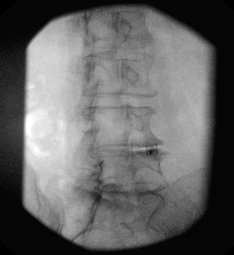
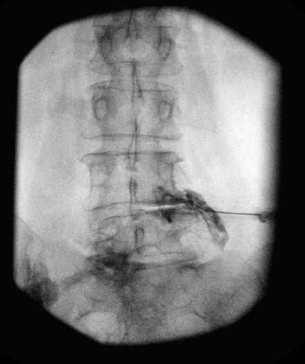
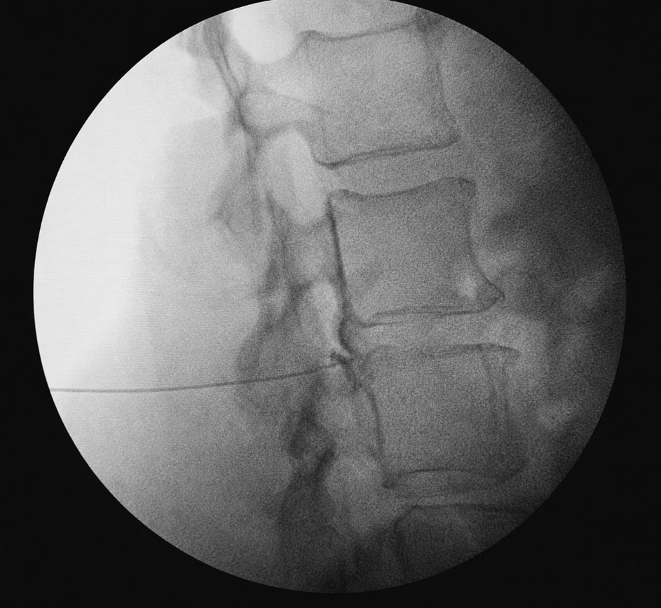
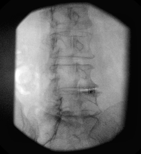
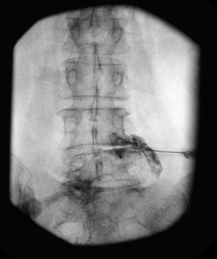
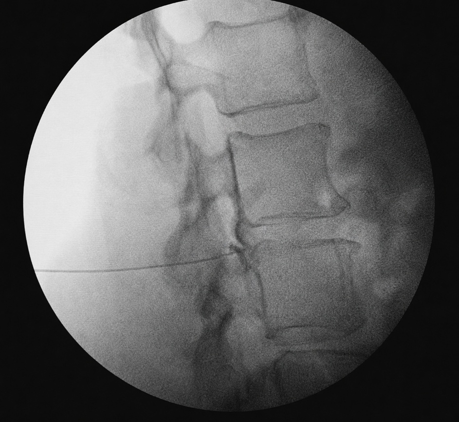
The infraneural approach may reduce the likelihood of encountering arterial structures in the superior portion of the neural foramen by positioning the needle below the exiting nerve root.13,14 The target is typically the inferior-posterior portion of the neural foramen, with the needle directed toward the region adjacent to the posterior disc margin and the superior endplate of the caudal vertebral body. From this position, contrast may spread along the exiting nerve root sleeve and into the ventral epidural space.
Another potential advantage is improved patient comfort. With a supraneural approach, the needle may contact or irritate the targeted exiting nerve root, causing dysesthesias or radicular pain during needle advancement.16 In contrast, the infraneural approach is designed to pass below the exiting nerve root, which may reduce direct nerve irritation and improve procedural tolerability. A randomized prospective study of 100 patients comparing the Kambin triangle approach to the supraneural approach found that neural contact occurred in 9 cases with the supraneural approach, whereas no patients experienced neural contact with the Kambin triangle approach.16
However, an important limitation is that the infraneural approach is also a retrodiscal approach and places the needle relatively close to the intervertebral disc.17 If the needle is advanced too aggressively, or if a large foraminal disc herniation narrows the available entry zone, there is a potential risk of intradiscal needle placement or intradiscal injection.17 Although uncommon, intradiscal injections are clinically important because they may increase the risk of discitis.17,18 This can usually be recognized during contrast injection when contrast is seen filling the disc space rather than spreading along the nerve root or into the epidural space.18
Kambin’s triangle is an important anatomic framework for understanding this technique.19,20 Originally described in the context of posterolateral endoscopic lumbar disc access, the triangle provides a working zone that can allow access to the disc and ventral epidural region while attempting to avoid direct injury to neural structures.19,20 However, its size varies by level and patient anatomy.21 It may be narrowed by foraminal stenosis, disc collapse, facet hypertrophy, osteophytes, scoliosis, spondylolisthesis, postoperative change, or large foraminal/extraforaminal disc herniations.21,22 Therefore, the infraneural approach requires individualized imaging review rather than reliance on a fixed fluoroscopic target.
Technique and Procedural Considerations
The infraneural TFESI is performed under fluoroscopic guidance with the patient prone. Preprocedural MRI or CT should be reviewed to confirm the symptomatic level, nerve root involved, degree of foraminal compromise, location of disc pathology, and presence of anatomic variants.4,23 The physician should also assess whether the infraneural corridor is accessible. Severe foraminal stenosis, advanced disc collapse, high-grade spondylolisthesis, or large foraminal disc extrusion may make the infraneural route technically difficult.4,7,21,22
Fluoroscopic techniques vary, but a common method begins with an anteroposterior view to confirm the level and optimize endplate alignment. The fluoroscope is then obliqued ipsilaterally to open the neural foramen.6,24 A survey of interventional pain physicians found that 60.9% preferred using a combination of AP, oblique, and lateral views for lumbar TFESIs.6 Compared with a traditional subpedicular approach, the needle is directed toward the inferior portion of the foramen rather than the superior subpedicular region. The final target is usually near the posterior vertebral body line and posterior disc margin.14,16,17,25 Lateral imaging is performed to confirm depth and to avoid excessive anterior advancement. Care should be taken not to advance the needle too deeply as this can cause the needle to enter the disc.17,18
Once the needle is positioned, aspiration may be performed but should not be relied upon as the primary safety measure. Negative aspiration does not exclude intravascular placement. A prospective study of 761 lumbar TFESIs demonstrated that using flash or blood aspiration to predict intravascular injection was 97.9% specific but only 44.7% sensitive, meaning that a negative aspiration does not reliably exclude intravascular placement.26
A small volume of contrast should be injected under fluoroscopy to confirm an appropriate pattern and spread and to rule out vascular uptake.27 Desired contrast spread includes perineural and epidural flow, ideally with extension into the epidural space.25 Intravascular uptake, intrathecal spread, subdural spread, or intradiscal contrast should prompt needle repositioning or termination of the procedure depending on the pattern and clinical context.17,24 Digital subtraction angiography may increase detection of vascular uptake in some settings and can be considered in some circumstances as an extra precaution.28
After appropriate contrast spread is confirmed, the therapeutic injectate is administered slowly. Many clinicians favor non-particulate steroid, particularly dexamethasone, for lumbar TFESI because of safety concerns related to particulate steroid embolization.9,29
Potential Advantages of the Infraneural Approach
Avoidance of Arterial Structures
The primary advantage of the infraneural approach is that it avoids the superior aspect of the neural foramen. Radicular and radiculomedullary arteries may course through the superior/anterior foramen.12,13,15 A retrospective review by Murphy et al. of 248 spinal angiograms found that the artery of Adamkiewicz was located in the superior half of the neural foramen in 97% of cases and was never identified in the inferior one-fifth of the foramen.15 Infraneural needle placement may theoretically reduce arterial encounters and minimize risk of arterial embolization or spasm during injections. This rationale is anatomically well-supported, although not definitively proven to reduce catastrophic neurologic complications as the rarity of spinal cord infarction makes comparative safety trials impractical.
Utility in Select Anatomic Scenarios
Infraneural TFESI may be useful when the conventional subpedicular route is difficult or undesirable. Clinical scenarios include patients with severe superior foraminal narrowing, prominent osteophytes beneath the pedicle, high-riding nerve root anatomy, or large transverse processes that obstruct subpedicular needle placement.4,7,16,23 It may also be considered when the operator wishes to avoid a needle trajectory close to the superior foraminal vascular zone. In some cases, the infraneural approach may be technically easier at mid-lumbar levels than at L5-S1, where the iliac crest, sacral ala, and facet anatomy can limit access.30
Reduced Direct Contact with the Exiting Nerve Root
The traditional supraneural approach places the needle near the exiting nerve root and dorsal root ganglion. In some patients, this may provoke severe shooting radicular pain during needle advancement. Approaching inferior to the exiting nerve, infraneural TFESI may reduce direct irritation of the exiting root in select patients. randomized prospective study of 100 patients directly comparing the Kambin triangle and supraneural approaches found that neural contact occurred in 18% of cases with the supraneural approach, whereas no patients experienced neural contact with the Kambin triangle approach.16 This may improve patient comfort and tolerance during the procedure, especially if the procedure is being done without sedation. However, this advantage is not universal as nerve irritation is still possible depending on anatomy and trajectory.
Value as Part of a Technical Toolbox
Perhaps the most practical utility of the infraneural approach is that it gives the interventionalist another option. No single TFESI trajectory is ideal for every patient. Foraminal anatomy, pathology location, vascular risk, patient symptoms, prior surgery, and operator experience all influence technique selection.4,7,13 Infraneural TFESI is best understood as a complementary method that can be selected when it offers a favorable balance of access, safety, and expected injectate spread.
Clinical Efficacy
The broader evidence base supports lumbar TFESI as a treatment option for lumbosacral radicular pain, particularly in the setting of disc herniation.1 Outcomes are generally strongest for acute or subacute radicular pain with concordant imaging and less severe fixed compression. Evidence for lumbar spinal stenosis is more mixed, with benefits often being modest and time limited.1
The literature specifically comparing infraneural or Kambin triangle TFESI with conventional subpedicular TFESI remains limited. Existing studies generally suggest that infraneural approaches can produce clinically meaningful pain relief and functional improvement.16,31 In comparative studies, outcomes are often similar between infraneural and supraneural techniques when appropriate epidural or periradicular contrast spread is achieved. A randomized prospective study of 100 patients found that both the Kambin triangle and supraneural approaches produced significant improvements in verbal numeric pain scale and Oswestry Disability Index scores at 2 and 12 weeks, with no significant differences between groups.16 This suggests that the final distribution of injectate may be more important than the named approach itself.16,31
The infraneural approach appears to be a reasonable approach in carefully selected patients and may be particularly attractive when there is concern about the superior foraminal route. However, current evidence does not prove that infraneural TFESI is clinically superior to conventional TFESI. Rather, it appears to provide similar therapeutic benefits with a potentially favorable anatomic safety rationale.
Limitations and Risks Unique to the Infraneural Approach
Technical Difficulty
The infraneural corridor may be narrow, particularly in patients with advanced degenerative disease. Foraminal stenosis, hypertrophic facet arthropathy, disc height loss, spondylolisthesis, and osteophytes can all reduce the available working space within Kambin’s triangle.19–22 A cadaver study found that only 17.6% of patients had a wide Kambin’s triangle, while 82.4% had a narrowed or absent working space within the triangle.21 These findings demonstrate that the infraneural approach requires individualized preprocedural imaging review rather than reliance on a fixed fluoroscopic target.23 Additionally, excessive needle manipulation within a narrowed foramen may increase patient discomfort or the risk of neural irritation. This limitation is not unique to the infraneural technique and may also occur with the supraneural approach.
Risk of Intradiscal Injection
Since the infraneural target is positioned near the posterior disc margin, inadvertent intradiscal needle placement or intradiscal contrast spread is a recognized risk. A retrospective review of 257 retrodiscal TFESIs found that inadvertent intradiscal injection occurred in 4.7% of cases.17 By comparison, a retrospective analysis of conventional subpedicular TFESIs found an intradiscal injection rate of only 1 in 402 (0.25%).32 Intradiscal injections are clinically important because it may increase the theoretical risk of discitis, although the overall incidence of infection following epidural steroid injections is extremely low.33 In a study by Levi et al. evaluating 257 retrodiscal injections, no cases of discitis were reported, despite an intradiscal injection rate of 4.7%.17 Fluoroscopic confirmation and careful contrast interpretation remain essential to detect intradiscal spread early and prompt needle repositioning or procedure termination.
Potential Traversing Root Irritation
Although the infraneural route avoids the exiting root superiorly, the traversing root lies medially. If the needle is advanced too medially or deeply, it may contact or irritate the traversing nerve root.7 Patient feedback during the procedure is important, and severe paresthesia should prompt immediate needle repositioning. Overall, this complication is uncommon, and the infraneural injection is typically better tolerated from a comfort standpoint than subpedicular access.16 However, the risk is not absent, particularly in patients with altered anatomy or large disc herniations that displace the traversing root posterolaterally
Conclusion
The infraneural approach to lumbar TFESI is a valuable technique in interventional pain medicine. Targeting the inferior portion of the neural foramen, often near Kambin’s triangle or the retrodiscal region, this approach may reduce reliance on the traditional subpedicular target and may theoretically decrease the likelihood of encountering vulnerable arterial structures as well as the exiting nerve root in the superior foramen. It can provide targeted delivery to the symptomatic nerve root and ventral epidural space and may be particularly useful in select patients with foraminal pathology or challenging superior foraminal anatomy.
However, the infraneural approach is not risk-free and should not be promoted as universally superior. It carries its own technical challenges, including potential intradiscal injection, traversing nerve root irritation, and variable contrast spread. Available evidence supports feasibility and likely comparable efficacy to traditional TFESI techniques, but definitive superiority in safety or outcomes has not been established. The greatest utility of the infraneural approach lies in individualized procedural planning. When performed by experienced clinicians with meticulous imaging guidance, contrast confirmation, thoughtful steroid selection, and careful patient selection, infraneural TFESI represents an important and practical option for the treatment of lumbar radicular pain.
Disclosure Statement
Artificial intelligence tools were used for grammar editing, language refinement, clarification of text, and minor image enhancement. All concepts, ideas, interpretations, and procedural content were developed and provided by the authors. One image was slightly adjusted to improve clarity, as noted in the corresponding figure legend.