
Introduction
About 5-10% of fractures fail to heal normally, resulting in additional surgeries, a rise in morbidity, more bills, and a lower quality of life.1 Other fracture disturbances occur “relatively often”, leading to additional treatments and costs.2 Repairing these defects and disturbances continues to be a major problem within the clinical setting, affecting both procedures and patient outcomes.3 While bone tissues can rapidly regenerate almost continuously, allowing the body to adapt to mechanical force, nonunions continue to plague patients. Nonunions are caused when WNT pathways are inhibited. Conversely, WNT pathways induce osteoblast differentiation, promoting bone and fracture healing; moreover, the pathway is also responsible for BMP2-induced bone formation. Down-regulation of this pathway suppresses osteoblast activity, causing further complications.4
The healing of fractures is a progressive operation that involves many components, including immune cells (macrophages, neutrophils, monocytes), hematopoietic cells, skeletal cells, and mesenchymal stem cells.5 These elements support osteoblastic activity by introducing inflammation, differentiating into osteoblasts, and activating TGF/BMP pathways.1,5–7 Osteoblasts are central in the process of fracture healing, forming when mesenchymal stem cells undergo six steps: migration, proliferation, chemotaxis, differentiation, inhibition, and extracellular protein synthesis.7 Osteoblasts are critical to fracture healing, as they can either promote or inhibit osteoclasts, controlling bone formation. Furthermore, osteoblast division increases during fracture healing, providing additional support to the process.8 Osteoblasts synthesize and mineralize the bone, as stem cells differentiate into osteoblasts, establishing the soft (cartilage) callus and subsequently the hard (bone) callus.1 The disruption of the osteoblastic lineage damages these crucial bone structures, leading to serious complications, such as a two-year follow-up period, higher costs for treatment, additional therapies, and aftereffects.2 While osteoblasts are similar to fibroblasts in structure, behavior, and morphology, osteoblasts can “induce mineralization of the secreted extracellular matrix”, providing a suitable environment for bone composition.4
Complement proteins are a key component of both the innate and adaptive immune systems. Their functions range from inducing opsonization, phagocytosis, and cell lysis to inflammatory responses.9,10 The system consists of more than 30 proteins in plasma and cell membranes, with a concentration of more than 3 g/L and accounting for more than 15% of the globulin fraction of plasma.10 The proteins are classified into 3 main types: pro-inflammatory mediators (anaphylatoxins), opsonins (opsonization-inducing proteins), and the membrane attack complex (MAC), which executes targeted lysis of the membrane of pathogens.10 For example, the C5b-9 MAC creates pores in the cellular membrane of pathogenic cells and bacteria, allowing ions and water to flow into the cell, disrupting homeostasis and completing cell lysis.9,10 C3a and C5a contribute to inflammatory responses as the anaphylatoxins activate mast cells, enhance phagocytosis, and promote adaptive immune responses.10 According to our literature review, much of our current evidence demonstrates that the relation between the cascade and bone fractures is derived from wet-lab environments.11 Therefore, this study aims to review the evidence and determine the effect of the C1q-C5 complement cascade on osteoblast activity and fracture healing in orthopedic models.
Methods
This systematic review of available literature addressing the effects of complement proteins on osteoblasts and fracture healing was conducted according to the Preferred Reporting Items for Systematic Reviews and Meta-Analyses (PRISMA) statement.12
On June 6, 2026, a literature search was conducted using the ML tool Elicit to perform an initial relevance ranking and semi-automated screening and extraction of search results. Elicit draws from multiple databases, including PubMed, Semantic Scholar, arXiv, and clinical trials.13 All records retained at this stage were then subjected to dual independent human title/abstract screening, followed by full-text review within the Covidence software and human screening of extracted data.
The literature search was conducted using a semantic search approach, in which the review question was entered directly into the search interface.14 The query used was: “How does activation or inhibition of complement components C1q–C5 influence osteoblast activity and fracture healing outcomes in orthopedic models?” The selected studies were evaluated according to study design, publication type, orthopedic relevance, complement component involvement, and model characteristics. Risk of bias assessment was conducted using the standard template derived from the Cochrane Risk of Bias version 1 tool, adapted for use among the heterogeneous types of studies presented in this review, given the absence of a single tool to facilitate this.
This review is distinguished by its integration of both fracture-model and non-fracture complement studies, allowing mechanistic findings from in-vitro and metabolic systems to be directly mapped onto in vivo healing outcomes. By synthesizing evidence across diverse experimental platforms, the review provides a uniquely comprehensive assessment of how complement signaling influences osteoblast behavior and skeletal repair.
Results
_flowchart_for_.jpg)
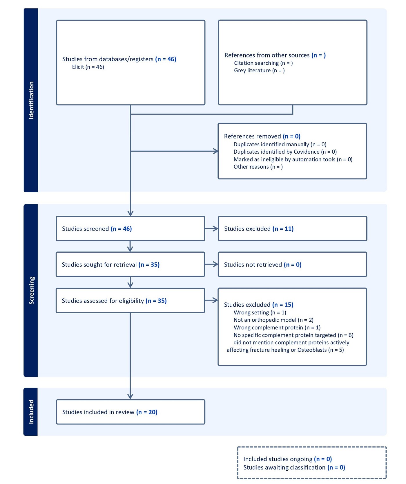
Figure 1 displays the semantic search conducted using the Elicit ML tool, which initially retrieved 4,981 records, from which the platform’s relevance ranking algorithm returned 46 studies for human screening. These 46 publications then went through both title and abstract review, after which 35 full texts were assessed for eligibility. Ultimately, 20 studies met all inclusion criteria and were incorporated into the final analysis. 11 studies were excluded during title and abstract screening, and 15 were excluded at the full-text phase, most commonly for lacking a specific complement target, failing to evaluate osteoblast or fracture-healing outcomes, or not using an orthopedic model. This review relied solely on Elicit’s database; no supplementary database or reference search was utilized to identify additional studies that may have suited our research question.
The 20 included studies encapsulated 8 fracture-model investigations, 9 non-fracture bone-metabolism or in vitro studies, and 3 doctoral theses/dissertations containing mixed mechanistic data. The fracture studies consistently used rodent models, predominantly C57BL/6 mice, with standardized femoral osteotomies stabilized by external fixation or intramedullary pinning in the single rat model.15–20 Osteotomy size was reported in some studies, such as the 0.4-mm gap model.15 The pathways investigated included C3, C5, C5a, C5aR1, and C5aR2, which were manipulated through genetic deficiency, pharmacologic antagonism (PMX53), osteoblast-specific receptor over-expression, or direct C5a stimulation. The ten non-fracture studies evaluated the effects of complement on osteoblasts, osteoclast precursors, or bone metabolism using primary murine osteoblasts, co-culture systems, ovariectomy-induced osteoporosis models, and aging models.21–28 The 2 theses and 1 dissertation contributed additional mechanistic and immunohistological data, but were judged as higher risk due to the non-peer-reviewed format and heterogeneous methodology.29–31
Figures 2 and 3 demonstrate that across osteoblast studies, C3/C3a/C3aR signaling was mainly pro-osteogenic. C3a increased ALP, Runx2, and OCN, and promoted osteoblast development.21,22,24 C3aR knockdown improved bone formation in aging mice,25 while C3 deficiency preserved osteoblast function in estrogen-deficient bone loss.23 C5a/C5aR1 signaling consistently induced a pro-inflammatory osteoblast phenotype with increased CXCL10, IL-6, and osteoclastogenic mediators.27 Osteoblast-specific C5aR1 overexpression disrupted normal bone-tissue organization and impaired osteoblast migration.17 C1q suppressed osteoblast differentiation in aging models.26
Figures 2 and 3 also show that complement manipulation produced distinct effects on fracture healing. C3-/- and C5-/- mice expressed divergent phenotypes, while C3 deficiency resulted in reduced early osteoid formation and delayed early healing, but callus parameters normalized by day 21.15 In contrast, C5 deficiency caused impaired mineralization, reduced callus volume, and diminished mechanical stiffness. C5aR1 antagonism displayed effects that were highly dependent on the inflammatory and physiological environment. In uneventful fractures, early C5aR1 blockade did not alter callus volume, BV/TV, or biomechanical strength.15 However, in fractures complicated by systemic inflammation induced by blunt chest trauma, C5aR1 inhibition restored callus quality and mechanical strength.19 Receptor-specific knockout models demonstrated that C5aR1 drives early pro-inflammatory responses, and C5aR2 modulates resolution, though both receptors contributed to overall repair.18 Osteoblast-specific C5aR1 over-activation impaired fracture healing by producing disorganized matrix, altered osteoblast migration, and delayed mineralization.17
Three mechanistic themes emerged from the analysis. The mediators regulate early inflammatory signaling that shapes osteoblast–osteoclast interactions.18,21 Complement receptors directly influence osteoblast differentiation and migration, with C3aR generally pro-osteogenic and C5aR1 pro-inflammatory.24,27 Complement effects vary by physiological setting: neutral in uneventful fractures,16 beneficial when inflammation is excessive,19 and detrimental in aging or estrogen-deficiency states.23,26 Collectively, these findings identify complement as a dynamic regulator of osteoblast behavior and fracture repair.
Figure 4 shows that the risk of bias across studies was generally low to moderate, with recurring gaps in randomization reporting, allocation concealment, and blinding. Genotype-based studies demonstrated low risk for sequence generation,15,18 whereas pharmacologic studies rarely described randomization.16,19 No fracture study reported randomization or blinding, and blinding of µCT, histology, or mechanical testing was largely unclear. Attrition was minimal, and peer-reviewed fracture studies often reported prespecified outcomes. Thesis-based studies showed unclear selective reporting. Additional concerns included single-strain and single-sex models, limited complement measurement, interference of trauma models, and non-physiologic in vitro dosing.
Discussion
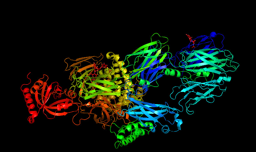
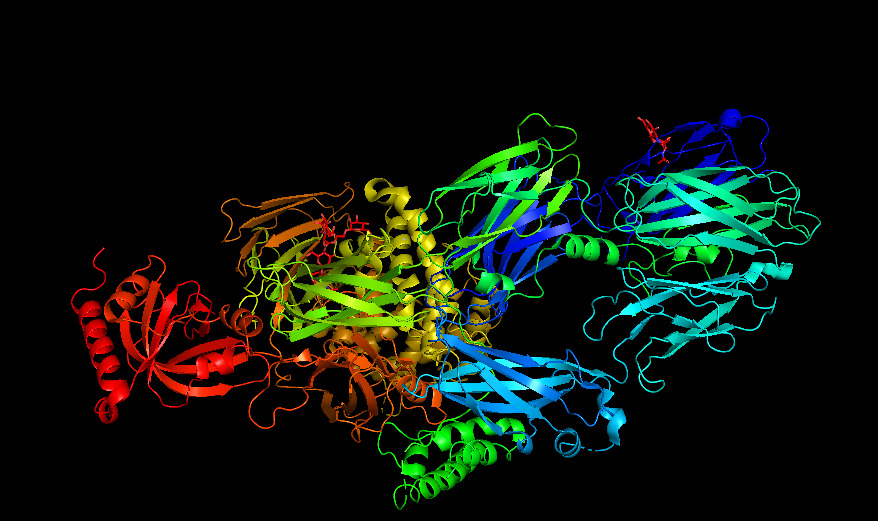
Complement signaling functions as a contextual regulator of fracture healing, and its effects depend on the ligand concentration, receptor engagement, and inflammatory context.15,16 Complement activation supports early fracture healing by coordinating immune–stromal communication. C3 and C5 cleavage generate C3a and C5a, which bind to their specific GPCR receptors (C3aR and C5aR1) located on osteoblasts and stromal cells, initiating intracellular signaling that promotes migration, differentiation, and early callus organization.15 Figure 4 shows the molecular structure of C3 in PyMOL software.
When the system is over-activated, such as during systemic trauma, the same ligand-receptor interactions shift toward pathologic inflammation, impairing osteoblast function and delaying healing.19 This conditional behavior reflects the complement proteins’ dual function as both a pro-regenerative signaling molecule and a potent amplifier of inflammation.
Non-fracture models such as osteoporosis studies are still highly informative because they isolate how complement signaling directly regulates osteoblast proliferation, differentiation, and OPG/RANKL balance outside the context of acute injury, providing mechanistic insight that supports the in vivo fracture findings.23,25 In vitro systems further clarify osteoblast-intrinsic responses to C3a and C5a, such as changes in AKP activity, apoptosis, and matrix production, which help explain the cellular behaviors observed in fracture models.20,22
Because the majority of available evidence comes from animal models and in vitro systems, the direct translation of these findings to human fracture biology remains uncertain.17,19,23 The limited availability of human data means that complement-mediated effects on osteoblast function and healing should be interpreted cautiously until validated in clinical studies.
Receptor Binding and Downstream Signaling
Mechanistic studies show that the complement system’s skeletal effects come directly from ligand–receptor engagement. C5a binds to C5aR1 on osteoblasts, activating Gαi-coupled signaling that reorganizes the cytoskeleton and promotes migration during early callus formation.20 This migration is essential for bridging the fracture gap.
When C5aR1 is over-activated, ligand binding drives excessive NF-κB activation, inflammatory cytokine release, and impaired matrix deposition, fundamentally disrupting regeneration.17
C5aR2, in contrast, appears to act as a regulatory receptor, modulating C5aR1-driven inflammation. Differential receptor activity during fracture-associated inflammation demonstrates that C5aR1 promotes pro-inflammatory signaling, whereas C5aR2 inhibits it.18 These receptor dynamics explain why the complement blockade is considered neutral in low-inflammation fractures16 but beneficial in trauma-induced systemic inflammation.19
Complement as an Amplifier of Osteoimmune Communication
Complement ligands also integrate with innate immune receptors to amplify osteoimmune signaling. C5aR1 physically and functionally cooperates with TLR2 on osteoblasts, and co-activation of these receptors increases CXCL10 production, a chemokine that strongly promotes osteoclast recruitment and differentiation.27
C3a and C5a enhance osteoclast formation and inflammatory cytokine release when combined with IL-1β, demonstrating that complement ligands act as signal amplifiers in inflammatory microenvironments.21 These interactions show complement as a mediator of osteoblast and osteoclast communication. This is consistent with the fact that bone is a direct target of complement signaling.24,29
Complement in Chronic Bone Physiology
Complement also influences chronic skeletal physiology through ligand–receptor interactions. Age-related increases in C1q impair osteoblast differentiation by binding to cell surface receptors and altering downstream signaling.26 C3aR signaling contributes to age-associated bone loss, and its inhibition activates YAP1/β-catenin pathways that restore osteoblast function.25 Estrogen-deficiency models further demonstrate that the cascade contributes to pathological bone resorption, as C3 deficiency protects against postmenopausal bone loss.23,32 These findings identify complement proteins as a shared framework connecting inflammation, aging, and osteoporosis.
Cellular Coupling Mechanisms
In vitro studies reveal that complement ligands function as paracrine coupling factors between bone-resorbing and bone-forming cells. Osteoclast-derived C3a directly stimulates osteoblast differentiation,22 while basal C3 expression appears necessary for osteogenic maturation.28 Histologic analyses confirm complement deposition and receptor expression in bone tissue,30,33 supporting its integration into skeletal homeostasis. Analyses of receptor activity demonstrate that C5aR1 and C5L2 both play a role in modulating bone metabolism.31
Clinical Implications for Complement Proteins
These mechanistic insights could have direct translational relevance. Inhibition of complement proteins may benefit poly-trauma patients, while excessive C5a signaling impairs healing.19 Complement modulation may also hold therapeutic value in osteoporosis, given the protective effects of C3 deficiency and C3aR inhibition.23,25 Complement profiling could act as a biomarker for predicting fracture outcomes, especially in cases where C5aR1 is over-activated.17 Complement-mediated osteoblast migration suggests potential applications in implant osseointegration, too.20
Future Directions Concerning Research in Complement
Future work could prioritize human studies, temporal mapping of complement activation, and receptor-specific interventions, as current evidence is dominated by animal models.15,17 Mechanistic research may perhaps further explore the system’s role in osteoblast–osteoclast coupling and TLR2 cooperation to improve understanding of the relationship.22,27 In the future, clinical trials evaluating complement antagonists in trauma-associated fracture impairment may give more insight into how fractures are treated in the clinical setting.19
Complement signaling is a central, tunable regulator of skeletal biology, supporting repair under physiologic conditions, but impairing regeneration when overactivated by trauma, aging, or receptor imbalance.15,17,26 These insights identify complement as a promising therapeutic and diagnostic target in trauma, osteoporosis, and age-related skeletal decline.
Emerging evidence suggests that complement activation profiles may have future applications in emergency medicine as prognostic biomarkers for trauma patients.34–36 Early alterations in complement components such as C3a, C5a, and soluble C5b-9 have been associated with injury severity and adverse clinical outcomes, while experimental studies indicate that dysregulated complement signaling also influences fracture healing.34–36 Although these findings are promising, further prospective clinical studies are needed before complement biomarkers can be incorporated into routine emergency care.34–36
Limitations
This study focused only on data readily available in the Elicit ML tool, which may have excluded other relevant publications. A comprehensive search utilizing other databases would provide a more detailed and accurate depiction of complement proteins in osteoblastic and fracture healing research. It is also important to consider the search strategy. This study used a semantic search strategy rather than a Boolean expression. A search method that uses stricter parameters across multiple databases could provide more accurate results.
Additionally, significant variability among study designs may limit interpretation. The evidence contains animal fracture models, in vitro tests, and various strategies for manipulating complement, such as C3/C5 deficiency,15 over-expression of C5aR1,17 receptor-specific knockouts,18 and pharmacological inhibition.16 Publication bias is also a potential limitation, as studies with negative or unexpected findings are less likely to be published. This tendency may skew the available evidence toward positive results and overestimate the apparent impact of complement modulation on bone biology.
In vitro experiments with C3a or C5a stimulation may also introduce additional variability. Measured outcomes and complement-analysis methods were reported inconsistently, varying from µCT and biomechanical testing to just histology, while complement activity was measured using different methods or not assessed at all. Certain studies merged fracture models with systemic injury or failed to clearly differentiate between open and closed fractures,16,20 making it challenging to assign complement-specific effects. The lack of a documented review protocol could possibly have led to potential methodological deviation. Future systematic reviews can overcome these limitations to produce a more comprehensive view of the literature in the field of the complement system in relation to bone fractures.
Conclusions
This systematic review shows that complement proteins exert a dual, context-dependent influence on osteoblast biology and fracture healing. Physiologic C1q–C5 activation supports osteoblast migration, differentiation, and early callus formation, whereas excessive C5a/C5aR1-driven inflammation disrupts matrix organization, enhances osteoclastogenic signaling, and impairs repair in trauma, aging, and estrogen-deficient states. These findings position complement as a tunable regulator of skeletal homeostasis and highlight the therapeutic potential of targeted complement modulation in inflammatory fracture environments. Further human studies and receptor-specific interventions are needed to translate these mechanistic insights into clinical practice.
Conflicts of interest
In compliance with the ICMJE uniform disclosure form, all authors declare the following
Payment/services info
All authors have declared that no financial support was received from any organization for the submitted work.
Financial relationships
All authors have declared that they have no financial relationships at present or within the previous three years with any organizations that might have an interest in the submitted work.
Other relationships
All authors have declared that there are no other relationships or activities that could appear to have influenced the submitted work.
Author Contributions
Concept and design: Latha Ganti, Tori M. Baer, Patrick R. Counts
Critical review of the manuscript for important intellectual content: Latha Ganti, Tori M. Baer, Patrick R. Counts
Supervision: Latha Ganti
Acquisition, analysis, or interpretation of data: Patrick R. Counts
Drafting of the manuscript: Patrick R. Counts