
Introduction
Thoracic disc herniations (TDHs) are rare pathologies that account for approximately 0.25–1% of all disc herniations, yet they carry disproportionate clinical significance due to their potential to cause spinal cord compression and progressive neurological decline.1,2 Calcification is present in up to 30–70% of cases and, when present, tends to make these lesions more complex and significantly more challenging to manage surgically.1,3 These calcified lesions are characterized by increased rigidity, a predominantly ventral location, and a greater tendency toward dural adherence, all of which substantially increase the complexity and risk of surgical intervention.4,5 These features distinguish calcified thoracic disc herniation from non-calcified disease and underscore the need for careful anatomical assessment and tailored surgical planning.
The surgical management of calcified TDHs remains challenging, with no universally accepted approach. Both anterior and posterolateral techniques have been widely described, each with distinct advantages and limitations depending on lesion location and degree of calcification.6,7 While anterior approaches allow direct access to ventral pathology, they are associated with higher morbidity, whereas posterolateral approaches may be less invasive but can be technically demanding in central or densely calcified lesions.7,8 Consequently, key decisions like the choice of surgical corridor and extent of decompression remain largely experience-dependent, which results in considerable variability in surgical strategies and outcomes.
Therefore, this narrative review aims to synthesize the current evidence on the surgical management of calcified thoracic disc herniations and to propose a structured, anatomy-driven framework to guide surgical decision-making.
Methods
A narrative literature review was performed on calcified thoracic disc herniation and its surgical management. Searches were conducted using terms such as “thoracic disc herniation,” “calcified thoracic disc herniation,” “giant thoracic disc herniation,” “thoracic discectomy,” “transthoracic approach,” “posterolateral approach,” “transpedicular approach,” “costotransversectomy,” “lateral extracavitary approach,” and “endoscopic thoracic discectomy.” Peer-reviewed clinical studies, case series, technical notes, systematic reviews, and contemporary review articles were considered. Relevant articles were selected if they discussed diagnosis, preoperative imaging, lesion morphology, surgical approach selection, outcomes, or complications. Additional studies were identified from the reference lists of selected articles. The findings were synthesized narratively to highlight practical factors that influence surgical decision-making.
Preoperative Evaluation & Decision Variables
In calcified TDH, preoperative evaluation is not performed merely to confirm diagnosis, but to determine which operative corridor can be used safely and effectively for a given lesion. Surgical planning is fundamentally based on the combined interpretation of magnetic resonance imaging (MRI) and computed tomography (CT), which together define the neural impact of the herniation, the degree and morphology of calcification, and the anatomic constraints relevant to exposure.3,5 Current literature suggests that approach selection should be individualized according to three interrelated domains. These include lesion characteristics (location, size, calcific burden, and suspicion of dural or intradural involvement), patient factors (neurological status and physiological tolerance for thoracic exposure), and the surgical environment, including available instrumentation and surgeon experience with thoracic open and minimally invasive techniques.9,10 These factors can be conceptualized as key elements of preoperative planning, as summarized in Table 1.
Lesion Characteristics
Among lesion-specific variables, location and canal occupancy constitute the first major determinants of operative corridor selection in calcified TDH. Farber et al., in a recent classification study designed specifically to standardize thoracic disc morphology, distinguished lesions according to both topography and size, separating central from paracentral discs and defining giant lesions as those occupying >40% of the spinal canal.11,12 This framework is useful because it reflects the core anatomic reality emphasized in earlier surgical series: central lesions are positioned directly ventral to the thoracic spinal cord and therefore offer little tolerance for displacement or manipulation during decompression.9,10 In contrast, more lateralized or paracentral lesions may preserve a limited eccentric working zone, even when symptomatic, and are therefore not equivalent to central lesions of similar volume.10,13 However, despite attempts to standardize morphology-based decision-making, Farber et al. did not demonstrate outcome superiority for specific approaches within each category, and subsequent clinical series continue to report variability in approach selection even for similarly classified lesions, suggesting that morphology alone may not fully dictate operative strategy.
Size modifies this relationship further. Quraishi et al. and Court et al. both emphasized that giant TDHs represent a distinct subgroup rather than simply a larger version of ordinary disease, precisely because increasing canal occupancy progressively reduces the residual space available around the cord.9,14 This has practical implications even before calcification is considered: a small central lesion may be technically less forgiving than a larger but eccentric disc, whereas a giant central lesion combines maximal ventral compression with minimal working space and therefore substantially narrows the range of acceptable operative corridors.10,11 Importantly, Farber et al. showed that when experienced spine surgeons were surveyed using their classification system, preferred access strategies shifted as lesions became both larger and more central, underscoring that these variables are not merely descriptive but directly influence preoperative planning.11 Nevertheless, this relationship is not universally prescriptive. While earlier surgical series and traditional teaching have favored anterior or anterolateral approaches for giant central lesions, more recent reports suggest that selected posterior or posterolateral techniques may achieve adequate decompression in carefully chosen cases, indicating that size and centrality, although critical, do not rigidly determine the operative corridor.15
If location and size define the spatial problem, calcification defines its technical severity.15 Quraishi et al. argued that giant thoracic discs require altered surgical strategy specifically when calcification is present, because the lesion becomes more rigid, more ventrally impactant, and more likely to lose a safe dissection plane from the dura.9 This point has been reiterated in later reviews: Bouthors et al. and Yurtluk et al. both emphasized that MRI and CT should be interpreted together in calcified thoracic disease, because MRI better delineates cord compression, cord signal change, and associated soft-tissue effect, whereas CT more accurately defines the extent, density, and morphology of calcification as well as the surrounding osseous anatomy.5,10,16 Farshad et al. likewise noted that CT is particularly important because the amount of calcification has direct consequences for resection strategy.17 In practical terms, a densely calcified lesion with sharp or beak-like ventral morphology on CT should be treated differently during planning from a softer or only partially calcified disc of similar size. At the same time, emerging imaging-based analyses suggest that calcification should not be viewed as a binary variable. Morphological features such as focal beak-like ossification or irregular ventral contours may carry greater operative significance than calcific presence alone, indicating that lesions of similar size and “calcified” classification may differ substantially in technical complexity.
The possibility of dural adherence or intradural extension further raises the threshold for caution. This should not be overstated, because preoperative diagnosis remains imperfect and the reported incidence of intradural thoracic disc herniation is low; however, the literature consistently identifies large, central, ventral, calcified lesions as the group in which this possibility must be actively considered.18,19 In an imaging comparison of intradural disc herniation versus large extradural extrusion, Nam et al. found that beak-like morphology, calcification or ossification, ill-defined lesion margins, abrupt PLL discontinuity, and the Y-sign were significantly associated with intradural disease, supporting the use of these findings as preoperative warning features rather than definitive diagnostic criteria.20 Accordingly, the preoperative aim is not simply to label a lesion as calcified, but to recognize when calcific burden, ventral morphology, and possible dural violation indicate loss of a safe cleavage plane and a materially higher-risk decompression. Once these features are present, particularly in a giant central lesion, they should be understood as factors that may exclude simpler corridors and demand a more deliberate operative strategy. Importantly, while such features have traditionally been interpreted as favoring more direct anterior exposure, recent posterior transdural and minimally invasive series have challenged this assumption in selected cases, further emphasizing that lesion characteristics must be integrated with surgical expertise rather than applied as absolute determinants of approach selection.
Patient Factors
Patient-specific variables play a decisive role in preoperative planning, as operative strategy in calcified TDHs is not dictated by lesion morphology alone, but by the interaction between anatomical constraints and the patient’s neurological and physiological reserve. Clinically, myelopathy represents the dominant presentation in thoracic disc disease, reported in approximately 60–70% of symptomatic patients, which reflects the propensity of these lesions, particularly central ones, to produce direct ventral cord compression.10,21 This has immediate implications for surgical planning. Progressive neurological decline, including gait disturbance, long-tract signs, or sphincter dysfunction, reduces tolerance for incomplete or indirect decompression and shifts the operative objective toward achieving reliable ventral cord decompression in a single stage.21–23 As emphasized by Sharma et al., the severity of neurological compromise is not only an indication for surgery but also a determinant of how aggressively decompression must be pursued, particularly in calcified lesions where the margin for safe manipulation is already limited.13
At the same time, neurological status must be interpreted alongside the patient’s physiological capacity to tolerate the planned exposure. Thoracic approaches, especially transthoracic and transpleural corridors, are inherently associated with violation of the thoracic cavity and increased perioperative burden, which may not be acceptable in patients with limited pulmonary reserve or significant comorbidity.24 Mulier et al. identified compromised respiratory function as a key limitation for transthoracic surgery, a concept that has been echoed in more recent reviews, where the expansion of posterolateral and minimally invasive techniques is partly attributed to efforts to reduce approach-related morbidity.24,25 Importantly, this introduces a practical tension in surgical decision-making: while severe myelopathy may favor more direct and extensive decompression, patient frailty or reduced physiological reserve may necessitate selection of a corridor with lower access-related burden. Accordingly, preoperative planning must balance the need for adequate neural decompression against the patient’s ability to tolerate the surgical approach, rather than considering either factor in isolation.
Surgical Environment & Expertise
Beyond lesion morphology and patient-specific considerations, the surgical environment represents a critical and often underappreciated determinant of operative strategy in calcified TDH.26,27 Despite decades of surgical evolution, no universally accepted approach exists, and contemporary literature consistently emphasizes that approach selection remains strongly dependent on surgeon experience, institutional resources, and familiarity with specific techniques. This variability is not trivial. In a systematic review and meta-analysis of surgical management for thoracic disc herniation, overall complication rates ranged widely from approximately 12% to 48%, with neurological complications reported in around 5% of cases, underscoring the heterogeneity of outcomes across different techniques and practice settings. In contrast, individual high-volume series using specific approaches have reported favorable outcomes, with symptom improvement exceeding 90% in selected cohorts, suggesting that results are closely linked to technical expertise and case selection rather than the inherent superiority of any single approach.26,28,29
This introduces a fundamental challenge in preoperative planning: even when lesion characteristics and patient factors are similar, operative decisions may differ significantly depending on the surgeon’s training and available infrastructure. Techniques such as thoracoscopic, endoscopic, or advanced posterolateral approaches require dedicated experience and are not universally reproducible, limiting their applicability outside specialized centers.13,25 Consequently, the “optimal” surgical corridor is often not defined in absolute terms, but rather as the most appropriate approach that can be executed safely and reliably within a given surgical environment. Recognizing this, preoperative decision-making must incorporate not only anatomical and clinical variables, but also the practical capabilities of the treating team, thereby acknowledging that variability in outcomes is, in part, a reflection of variability in expertise. This lack of standardization further reinforces the need for a structured, anatomy-driven framework to guide approach selection in a consistent and reproducible manner.
Surgical Approaches: Evidence, Indications, and Limitations
The surgical management of calcified TDH remains characterized by the absence of a universally accepted approach, with multiple operative corridors described and none demonstrating clear superiority across all clinical scenarios.7,27,30 Accordingly, the choice of surgical approach is best understood not as a fixed algorithm, but as a decision-making process guided by the anatomical and clinical variables outlined above. The following section therefore examines the major surgical strategies in the context of their indications, strengths, and limitations, with emphasis on how each approach aligns with specific lesion and patient profiles. The principal technical variations within each surgical corridor and their distinguishing characteristics are summarized in Table 2.31–42
Anterior and Anterolateral Approaches
Anterior and anterolateral approaches are most commonly indicated for lesions characterized by central location, large canal occupancy, and dense calcification, where direct ventral access allows decompression of the spinal cord with minimal manipulation. This rationale is consistently supported across the literature.43–46 In a systematic review of 11 studies including 164 patients, Gong et al. reported that approximately 60% of giant calcified TDHs were managed through transthoracic approaches, with neurological improvement or stabilization achieved in up to 96% of cases, reinforcing the role of ventral access in high-risk morphologies.46 Similarly, Fujimura et al., in a series of 33 patients undergoing anterior decompression and fusion via an extrapleural approach, demonstrated favorable neurological recovery in patients with myelopathy, further supporting anterior strategies for ventral cord compression.47
From a technical perspective, the principal advantage of anterior approaches lies in direct visualization and removal of ventral pathology, which is particularly relevant in calcified lesions where a safe dissection plane is often absent. This benefit has been maintained in less invasive adaptations of ventral surgery. Quint et al., in a prospective cohort of 167 thoracoscopic microdiscectomies, reported an overall complication rate of 15.6%, demonstrating that thoracoscopic approaches can achieve effective decompression with reduced morbidity compared to traditional thoracotomy in experienced centers.48 However, this advantage must be interpreted in the context of operative burden.49 In comparative studies, anterior approaches have been associated with longer operative time, greater blood loss, and prolonged hospital stay. Arts and Bartels, in a cohort comparing mini-transthoracic and posterior transpedicular approaches, found that anterior surgery carried significantly higher surgical burden without clear superiority in all outcome measures.6 Similarly, registry-level data from Kerezoudis et al. suggest that anterior approaches are associated with longer length of stay and a trend toward higher complication rates, highlighting the physiological cost of thoracic exposure.8
Importantly, the role of anterior approaches remains a subject of ongoing debate. While traditional surgical paradigms have favored ventral exposure for giant central calcified lesions, more recent posterior and minimally invasive series have challenged the necessity of anterior access in all such cases. Furthermore, even within the anterior category, controversy persists regarding the optimal technique, with open thoracotomy, mini-thoracotomy, and thoracoscopic approaches offering differing trade-offs between exposure and morbidity. Accordingly, anterior and anterolateral approaches should not be viewed as universally required for central calcified TDHs, but rather as strategies that provide the most direct and controlled decompression when ventral pathology is severe and when patient physiology and surgical expertise permit their safe execution.
Posterolateral Approaches
Posterolateral approaches, including transpedicular, transfacet, costotransversectomy, and lateral extracavitary techniques, have historically been indicated for paracentral or lateral thoracic disc herniations, where access to the ventral canal can be achieved without traversing the thoracic cavity.50 However, their role has progressively expanded beyond these traditional indications. In a series of 51 patients treated using a modified transfacet pedicle-sparing approach, Carr et al. reported significant improvement in both pain and myelopathy with a major complication rate of 3.9%, supporting the safety and efficacy of this technique in appropriately selected cases.51 Similarly, Kashyap et al., in a larger cohort of 86 patients, demonstrated symptom resolution or improvement in approximately 91%, further establishing posterolateral strategies as viable alternatives to more invasive ventral approaches.29 These outcomes highlight one of the principal strengths of posterolateral surgery: the ability to achieve effective decompression while avoiding thoracic cavity violation, thereby reducing access-related morbidity and making these approaches particularly attractive in patients with limited pulmonary reserve.
Despite these advantages, posterolateral approaches are inherently constrained by their indirect trajectory to ventral pathology, which may limit visualization and control in central, giant, or densely calcified lesions.52 This limitation has traditionally led to the view that such morphologies are better managed through anterior or anterolateral corridors. However, this paradigm is increasingly being challenged. Shedid et al., in a study of posterior minimally invasive transpedicular surgery for TDH, reported successful management of central lesions with a mean canal compromise of 61%, minimal blood loss (around 250 mL), and no postoperative neurological deterioration, which suggests that even lesions that were previously considered as unsuitable for posterior access may be addressed safely in experienced hands.53
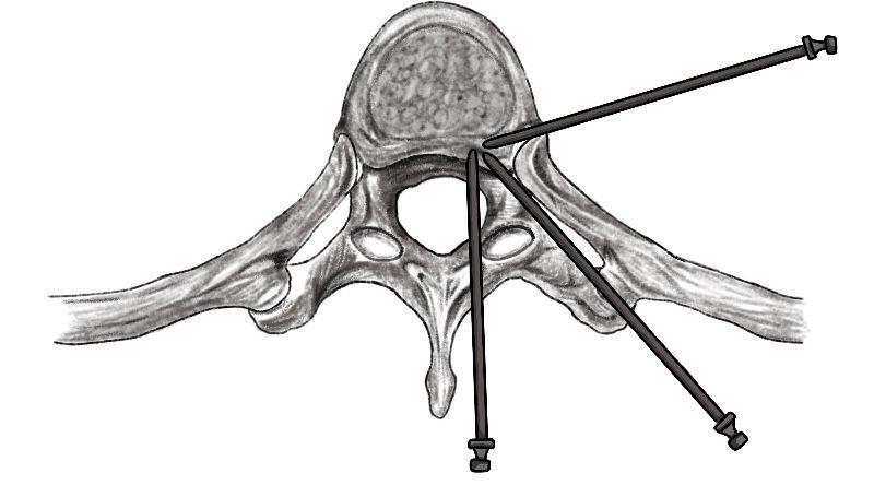
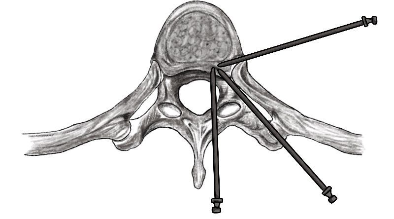
Additional evidence also challenges assumptions regarding the extent of exposure required for calcified disease.30,54 In a comparative study of 65 patients undergoing costotransversectomy, transpedicular, or transfacet approaches for calcific TDH, Corazzelli et al. found no significant differences in neurological recovery, complication rates, or extent of resection among the techniques, despite substantial differences in surgical exposure and operative time.30 These findings suggest that larger or more invasive exposures do not necessarily translate into superior outcomes, emphasizing the importance of tailored, anatomy-driven strategy rather than routine escalation of surgical corridor.55,56 Taken together, posterolateral approaches should not be viewed merely as alternatives to anterior surgery for less complex lesions, but as a flexible and evolving set of techniques capable of addressing a broad spectrum of thoracic disc pathology, provided that their limitations in ventral control are recognized and balanced against their lower physiological burden. These posterior and posterolateral trajectories are illustrated schematically in Figure 1.
Minimally Invasive and Endoscopic Approaches
Minimally invasive and endoscopic approaches to TDH have emerged as strategies aimed at reducing the morbidity associated with both anterior thoracic exposure and extensive posterior bony resection, while still achieving effective neural decompression.57–59 These techniques are most consistently indicated for soft, paracentral or lateral lesions, where a transforaminal or extraforaminal corridor permits access to the pathology with minimal disruption of surrounding structures.60–62 Bae et al. reported that transforaminal endoscopic thoracic discectomy provides favorable clinical outcomes in such cases, reinforcing its role as a minimally invasive alternative in carefully selected patients.63 Similarly, Ruetten et al. demonstrated that full-endoscopic uniportal techniques can be applied to both soft and partially calcified thoracic disc herniations, emphasizing that successful application depends on individualized planning based on lesion morphology and surgeon experience.64
From an outcomes perspective, available series suggest that endoscopic approaches can achieve substantial and durable clinical improvement in selected cohorts. Houra et al., in a study with 5-year follow-up of 16 patients, reported greater than 50% reduction in VAS pain scores and ODI in 15 of 16 patients, with 62.5% of cases involving partially or totally calcified discs, indicating that favorable outcomes are achievable even beyond purely soft-disc pathology.65 These findings support the principal advantages of minimally invasive approaches, including reduced tissue disruption, lower blood loss, shorter recovery, and avoidance of thoracic cavity violation. However, these benefits must be interpreted within the context of strict indications and technical demands. Multiple authors, including Choi and Lee, have emphasized that thoracic endoscopic surgery carries a steep learning curve and requires precise anatomical understanding, limiting its widespread reproducibility outside experienced centers.66–68
Importantly, the role of minimally invasive and endoscopic approaches in calcified, central, or giant TDHs remains controversial. While emerging reports, including those by Farshad et al. and recent case-based series, demonstrate that even giant or centrally located calcified lesions may be approached endoscopically using advanced techniques such as foraminoplasty, these applications remain highly selective and are not yet supported by robust comparative data.17 Accordingly, minimally invasive and endoscopic approaches should be understood as highly effective in well-selected lesions and experienced hands, but not as universally applicable alternatives to open anterior or posterolateral strategies, particularly in the setting of extensive calcification or complex ventral pathology. The principal controversies and evolving concepts in approach selection for calcified TDH are summarized in Table 3. Based on these considerations, commonly encountered patterns of anatomical and clinical features that tend to favor specific operative corridors are outlined in Table 4.
Conclusion
Taken together, the available evidence underscores that no single surgical approach is universally optimal for calcified TDH, as each strategy involves distinct trade-offs between exposure, safety, and physiological burden. Anterior and anterolateral approaches provide the most direct access to ventral pathology but at the cost of greater invasiveness, whereas posterolateral techniques offer lower access-related morbidity with evolving capability in more complex lesions. Minimally invasive and endoscopic strategies further reduce surgical trauma but remain highly dependent on case selection and surgeon expertise. Accordingly, optimal management requires integration of lesion characteristics, patient-specific factors, and surgical environment, rather than adherence to a fixed hierarchy of approaches.
Acknowledgements
None
Author Contributions
All authors contributed to the study conception and design. Material preparation, data collection and analysis were performed by Stylianos Kapetanakis, Paschalis Tsioulas, and Lazaros Poultsides. The first draft of the manuscript was written by Benjamin Cappin and all authors commented on previous versions of the manuscript. All authors read and approved the final manuscript.
Funding
The authors declare that no funds, grants, or other support were received during the preparation of this manuscript.
Competing Interests
The authors have no relevant financial or non-financial interests to disclose.
Previous presentations
This paper is not based on any previous communication to a society or meeting.