
Introduction
Subtrochanteric femur fractures are challenging fractures to manage due to the strong deforming muscular attachments of the proximal femur. The estimated incidence of these fractures is 15-20 per 100,000 individuals and they are commonly due to high energy mechanisms in young patients and falls from standing in the geriatric population.1–3 Antegrade, reamed intramedullary nailing is the gold standard treatment of these injuries, but the optimal nail design and subsequent entry portal has been debated over the years.1,4,5 Piriformis entry nails provide the advantage of a straight nail in the coronal plane which simplifies fracture reduction and avoids iatrogenic varus malreduction.6 However, technical difficulty with obtaining the piriformis entry portal on larger patients, abductor weakness and increased risk of injury to the medial femoral circumflex artery have led to the popularization of greater trochanteric entry nails.1,4,5,7–9
A drawback of trochanteric nails for subtrochanteric femur fractures is the propensity for varus malreduction if the entry portal is too lateral, presence of fracture site comminution or eccentric medial cortical reaming.6,10 Varus malreduction is poorly tolerated due to the high strain environment of the proximal femur and greater than 10° of malalignment at the fracture site increases the risk of delayed union and nonunion.9,11 Therefore, obtaining an adequate, functional reduction and maintaining it with a stable implant is crucial to optimize outcomes in subtrochanteric femur fractures. This goal becomes more difficult to achieve in proximal or comminuted subtrochanteric femur fractures that involve the meta-diaphyseal junction or in a patient with poor bone quality resulting in decreasing nail canal fill. Even if the fracture is anatomically reduced, nail-canal mismatch can lead to varus malreduction during nail passage or progressive varus collapse in poor bone quality.12
Defining the ideal starting point during trochanteric nailing of subtrochanteric femur fractures is difficult due to the significant degree of greater trochanteric anatomical variation and variability in the proximal valgus bend of cephalomedullary nail designs.12–15 The ability to preoperatively template and thus predict varus malreduction would greatly benefit surgeons so they could consider an alternative entry portal location or nail design.12 Preoperative templating of the uninjured femur has been advocated to achieve this goal but may not be feasible in the acute trauma setting where patient positioning and radiographic technique can limit image quality.1,12 Therefore, we seek to predict varus malalignment based on the anteroposterior femur injury radiographs and diameter of the nail utilized. The primary aim our study is to determine if the width of the canal at the fracture site minus nail diameter correlates with final proximal femur neck shaft angle. We hypothesize that as the difference between fracture site canal width and diameter of the nail increases so will the risk of varus malalignment.
Methods
Ethical Considerations
This study was reviewed and deemed an exempt retrospective study by our institution’s local Institutional Review Board (IRB-01). Study approval number IRB202301992. Data was stripped of patient identifiers once the data collection was completed by the study team. It was stripped prior to data analysis. The data was password protected and secured on an institutional approved server.
Detailed Methods
Patients who underwent intramedullary nailing for subtrochanteric femur fractures from January 1st, 2016 to July 1st, 2023 at an academic level one trauma center were identified by querying the medical record for Current Procedural Terminology (CPT) code 27245 (treatment of intertrochanteric, peritrochanteric or subtrochanteric femoral fracture with intramedullary implant). The subtrochanteric region was defined as within 5cm from the lesser trochanter. Patients were excluded if they were younger than 18 years old, older than 89 years old, had less than 6 weeks of postoperative follow-up or had fractures that involved the intertrochanteric region.
A total of nine surgeons were involved in the study (seven fellowship trained in trauma and two fellowship trained in sports medicine) and worked with resident surgeons as assistants. The choice of cephalomedullary nail (CMN) to utilize was left to the discretion of the surgeon. The two nails analyzed in the study were the TFN-Advanced Proximal Femoral Nailing system (TFNA) and the FRN-Advanced Femoral Recon Nailing System Greater Trochanteric entry nail (GTE) (Depuy Synthes, West Chester, PA). Both nails have an equivalent 5o lateral bend. The GTE nail has a screw to shaft angle of 130o.
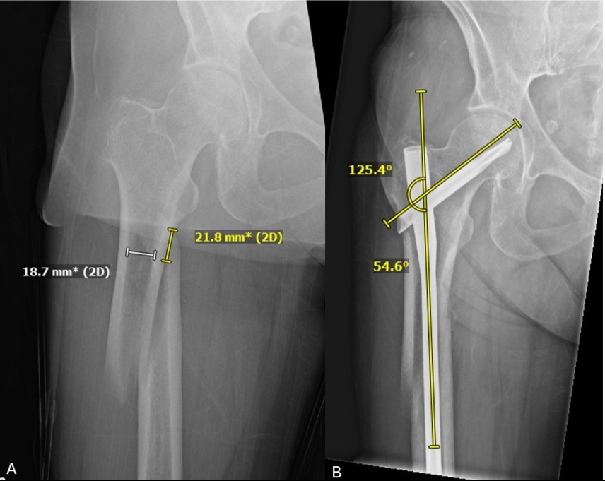
The following baseline patient characteristics were then retrospectively collected from the medical record: age, sex, body mass index (BMI), tobacco use, presence of diabetes mellitus, follow-up duration in months, mechanism of injury, AO/OTA fracture classification, and presence of open fracture. The following fracture and implant characteristics were retrospectively collected by reviewing radiographs and operative reports: fracture location distal to the lesser trochanter, width of canal at the fracture site, nail diameter utilized and neck shaft angle at most recent follow-up. The canal width at the fracture site minus the diameter of the nail (nail-canal mismatch) was calculated to represent the extent of canal fill of the nail (Figure 1). The following fracture related complications were also recorded: nonunion, varus malunion (femoral neck shaft angle <120°), fracture site shortening greater than 2mm, delayed union (fracture union greater than 9 months) and posttraumatic arthritis.
_anteroposterior_(ap)_injury_radiograph_of_a_right_proximal_femur_demonstrating_how_the.jpeg)
Statistical Analysis
CMNs were initially analyzed together and then divided into TFNA and GTE cohorts. Descriptive statistics were calculated using the mean with standard deviation (SD) for continuous variables and total number (n) with percentage for categorical variables. Univariate analysis of the descriptive statistics was then performed to compare baseline patient characteristics, injury characteristics, fracture characteristics, outcome data and complications between the cohorts. Two-independent samples t-test and Chi-square tests were used to evaluate the continuous and categorical variables, respectively. Pearson correlation coefficient (r) was utilized to determine the correlation between canal width at the fracture site minus the diameter of the nail with final neck shaft angle. An alpha of 0.05 was used for statistical significance. These analyses were completed using Excel 2022 (Version 2408 Redmond, WA, USA).
Blue Sky Statistics was utilized to perform regression analysis. Linear regression was used to evaluate the relationship between fracture site to nail difference and final neck-shaft angle as a continuous variable. Logistic regression was used to evaluate the relationship between fracture site to nail difference and the risk of varus malreduction, defined as neck-shaft angle <1200. Analysis performed separately for all nails, GTE nails, all TNFA nails, 1300 TNFA nail, and 1250 TNFA nail.
Results
One hundred and fifteen subtrochanteric femur fractures were reviewed and 71 met the inclusion criteria. 35 were fixed with a TFNA and 36 with GTE nail. Baseline characteristics were similar between the cohorts in terms of sex, BMI, tobacco use, presence of diabetes mellitus, follow up and presence of open fractures. The TFNA cohort was significantly older (mean age 63.8 years vs 40.97 years, p<0.001) and sustained more low energy injuries compared to the GTE cohort (p<0.001). The fracture AO/OTA fracture classification distribution between the groups was similar except for more 32B3 fractures occurring in the GTE cohort (0% vs 22%, p=0.003) (Table 1).
In terms of fracture and implant characteristics, there was no difference in mean width of the intramedullary canal at the fracture site, canal width at the fracture site minus the diameter of the nail or final mean neck shaft angle. There were significant differences in mean fracture location measured from the lesser trochanter (7.18mm vs 15.93mm, p=0.016) and frequency of 10mm diameter nails utilized (28% vs 58%, p=0.011) (Table 2).
When considering all CMNS, there was a weak negative correlation with the canal width at the fracture site minus the diameter of the nail and final neck shaft angle (Pearson correlation coefficient (r) -0.4357, p=<0.001). There was not a significant correlation between canal width at the fracture site minus the diameter of the nail with final neck shaft angle for GTE nails ((r) -0.1874, p=0.274). However, there was moderate negative correlation for TFNAs ((r) -0.5564, p=<0.001). When TFNAs were subdivided into 125° and 130° cohorts, there was a stronger negative correlation for 125° TFNAs ((r) -0.6781, p=0.001) compared to the 130° TFNAs ((r) -0.5149, p=0.041).
Linear regression analysis demonstrated a significant negative association between fracture site to nail difference and neck shaft angle for all nails. Significant associations were present for all nails except for the individual analysis of GTE. The strongest effect was for the 1250 TFNA (β=-0.757, R2=0.46, p=0.001). For the 1250 TFNA nail, every 1 mm increase in fracture site-nail difference decreases the final neck shaft angle by 0.7570. Logistic regression analysis found a similar relationship. The greater the fracture site to nail difference, the increased odds of varus malreduction. Significant findings were found for all nails and the 1250 TFNA nail. The 1250 TFNA nail had the strongest effect (OR 1.33, 1.06-1.81). For the 1250 TFNA, each 1 mm increase in fracture site-nail difference is associated with a 32% increase in the odds of varus malreduction. In terms of fracture related complications, there was no significant difference in nonunion, varus malunion, fracture site shortening, delayed union and posttraumatic arthritis between the cohorts (Table 3).
Discussion
The primary aim of this study was to determine if cephalomedullary nail (CMN) canal fill influences final alignment of the proximal femur in subtrochanteric femur fractures. We sought to measure this by correlating the canal width at the fracture site minus the diameter of the nail (termed nail-canal mismatch) with the final neck shaft angle. Our results found no significant correlation when greater trochanteric entry recon nails (GTE) were utilized but there was a significant negative correlation with CMNs with sliding blade/screws (TFNA). In other words, for the TFNA nails, as the nail-canal mismatch increased, the risk of a final varus neck shaft angle increased. This correlation was most significant for the 125° TFNAs where the linear regression analysis demonstrated for every 1 mm increase in nail-canal mismatch, there was a decrease in the final neck shaft angle by 0.7570. To our knowledge, this is the first clinical study that demonstrates the significance of nail-canal fill at the fracture site with final coronal alignment in subtrochanteric femur fractures when CMNs are utilized. Adjacent evidence from pertrochanteric hip fractures indicates that utilizing a CMN with an angle less than the native neck shaft angle is associated with more varus malreductions and fracture displacement.16 This is in contrast to prior studies on intramedullary nailing of femoral shaft fractures where it has been demonstrated that nail-canal difference is not significantly correlated with time to union or nonunion.17–19
The literature comparing clinical outcomes of cephalomedullary to reconstruction nails for subtrochanteric femur fractures is sparse. Yoon et al retrospectively compared cephalomedullary nails to reconstruction nails in sixty patients greater than 65 years old and found no statistical difference in malalignment but noted the reconstruction nail trended towards less varus (10% vs 30%) and anterior angulation (23% vs 37%) compared to cephalomedullary nails which is similar to our findings. Average time to boney union and Merle d’Aubigne-Postel scores were also similar between the constructs.20 This study contrasts with our study in that it included twenty atypical subtrochanteric femur fractures and only a geriatric population. Huang et al performed another retrospective cohort study comparing ten reconstruction nails to thirteen cephalomedullary nails and found longer operative time and more blood loss in the reconstruction nail group. They also demonstrated more postoperative change in the neck shaft angle in the recon group versus the cephalomedullary nail group (8.8° vs 4.7°) with more varus malreductions seen in the reconstruction nail cohort.21 Both implants demonstrated similar time to union, nonunion and functional results. While the patient cohorts are similar to our study, it should be noted the reconstruction nail utilized was composed of 316L stainless steel as opposed to a titanium alloy which may explain some of the discrepancies between our study. However, the sample size is too small to make any strong conclusions from their findings.
There have been multiple biomechanical studies comparing CMNs and reconstruction nails for subtrochanteric femur fractures. McKeeman et al demonstrated that the DePuy Synthes TFN-ADVANCED withstood significantly higher maximum force and approached higher energy to failure compared to the DePuy Synthes FRN-ADVANCED. It also withstood more proximal segment motion before failure compared to the FRN-ADVANCED. There was no difference in osteotomy site motion or construct stiffness.22 Yoon et al performed a similar biomechanical study but utilized an osteoporotic fracture model and still found a higher load to failure in the cephalomedullary nail group (Synthes TFNA) compared to reconstruction nails (Synthes A2FN, Expert Asian Femoral Nail) but with similar stiffness.23 Implants in both studies sustained much higher loads to failure than is required for normal walking in a 75-kg individual and are unlikely to be clinically relevant differences.22,23 While optimizing stiffness is essential to creating a healthy bone healing environment, these studies were not designed to assess how implant design and fit influence final fracture alignment which is critical to successful outcomes in subtrochanteric femur fractures.
The rate of varus malunion in our study was 7% with no significant difference in the TFNA cohort (11%) versus the GTE cohort (3%). However, the logistic regression analysis in the 1250 TFNA cohort demonstrated that each 1 mm increase in nail-canal mismatch is associated with a 32% increase in the odds of varus malreduction. Avoiding varus malalignment, especially in subtrochanteric femur fractures, is essential as it is well established that it has been shown to increase the risk of nonunion, lag screw cut-out, complication rates, and worse functional outcomes scores.9,24–27 Panteli et al retrospectively reviewed 309 subtrochanteric femur fractures fixed with cephalomedullary nails and found malreduction of 5-10° of varus and greater than 10° of varus was associated with 3.25 and 6.76 greater odds of reoperation respectively.27 Varus malalignment combined with medial cortical comminution was associated with a 53% risk of nonunion in a retrospective review by Krappinger at al.24 In a risk assessment model performed by Wang et al, avoiding varus and reducing the medial cortex has been demonstrated to be protective against nonunion.26
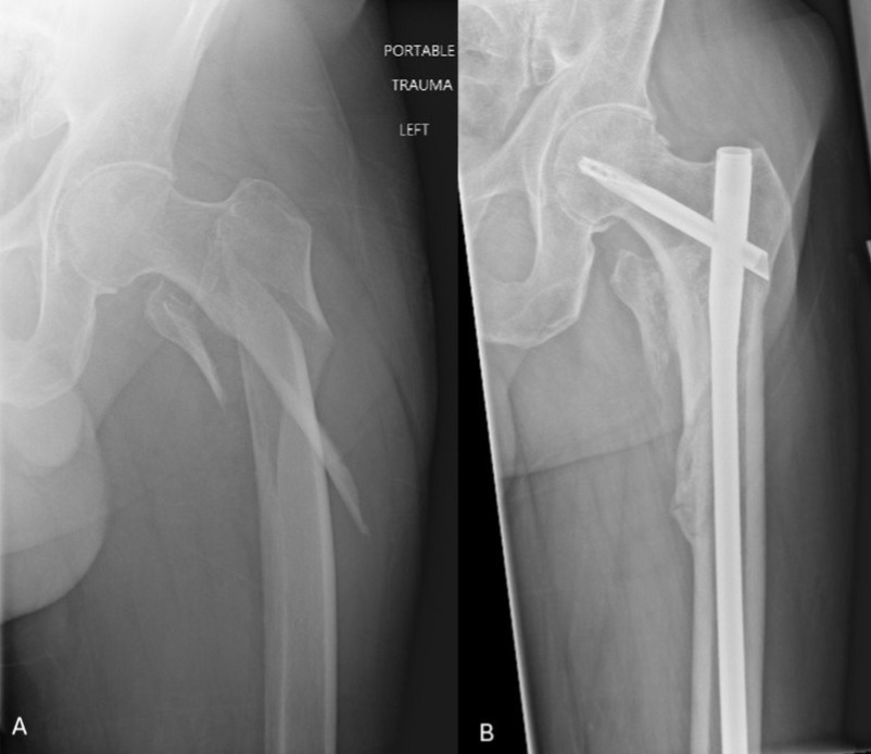
A thorough understanding of proximal femur osteology, deforming forces, fracture pattern characteristics and how these interplay with intramedullary nail design are essential elements that must be accounted for to avoid varus malreduction.24 First, subtrochanteric femur fractures can produce short proximal segments and extend into the intertrochanteric region where the medullary canal broadens at the metaphyseal segment of the bone which facilitates varus malalignment due to lack of cortical fit with the nail and deforming force of the hip abductors.9,28,29 We sought to quantify and predict this by correlating nail-canal mismatch with varus malreduction. This correlation was only significant for the 1250 TFNA cohort where each 1 mm increase in nail-canal mismatch was associated with a 32% increase in the odds of varus malreduction. Therefore, based on the results of our study, one should consider using a 1300 TFNA if a CMN is desired in patients with very proximal fracture patterns or capacious proximal femur anatomy due to osteoporosis (Figure 2).
_anteroposterior_(ap)_injury_radiograph_of_a_left_proximal_femur_demonstrating_a_subtro.jpeg)
Variations in proximal femur anatomy and bone quality can make determining the ideal entry portal challenging but most would agree that utilizing a starting point just medial to the tip of the greater trochanter to account for the valgus bend of cephalomedullary nails is ideal to avoid varus30-32. The differences in proximal body nail design should be considered when deciding which implant is best suited for a subtrochanteric femur fracture. Within the context of our study, the Synthes TFNA has a larger proximal diameter measuring 15.66 mm compared to 13 mm proximal diameter in the FRN-Advanced Femoral Recon Nail which may influence the final reduction once the nail is fully seated.22,23 This difference is likely to be more influential when the fracture site is more proximal, which was seen in our TFNA cohort, or when a lateral starting portal is utilized.23
This study has several notable limitations. First, it is a retrospective study that cannot control surgeon bias when selecting implants, methods of reduction or provisional fixation. In addition, it lacks patient reported outcome measures as this data is not routinely collected. Second, the cohorts also have notable differences in their characteristics. The TFNA cohort was older, sustained more low energy mechanisms of injury and was utilized to fix more proximal fractures. Statistical matching techniques were unable to be utilized to account for these differences due to the small samples size. Additionally, fracture location, canal width, and neck-shaft angle were measured retrospectively from radiographs, which are easily influenced by hip rotation, flexion and radiograph quality. Moreover, additional factors such as bone quality and intraoperative reduction quality can not be controlled for and may have influenced both implant choice and outcomes.
Conclusion
Overall, GTE femoral recon nails and TFNAs demonstrated similar rates of varus malalignment and nonunion for subtrochanteric femur fractures. However, increased nail-canal mismatch was associated with increased risk of varus neck shaft angles for the 125° TFNAs. These findings emphasize the importance of understanding nail geometry and canal fill, particularly in patients with capacious canals and proximal fractures, in order to avoid varus malalignment.
Author Contributions
Momin Ahmed BS: contributed to data acquisition, reviewing the manuscript draft, has approved of final version of manuscript and is agreeable to be accountable for all aspects of the study.
John Mickley MD: contributed to data analysis, reviewing the manuscript draft, has approved of final version of manuscript and is agreeable to be accountable for all aspects of the study.
Andres Lopez MD: contributed to data acquisition, reviewing the manuscript draft, has approved of final version of manuscript and is agreeable to be accountable for all aspects of the study.
Patrick Baki MD: contributed to data acquisition, reviewing the manuscript draft, has approved of final version of manuscript and is agreeable to be accountable for all aspects of the study.
Alex Moses MD: contributed to study design, data acquisition, reviewing the manuscript draft, has approved of final version of manuscript and is agreeable to be accountable for all aspects of the study.
Porter Young MD: contributed to design and conception, reviewing/editing the manuscript draft, has approved of final version of manuscript and is agreeable to be accountable for all aspects of the study.
Disclosures
All of the above authors have no declarations of interest to disclose.
Further Information
No funding was utilized for this project.
This project was presented as a podium presentation at the 2026 Southern Orthopaedic Association Annual Meeting in White Sulphur Springs, WV, as a poster at the 2026 Florida Orthopaedic Society Annual Meeting in Orlando, FL and as a poster at the 2026 University of Florida College of Medicine-Jacksonville Celebration of Research in Jacksonville, FL.