Introduction
Lipomas are common benign soft-tissue tumors that usually present as solitary, soft, mobile subcutaneous nodules on the trunk and proximal limbs.1–3 When multiple lesions are present, clinicians should consider inherited and syndromic lipomatosis, including familial multiple lipomatosis (FML), multiple symmetric lipomatosis (MSL; Madelung’s disease), Dercum’s disease, and Gardner syndrome.2,4–6 FML is a rare hereditary disorder characterized by multiple, well-encapsulated subcutaneous lipomas predominating on the trunk and extremities, often sparing the head and neck.1,3,4,7,8 Although benign, lesions may cause pain, cosmetic concern, and functional limitation. Diagnosis remains largely clinical and hinges on distribution and a careful pedigree, while management is guided by symptoms and the need to exclude atypical lipomatous tumor/well-differentiated liposarcoma (ALT/WDL) in selected lesions.4 We report a limb-predominant case of FML in a 36-year-old male treated with open en bloc excision and provide a focused review relevant to orthopedic and soft-tissue surgical practice.
Case Presentation
Ethical Considerations
Written permission to publish clinical details and images was obtained from the patient. A copy of the signed permission form will be uploaded as a supplementary file.
Timeline
The first lump was noted approximately 6 months prior to presentation, followed by additional nodules in both arms and thighs. Symptoms remained mild, but the number and size increased, prompting outpatient assessment and elective surgery.
Patient Information
A 36-year-old male presented with multiple gradually enlarging swellings over both upper and lower limbs for 6 months. The lesions caused mild discomfort without functional limitation. He denied fever, weight loss, night sweats, fatigue, or constitutional symptoms. He had no known comorbidities, no prior surgeries, and no history of tobacco, alcohol, or illicit drug use. Initial history did not reveal a family history of similar lesions.
Drug History and Allergies
He was not taking regular medications and reported no known drug allergies.
Family History
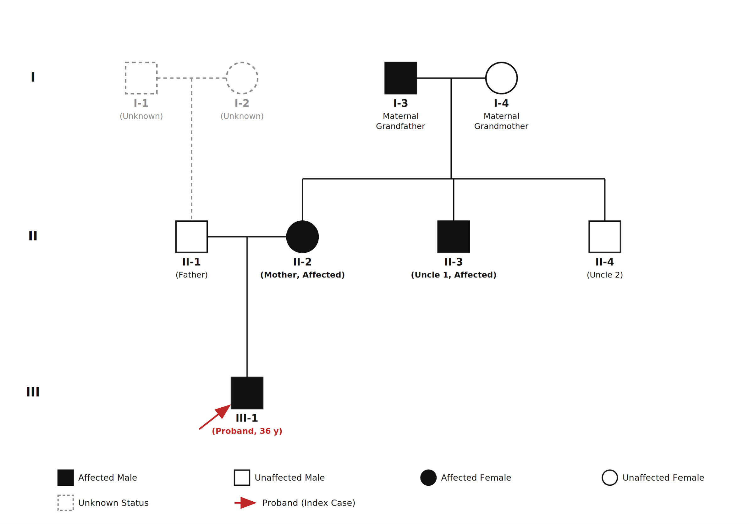
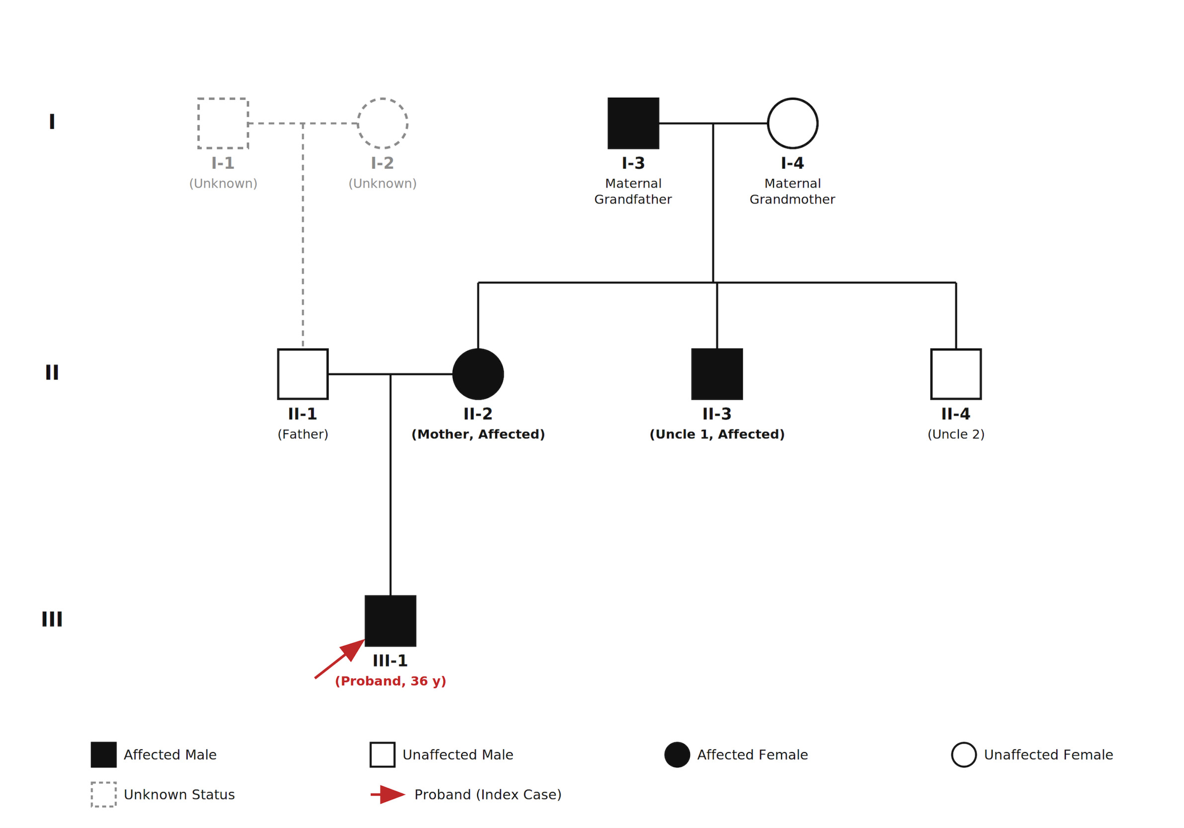
History revealed affected maternal relatives, including the patient’s mother and maternal uncle.
Social History and Review of Systems
He lived independently and continued manual work. Review of systems was otherwise unremarkable.
Clinical Findings
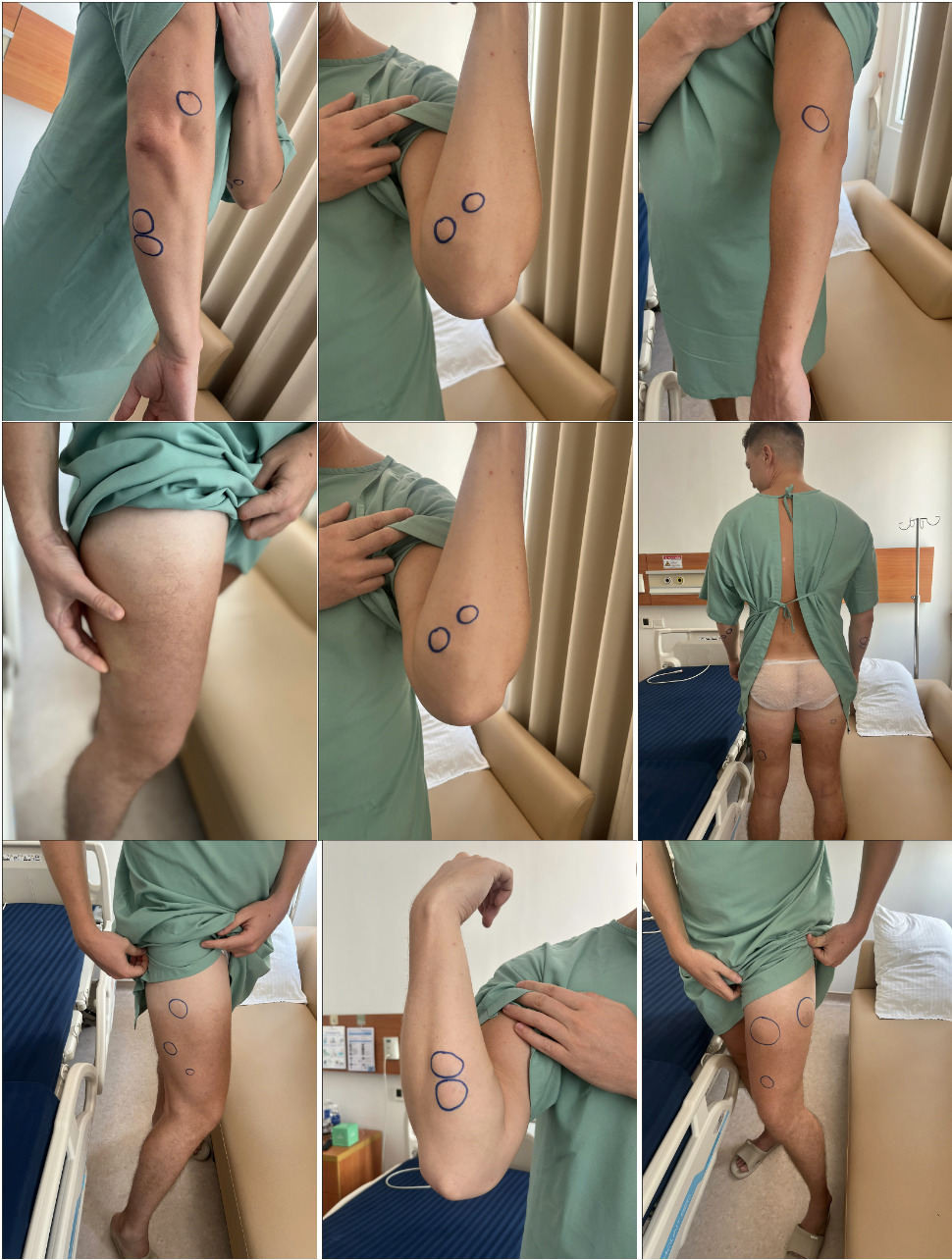
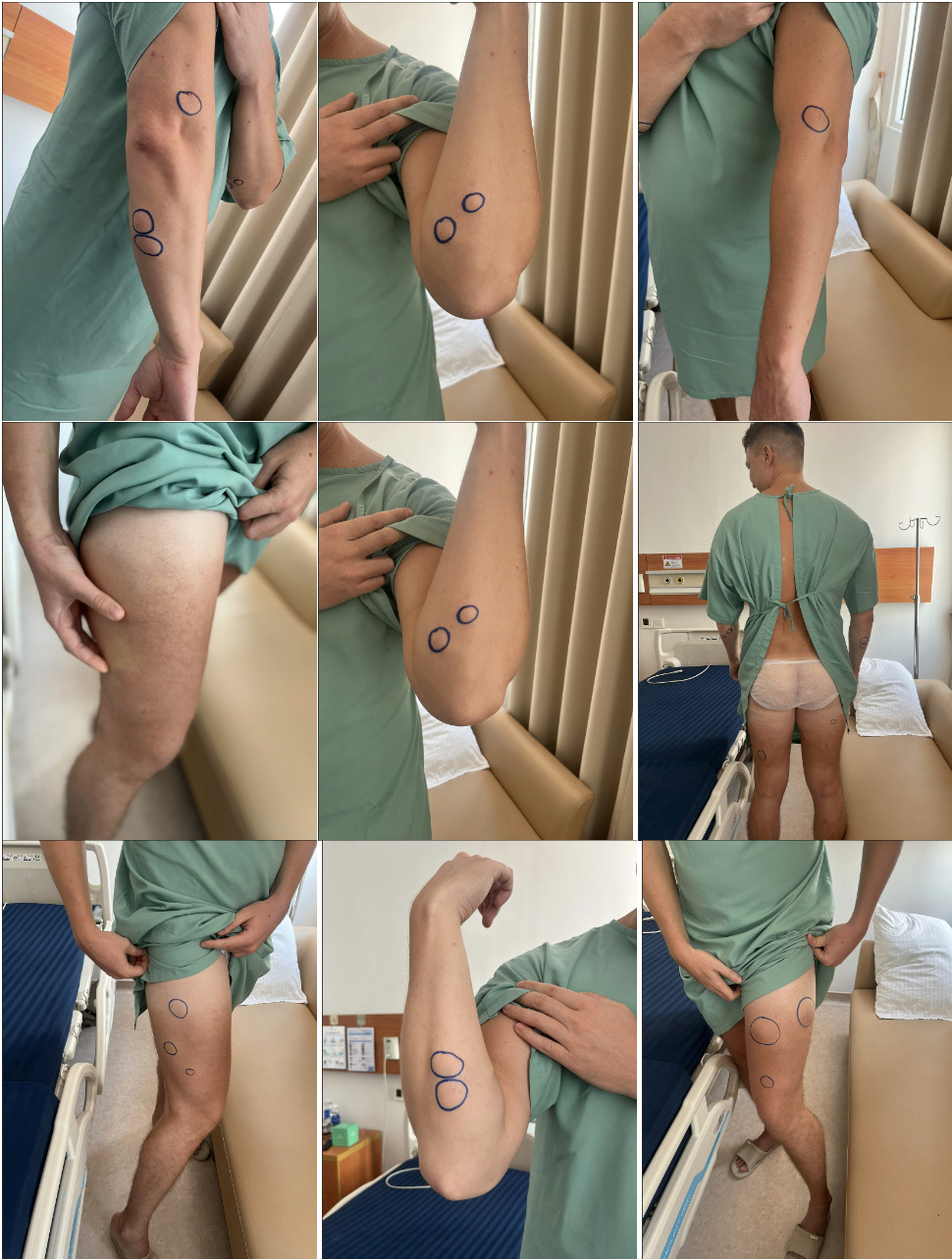
Examination demonstrated multiple well-defined soft-tissue masses in both arms and thighs. The largest lesion was in the left thigh and measured approximately 6 × 7 cm. All masses were soft, mobile, mildly tender, and confined to the subcutaneous plane. The overlying skin was normal without discoloration, ulceration, or adherence. The range of motion of the shoulder, elbow, hip, and knee was full and pain-free, and neurovascular examination of all four limbs was normal. No masses were detected in the head, neck, or trunk.
Imaging
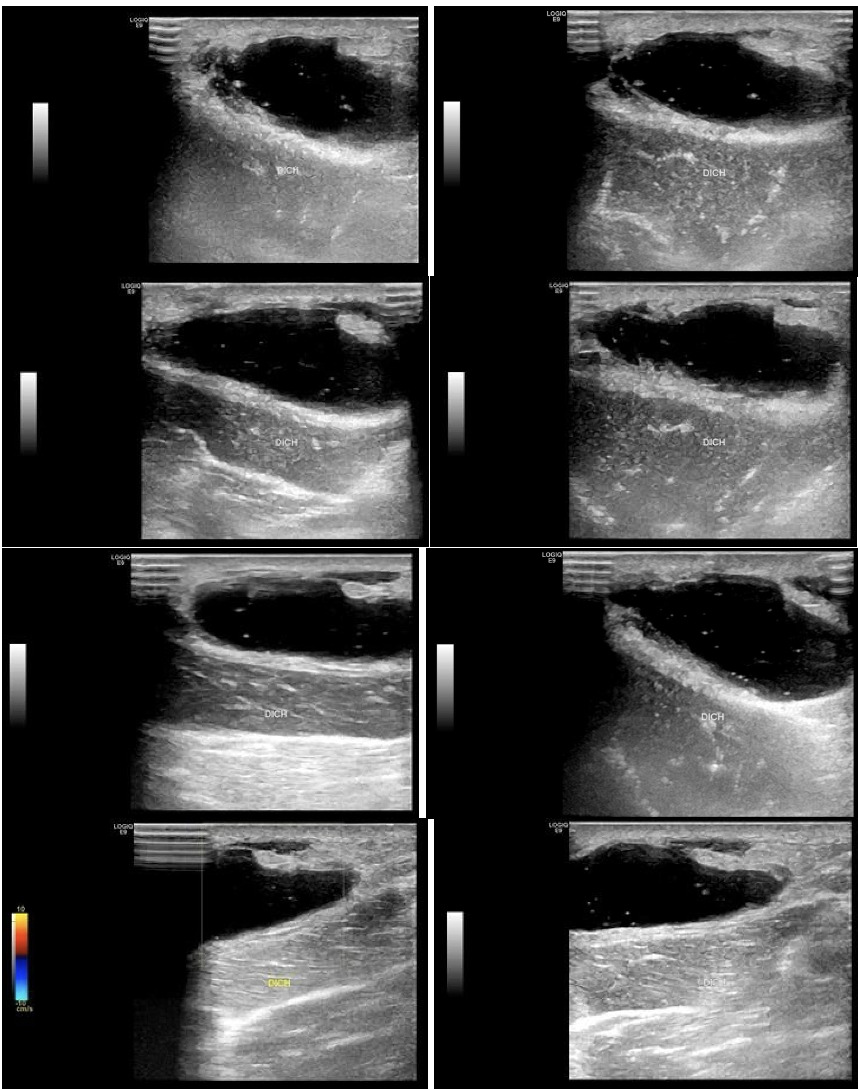
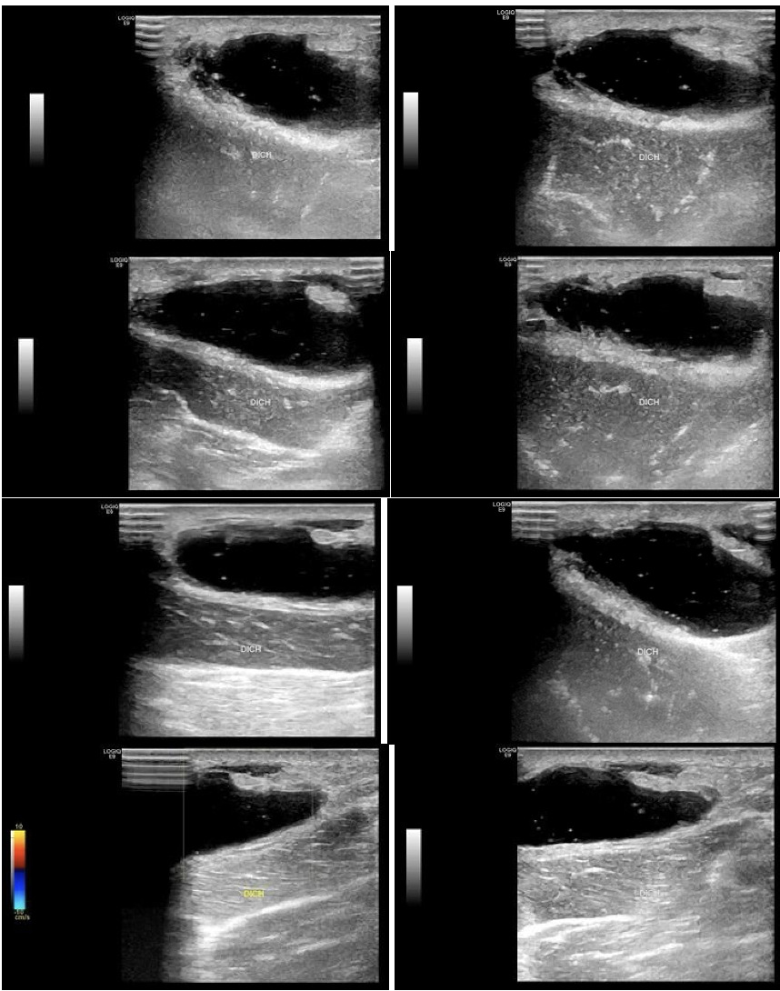
Ultrasonography demonstrated a well-defined, heterogeneous hypoechoic subcutaneous lesion without internal vascularity, consistent with a benign lipoma (Figure 2). MRI was not obtained because of resource constraints and the clinically benign appearance of the lesions.
Diagnostic Considerations
The working diagnosis was multiple lipomas with a strong suspicion for FML. Differential diagnoses included MSL, Dercum’s disease, multiple angiolipomatosis, and ALT/WDL. Limb-predominant, well-circumscribed subcutaneous nodules and absence of cervicothoracic fat deposition favored FML over MSL; modest pain severity and lack of systemic features made Dercum’s disease less likely.2,4–6,8 Furthermore, the diagnosis of FML is strongly supported by the patient’s family history, as systematically documented in the three-generation pedigree. Given multiplicity and the presence of a larger thigh lesion, complete excision with histopathologic evaluation was planned.
Patient Perspective
The patient’s primary concern was a progressive increase in the number and size of lumps and fear of malignancy; he requested a definitive diagnosis and removal for comfort and appearance.
Management and Outcomes
Surgical Strategy and Rationale
All lesions were excised in a single session under general anesthesia. Minimally invasive options (liposuction and minimal-incision techniques) have been described for selected FML presentations,8 but our priorities for limb disease were: (1) capsule-complete removal within a clear anatomic plane; (2) protection of neurovascular structures; and (3) procurement of an intact specimen for histopathologic confirmation.2 Open excision facilitates circumferential dissection along the capsular plane and en bloc removal, which is the most reliable technical determinant of durable local control for encapsulated lipomas.9 By contrast, liposuction or chemical/laser lipolysis may leave residual capsule or satellite nodules and typically yields fragmented tissue, limiting assessment for atypia.8 The largest thigh lesion and the heterogeneous ultrasound appearance further supported an approach that preserves oncologic safety through definitive pathology.
Operative Technique
Incisions were aligned with Langer’s lines. A tumescent solution containing lidocaine and adrenaline was infiltrated to reduce bleeding and facilitate hydrodissection. Dissection proceeded to the capsule, followed by circumferential mobilization and en bloc removal without rupture. Cavities were inspected for satellite nodules, hemostasis was secured, dead space was managed selectively (quilting sutures and/or drains), and layered closure with compression dressing was applied.
Pathology and Clinical Outcome
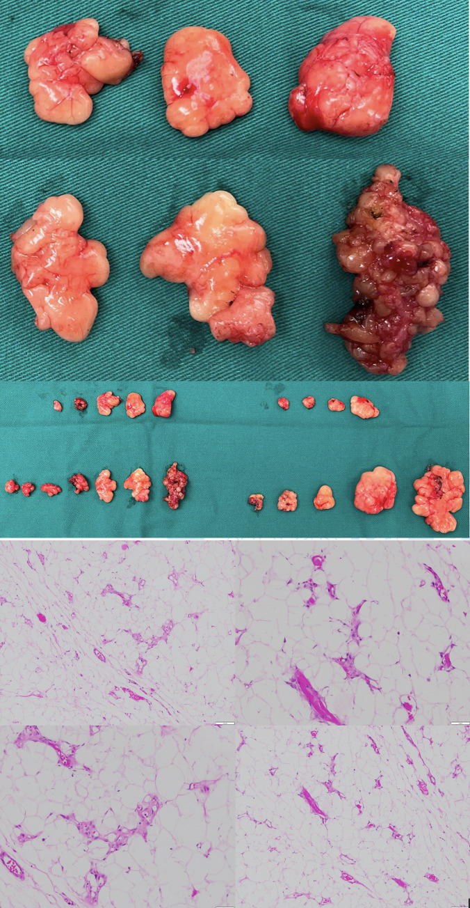
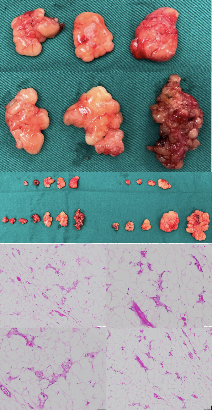
Grossly, specimens were soft, yellow, homogeneous, and encapsulated. Microscopically, tumors were composed of mature adipocytes in lobules separated by thin fibrous septa with branching capillary-sized vessels, without atypia, necrosis, lipoblasts, or increased mitotic activity—consistent with benign lipoma. No specimen showed ALT/WDL features.
Follow-up and Outcomes
Recovery was uneventful. The patient received routine analgesia and was advised to continue compression to reduce edema and maintain contour. Wounds healed satisfactorily; discomfort resolved and cosmetic outcome was acceptable. No early recurrence was observed within available follow-up.
Family History and Genetic Considerations
Postoperatively, a more detailed pedigree identified multiple affected maternal relatives, supporting autosomal dominant inheritance with variable penetrance.4,7,10–13 Genetic testing for HMGA9, PTEN, PALB9, or related genes was not pursued due to cost and limited local availability. The patient was counseled regarding familial risk and the potential role of genetic evaluation, particularly if additional relatives are affected or if cancer-risk features emerge.12–15
Discussion
Summary of Key Findings
This report describes limb-predominant, encapsulated subcutaneous lipomas in a young adult male with subsequent confirmation of familial clustering on the maternal side, consistent with FML. The case underscores two practical points: (1) the diagnostic value of systematic pedigree taking, and (2) the role of open en bloc excision for symptomatic lesions when definitive pathology is required.
Relevant Literature
FML is typically inherited in an autosomal dominant pattern with variable penetrance and is often underrecognized without systematic family history.4,7,10–13 The main clinical differentials include MSL (diffuse, non-encapsulated cervicothoracic fat accumulation often linked to alcohol use), and Dercum’s disease (disproportionate pain with systemic features).4,6,10,12 Angiolipomatosis may appear similar clinically but is defined histologically by prominent vascular channels.5
Genetic and Molecular Considerations
Molecular findings in lipomas commonly involve HMGA9 alterations; PTEN-pathway dysregulation is relevant to hamartoma tumor syndromes with lipomatous features.13–16 In selected families, PALB9 variants have been reported with multiple lipomas and potential cancer predisposition, supporting genetic heterogeneity and the role of counseling when clinically indicated.
Future Implications
For large, deep, or clinically atypical lesions—particularly in the thigh—an oncologic approach is prudent: obtain definitive histopathology and avoid techniques that compromise specimen integrity when ALT/WDL cannot be confidently excluded.17,18 In settings with limited access to MRI and genetic testing, careful examination, pedigree assessment, and sound operative planning remain the cornerstone of safe care.
Take Away Lessons
Multiple limb lipomas warrant a structured differential diagnosis and at least a three-generation pedigree. Open en bloc excision offers reliable symptom relief, capsule-complete removal, and intact tissue for confirmation. Long-term care should include family counseling and follow-up for new or changing lesions.
Follow-up should be individualized. In practice, we advise reassessment when new lesions appear, when a previously stable mass enlarges rapidly, becomes firm/fixed, or develops atypical pain. For deep, large, or clinically atypical lesions, MRI improves preoperative characterization and guides margin planning, while core needle biopsy can be considered in lesions with concerning imaging features to exclude ALT/WDL before definitive surgery.17,18
Strengths and Limitations
Strengths include structured operative planning and intact specimen submission for definitive pathology. Limitations are the absence of MRI and genetic testing due to resource constraints and limited follow-up, which may underestimate late recurrence.18
Conclusion
FML may be overlooked without systematic pedigree assessment. This limb-predominant case supports open en bloc excision for symptomatic lesions to achieve capsule-complete removal and definitive histopathology, with ongoing follow-up and family counseling.
Figures/Tables