
1. Introduction
Quadriceps tendon rupture is an uncommon but serious injury of the knee extensor mechanism that can lead to profound functional impairment, inability to ambulate, and loss of active knee extension if not diagnosed and treated promptly.1 While unilateral quadriceps tendon rupture is itself relatively rare, simultaneous bilateral quadriceps tendon rupture represents an exceptionally uncommon clinical entity, with previous reports estimating its incidence between 0.17% and 2.5% and accounting for less than 5% of all quadriceps tendon ruptures reported in the literature.2,3
The injury typically affects middle-aged and elderly patients and has been reported more frequently in male individuals and is also frequently associated with underlying systemic disorders that compromise tendon integrity.4,5 Chronic renal failure, secondary hyperparathyroidism, diabetes mellitus, obesity, gout, rheumatoid arthritis, systemic lupus erythematosus, and long-term corticosteroid exposure have all been implicated as important predisposing factors.2,4,6 In such patients, chronic metabolic derangements, collagen degeneration, vascular compromise, and altered tendon-bone junction mechanics may weaken the extensor mechanism and predispose it to rupture even after low-energy trauma or minor eccentric loading.2,7
Despite its characteristic clinical findings—including acute pain, inability to perform straight-leg raise, palpable suprapatellar defects, and extensor lag—the diagnosis is often delayed or missed, particularly because bilateral involvement may obscure side-to-side comparison and mimic neurological or generalized weakness conditions. Previous reports suggest that more than half of cases may be initially misdiagnosed, potentially resulting in delayed treatment and inferior functional outcomes.4
Early recognition followed by timely surgical repair and structured rehabilitation is considered the cornerstone of management, with most patients achieving satisfactory restoration of range of motion and ambulatory function when treated promptly. Given the rarity of the condition, each additional case contributes valuable information regarding mechanisms of injury, associated risk factors, surgical techniques, and postoperative recovery.
In this report, we present a case of simultaneous bilateral quadriceps tendon rupture in an elderly patient without recognized predisposing factors other than age, highlighting the diagnostic challenges, management strategy, and relevant review of the current literature.
2. Case Presentation
A 73-year-old male presented to the Emergency Department after falling while descending stairs and landing directly onto both knees. The patient reported immediate bilateral knee pain accompanied by an inability to stand or ambulate following the injury. A detailed history revealed no preceding symptoms, including knee pain, weakness, or prior trauma. His past medical history was unremarkable, with no known history of chronic renal failure, diabetes mellitus, inflammatory arthropathy, corticosteroid use, or other systemic conditions associated with tendon degeneration.
On clinical examination, both knees demonstrated swelling and tenderness over the suprapatellar region. Palpation revealed a palpable suprapatellar defect bilaterally. The patient was unable to actively extend either knee or perform a straight leg raise. Passive range of motion of both knees was preserved but painful. Distal neurovascular examination was normal.
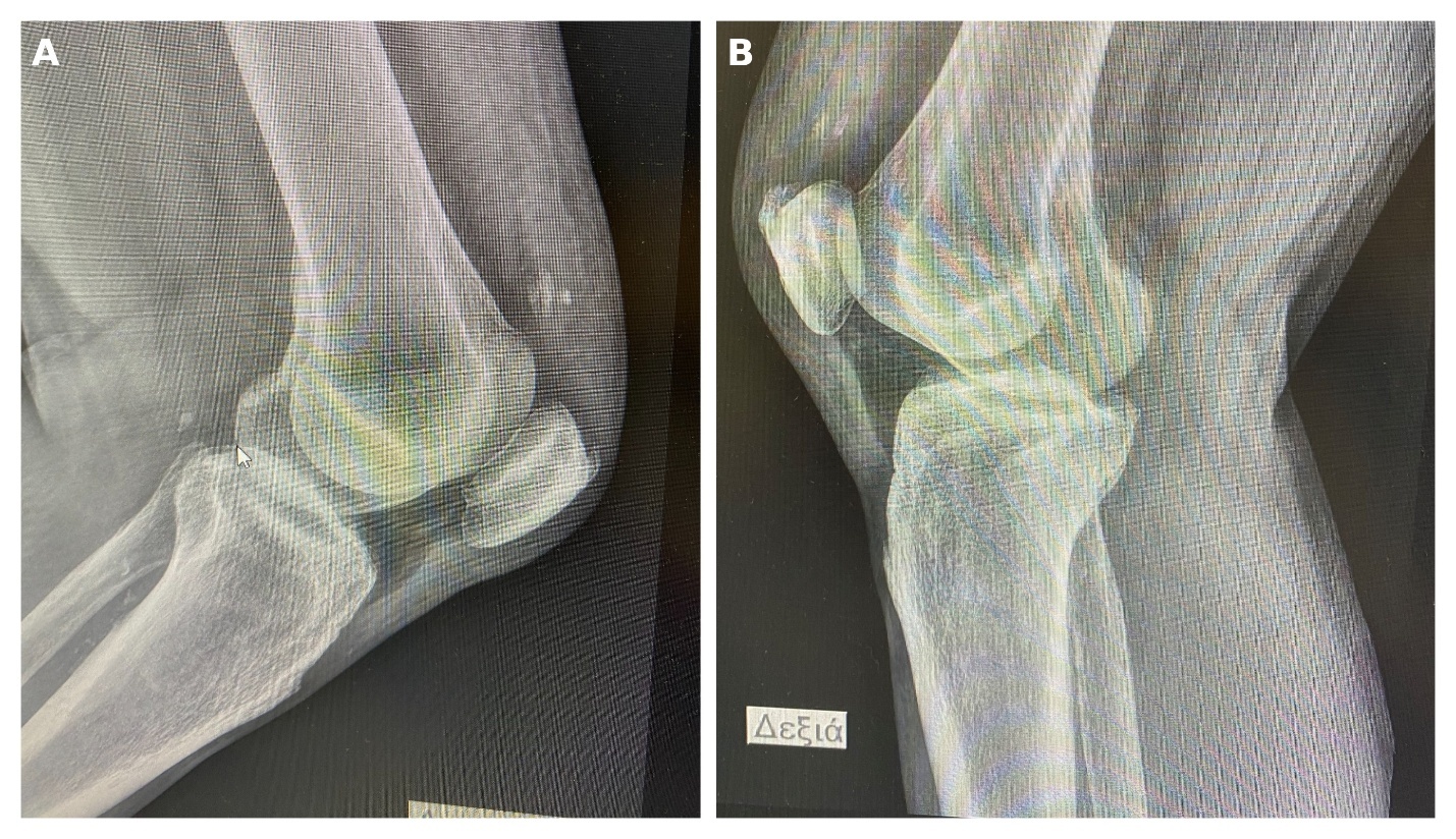
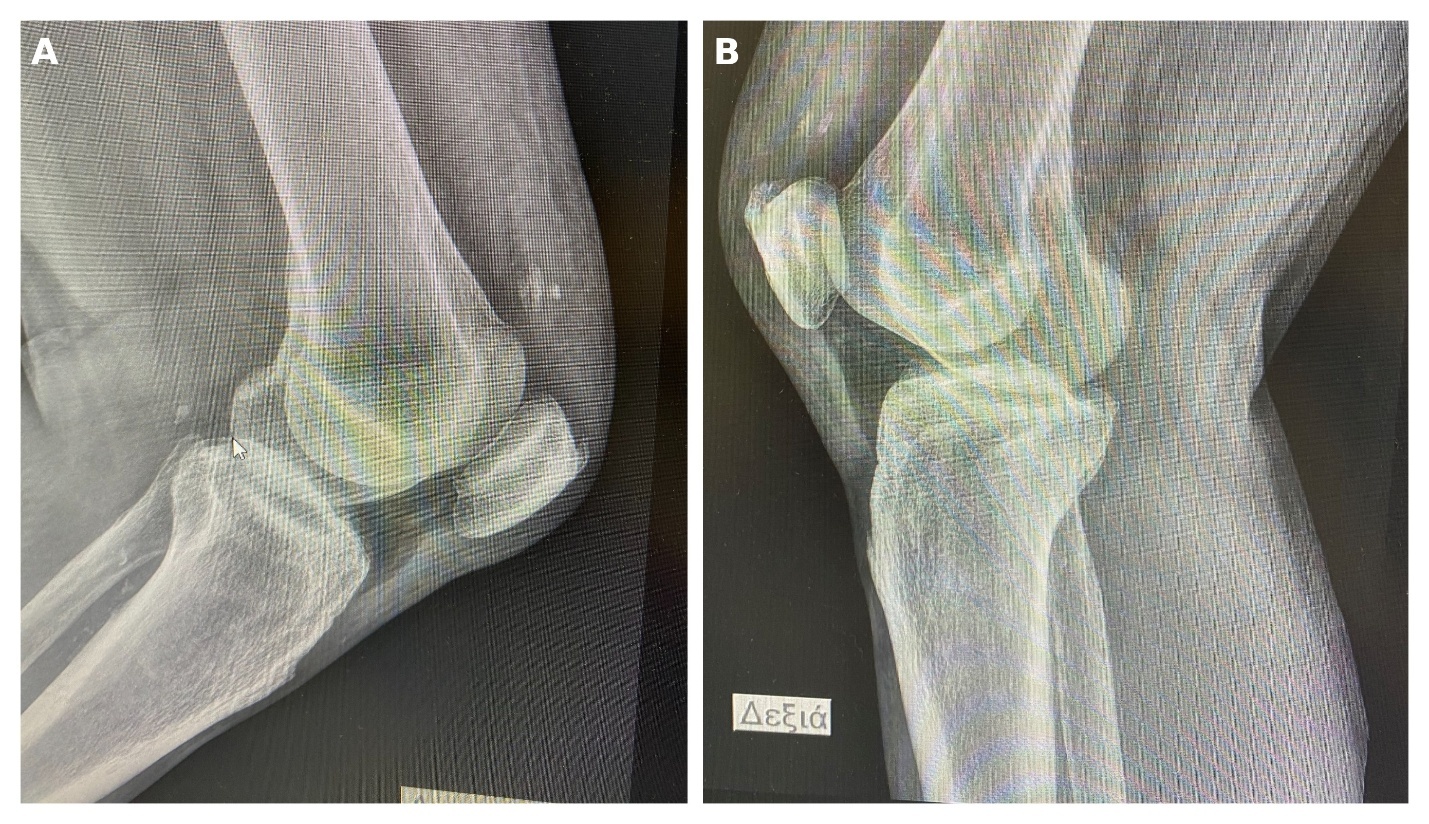
Preoperative plain radiographs of both knees were obtained to evaluate the extensor mechanism (Figure 1), demonstrating bilateral patella baja with a central calcification in each knee. Based on clinical findings, the diagnosis of simultaneous bilateral quadriceps tendon rupture was established, and the patient was scheduled for urgent surgical repair. Given the evident clinical diagnosis, further evaluation with magnetic resonance imaging was not considered necessary.
Surgical Technique
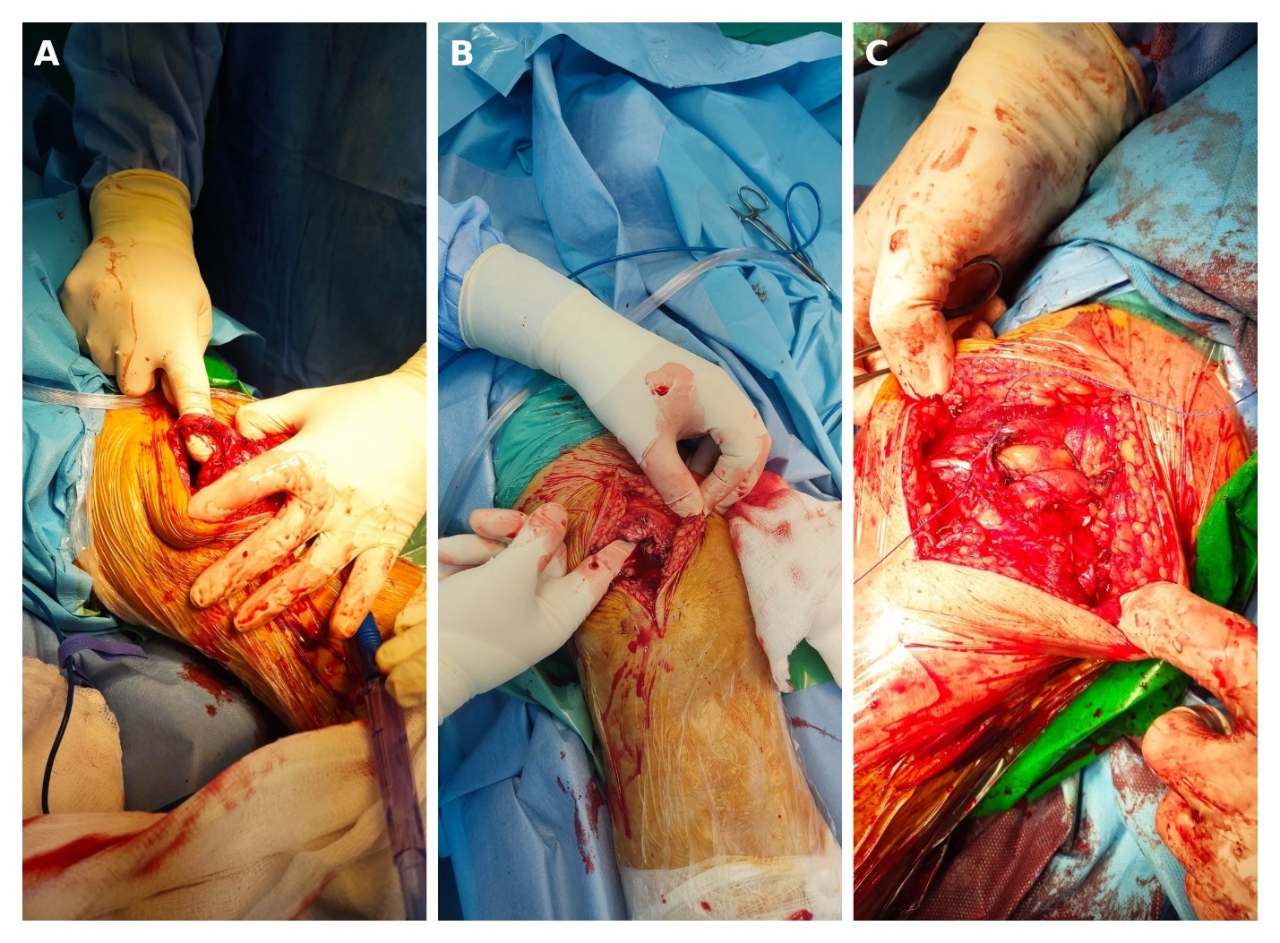
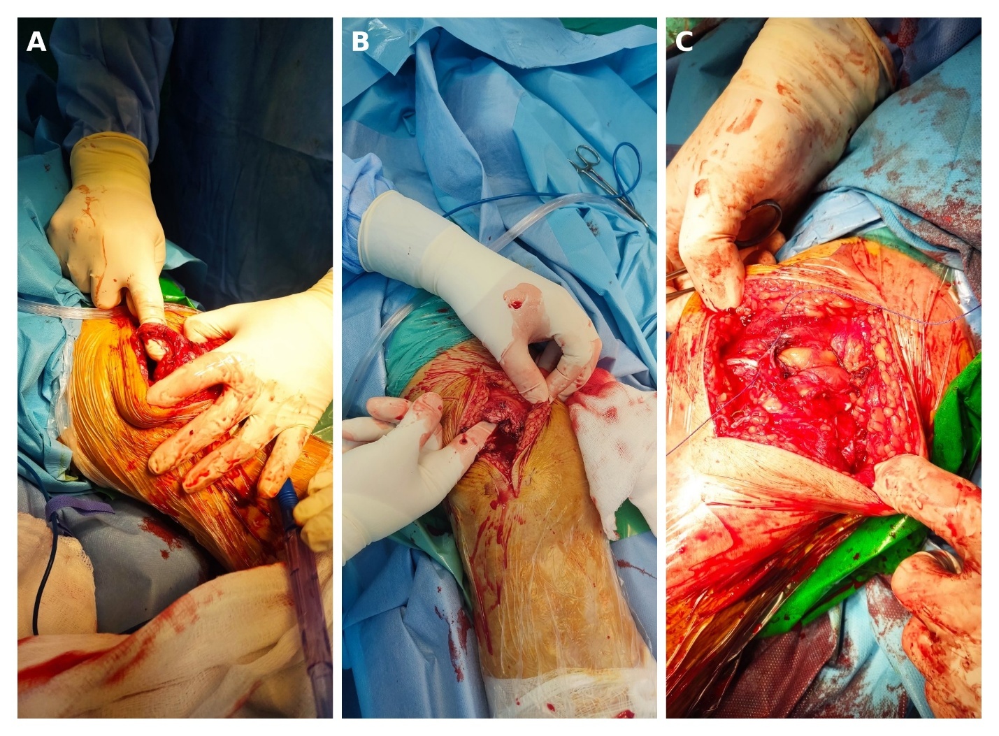
Surgery was performed under epidural anesthesia with the patient in the supine position. A longitudinal midline incision was made over each knee extending proximally from the superior pole of the patella. Dissection through the subcutaneous tissues exposed the ruptured quadriceps tendons bilaterally, which were found to be completely detached from the superior pole of the patella. The tendon ends were debrided and prepared for repair. A transosseous fixation technique was utilized. Four longitudinal bone tunnels were created through the patella using a 2.5 mm drill bit. The quadriceps tendon was secured to the superior pole of the patella using No. 5 nonabsorbable Ethibond sutures (Ethicon, Johnson & Johnson, Somerville, NJ, USA) placed in a locking Krackow configuration. The sutures were passed through the patellar tunnels and tied over the inferior pole of the patella, restoring the continuity of the extensor mechanism. Additional reinforcement sutures were placed to augment the repair and restore the medial and lateral retinacula where necessary (Figure 2). The stability of the repair was assessed intraoperatively with gentle knee flexion. Layered wound closure was subsequently performed and sterile dressings were applied.
Postoperative Rehabilitation
Postoperatively, both knees were immobilized in a hinged knee brace with an adjustable range-of-motion dial. The brace was initially locked in full extension for approximately two weeks. A structured rehabilitation protocol was initiated thereafter. Gradual passive range of motion exercises were introduced, with progressive increases in flexion using the hinged brace. Flexion advanced progressively, reaching approximately 90° by the sixth postoperative week. Weight-bearing status progressed according to tolerance. Partial weight bearing with crutches was allowed early postoperatively, progressing to full weight bearing at approximately six weeks. Quadriceps strengthening and functional rehabilitation exercises were gradually introduced under physiotherapy supervision.
Follow-Up and Clinical Outcome
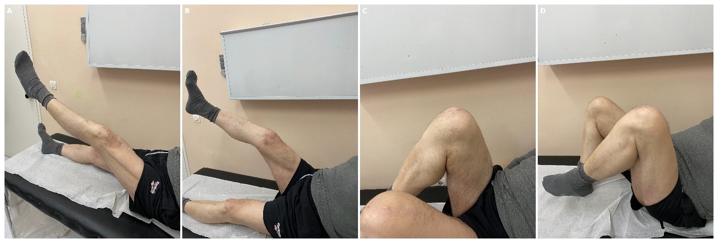
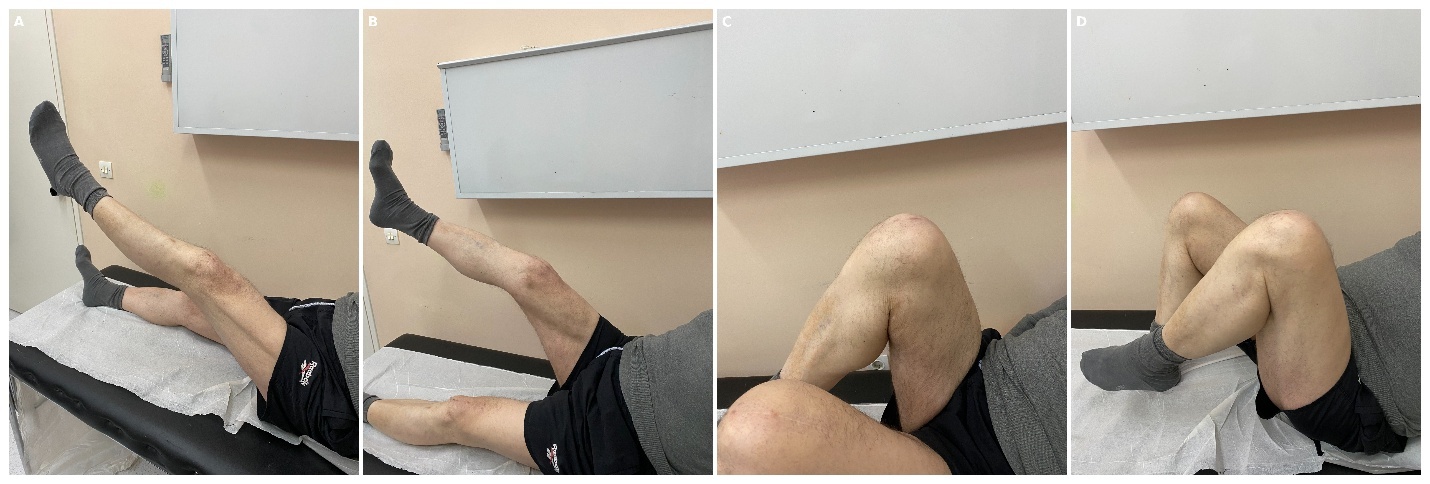
At the 18-month follow-up examination, the patient reported no pain and had independent ambulation without the need for assistive devices. He was able to perform activities of daily living without functional limitations.
Clinical examination demonstrated satisfactory bilateral knee function, with full knee extension and flexion to approximately 130° in both knees. The patient reported no subjective functional impairment and expressed high satisfaction with the surgical outcome.
Discussion
Simultaneous bilateral quadriceps tendon rupture is an exceptionally rare injury of the knee extensor mechanism and represents a diagnostic and therapeutic challenge. Most reported cases involve middle-aged or elderly male patients and are frequently associated with systemic disorders that compromise tendon quality, including chronic renal failure, secondary hyperparathyroidism, diabetes mellitus, obesity, gout, rheumatoid arthritis, and chronic corticosteroid exposure.2,4 In contrast, the present patient had no known history of metabolic or inflammatory disease, highlighting that this injury may also occur in the absence of overt systemic predisposition.
The usual mechanism of quadriceps tendon rupture involves sudden eccentric loading of the extensor mechanism with the knee in a flexed position. However, in tendons affected by age-related degeneration, chronic tendinopathy, or calcific changes, relatively low-energy trauma may be sufficient to precipitate rupture.7,8 In the present case, the injury occurred after a fall while descending stairs, with direct impact on both knees. Preoperative radiographs demonstrated bilateral patella baja and central calcific densities within the extensor mechanism, findings suggestive of pre-existing degenerative tendon changes that may have contributed to tendon failure.
Early diagnosis remains essential, as delayed recognition may lead to tendon retraction, scarring, prolonged disability, and inferior functional outcomes.6 Bilateral injuries are particularly prone to missed diagnosis because comparison with the contralateral side is not possible, and the presentation may be mistaken for generalized weakness or neurological impairment. Previous reports have shown that a substantial proportion of bilateral extensor mechanism ruptures are initially overlooked.4,5,8 In our patient, the diagnosis was established clinically based on inability to actively extend both knees and disruption of the extensor mechanism, while plain radiographs provided supportive findings. Although magnetic resonance imaging remains highly sensitive in equivocal cases, diagnosis in our patient was clinically evident and sufficiently supported by plain radiographs, allowing prompt surgical management without unnecessary delay.9
A previous systematic review identified 44 bilateral extensor mechanism ruptures, including 37 bilateral quadriceps tendon ruptures, highlighting the rarity of this condition.4 For the purposes of Table 1, we selected representative reports of simultaneous bilateral quadriceps tendon rupture with sufficient clinical, surgical, and outcome data to allow comparison with the present case.
Various surgical techniques have been described for quadriceps tendon repair, including transosseous tunnel fixation, suture anchor repair, augmentation with cerclage wiring, and allograft reconstruction in chronic or complex cases.16 Owing to the rarity of simultaneous bilateral injuries, no clear consensus exists regarding the optimal fixation method. In acute ruptures with adequate tissue quality, both transosseous and suture anchor techniques have demonstrated satisfactory outcomes.4,7,12 In the present case, surgical repair was performed using a robust primary fixation construct, allowing restoration of the extensor mechanism and subsequent rehabilitation. The chosen technique provided stable fixation, reproducibility, and favorable functional recovery.
Postoperative rehabilitation is equally important and must balance protection of the repair with progressive restoration of motion and quadriceps strength.17 Early controlled mobilization has been associated with improved range of motion and reduced stiffness, whereas prolonged immobilization may compromise functional recovery.10,11 At the 18-month follow-up, our patient reported no pain, independent ambulation without assistive devices, unrestricted activities of daily living, full knee extension, and flexion to approximately 130° bilaterally. These findings compare favorably with previously published reports, in which timely repair generally results in satisfactory restoration of function.2,4,7,18
This report has limitations inherent to any single-case study. The rarity of the condition limits broad generalization, and no validated patient-reported outcome measures were collected. Nevertheless, the case adds to the limited body of literature on simultaneous bilateral quadriceps tendon rupture in patients without major systemic comorbidity and demonstrates that prompt diagnosis followed by early surgical repair can lead to excellent mid-term outcomes.
3. Conclusion
Simultaneous bilateral quadriceps tendon rupture should be considered in elderly patients presenting with acute bilateral knee pain and inability to actively extend the knees after even low-energy trauma. Plain radiographs may reveal supportive findings such as patella baja and tendon calcification, facilitating rapid diagnosis. Early operative repair combined with structured rehabilitation can provide excellent functional recovery and high patient satisfaction.
4. Ethical Considerations
This study is a single anonymized case report describing standard clinical care. Formal ethical committee approval was not required according to institutional policy. Written informed consent for publication of clinical data and images was obtained from the patient.
5. Conflict of Interest
The authors declare no conflict of interest.
6. Funding
No funding or financial support was received for the preparation of this manuscript.