
Introduction
Subungual exostosis (SE) is a rare condition characterized by the formation of a bony outgrowth arising from the distal phalanx, prevalently of the great toe, occurring in young adults.1,2 Diagnosis is typically established through plain radiographs, while the treatment of choice is surgical excision. This condition is frequently associated with a delay in diagnosis, as it can be easily misdiagnosed as other nail disorders.3,4
We present a case report of hallux subungual exostosis presenting like infection in a female patient.
Case Report
We report a case of a 42-year-old woman presenting to our orthopaedic department with pain localized to the great toe. Her medical history revealed a previous blunt trauma to the toe approximately one year earlier, without any subsequent treatment. Over the preceding two months, she had experienced progressive pain associated with swelling, which had worsened to the point of preventing her from wearing shoes. The patient therefore underwent conservative treatment by a podiatrist, without clinical improvement. Due to symptoms persistency and functional limitation, surgical intervention was indicated.
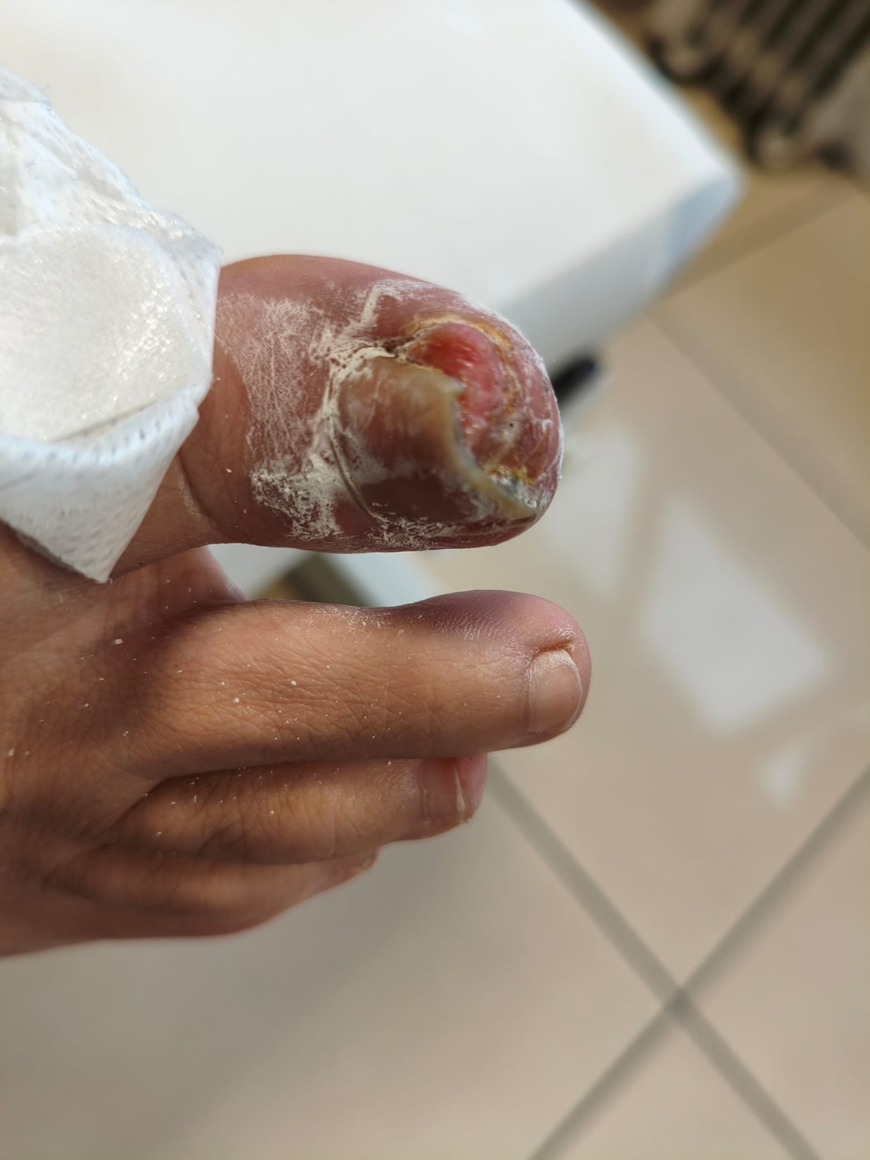
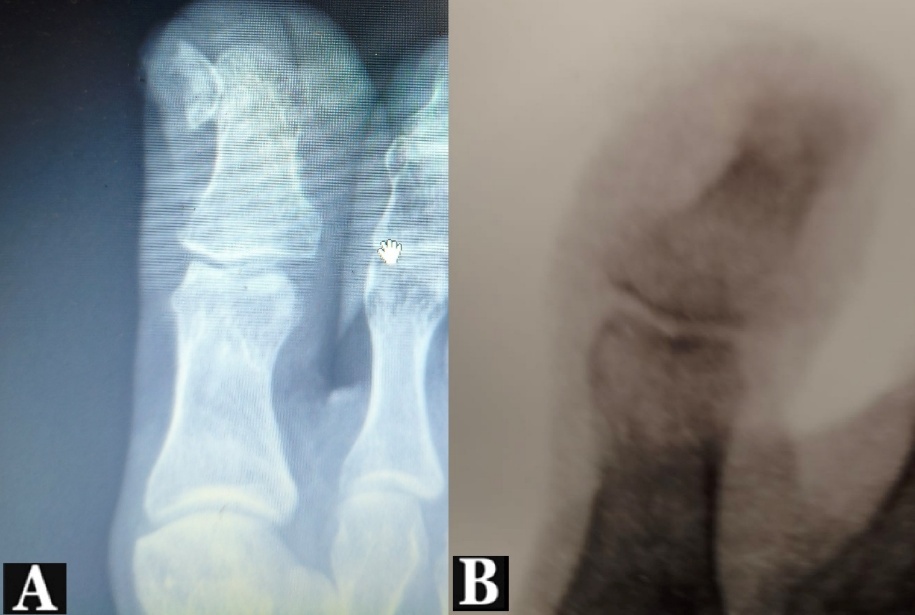
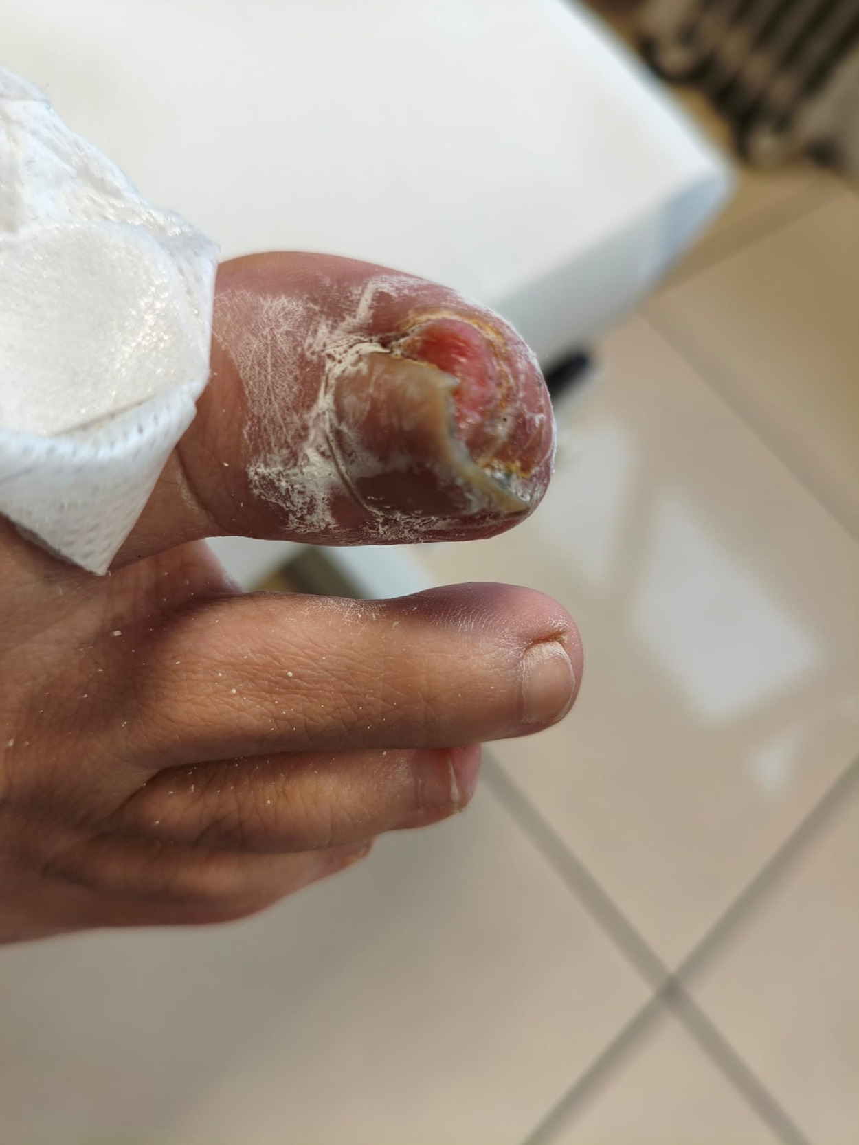
On physical examination, nail dystrophy was observed, with clinical suspicion of a concomitant infection (Figure 1). Plain radiographs demonstrated the presence of a lesion arising from the distal phalanx of the great toe, which was diagnosed as subungual exostosis (Figure 2A).
_and_the_post-excision_appearance_(b).jpeg)
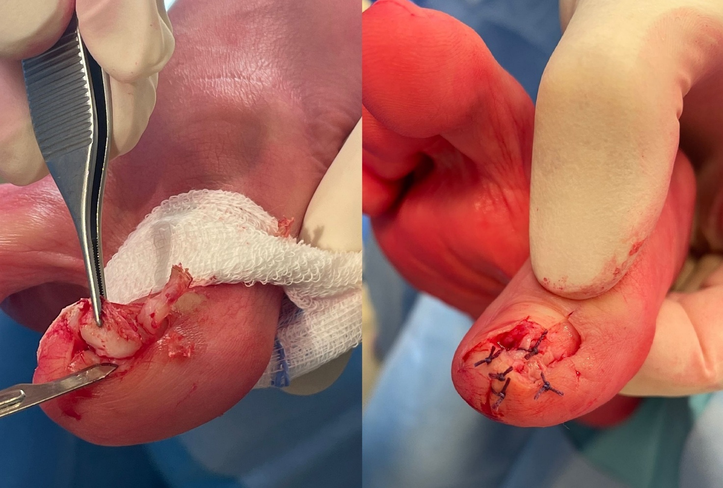
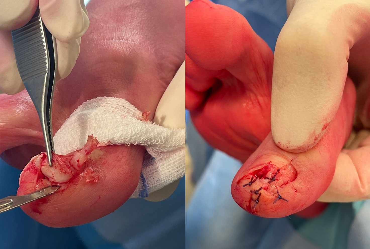
The procedure was performed under local anaesthesia. A skin flap was elevated, followed by partial nail removal. Careful dissection was carried out in layers, allowing identification and complete excision of the exostosis, measuring 0.5 × 0.4 cm. Thorough curettage was then carried out down to the cancellous bone. Intraoperative fluoroscopy confirmed complete excision of the lesion (Figure 2B). The damaged nail bed was repaired with sutures (Figure 3), and a sterile dressing was applied.
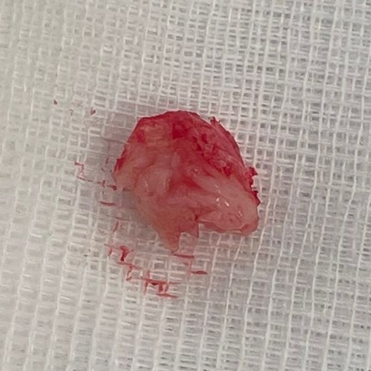
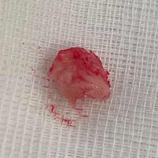
The mass was sent to the pathological anatomy lab (Figure 4); histopathological examination revealed mature trabecular bone covered by a fibrocartilaginous cap, confirming the diagnosis of subungual exostosis.
Postoperatively, wound care and dressings were maintained for 20 days. At the 3-month follow-up, the patient showed complete healing with resolution of symptoms. At the 9-month follow-up, she remained asymptomatic, with no evidence of recurrence and optimal cosmetic and functional recovery of daily living activities.
Discussion
Subungual exostosis is a benign osteocartilaginous lesion arising beneath the nailbed most commonly of the distal phalanx of the great toe.4
It can be classified into 4 types, where type I is a mild deformity of the nail plate, type II is an exostosis distal to the nail plate, type III is considered an exostosis under the nail plate and type IV is an exostosis involving the medial or lateral condyle.5
The aetiology of subungual exostosis is still unknown, although it is frequently related to local trauma and infection. The most widely accepted pathogenetic hypothesis is reactive growth of fibroblasts leading to cartilage metaplasia and ossification following trauma or infection. However, it can be thought as a benign nail tumour due to its association with the translocation t(X;6)(q22;q13–14) which leads to a deregulation of insulin receptor substrate 4 (IRS4) expression. Nonetheless, there is no full evidence in favour of a single pathogenesis.4
Diagnosis is based on a combination of patient’s medical history and symptoms, physical examination and plain radiographs. Clinically it manifests with pain, erythema and discomfort affecting daily living activities. At examination it presents as a firm, fixed nodule with hyperkeratotic surface. The average age of presentation is around 26 years with a female-to-male ratio reported to be 1:1 (6). Radiologically it appears as radiopaque pedunculated calcifications contiguous with the cortex of the distal phalanx usually of the great toe.2,6
The diagnosis confirmation is provided through histological examination revealing a fibrocartilaginous cap growing on a base of trabeculated bone with bone deposition in mid and lower dermis. Differently from true exostosis, the fibrocartilaginous cap is not covered by periosteum, but it blends with the overlying fibrous connective tissue.7
The differential diagnosis includes osteochondroma, wart, fibroma, dermatophytoma, subungual glomus tumor, pyogenic granuloma, amelanotic subungual melanoma, fibro-osseous pseudotumor (FOPD).8 Unlike FOPD, characterized by intramembranous ossification without connection to the underlying bone, exostosis develops from endochondral ossification and arises from the underlying phalanx. Another common misdiagnosis could be subungual verruca vulgaris, which as a higher incidence of presentation in paediatric patients.8 SE can be distinguished from osteochondroma due to its lack of communication with the medullary cavity and its location on the great toe.9
The treatment of choice is complete surgical excision with a success rate > 90%.10 Conservation of nail anatomy is recommended, if possible, sparing the nail matrix. Curettage down to normal trabecular bone should be optimal to prevent recurrence.3 Partial matrixectomy could be indicated in case of exostosis expansion under the nail matrix, but it can lead to poor aesthetic and functional results.5 Surgical approaches most frequently used are a direct dorsal surgical incision and fish-mouth-type of incision.10
Recurrence, which is reported to be around 4%, could take place with a more rapid and bigger growth of the exostosis, that is why it is of the utmost importance to follow up patients after surgery.7 Another possible complication following surgery is nail deformity secondary to nail bed damage.10
Conclusions
Subungual exostosis is a rare condition that may develop following trauma. When conservative management fails, surgical excision with a deep curettage of the bony lesion represents an effective treatment, leading to excellent clinical outcomes and symptom resolution.
Correspondence
Correspondence should be addressed to: Hassan Zmerly MD PhD, Orthopaedic Department, Villa Erbosa Hospital, Via Dell’Arcoveggio 50/2, 40129, Bologna, Italy
Tel 0039 0516315000
Author Contributions
Conceptualization, H.Z. and M.R.; methodology, A.V. and H.Z.; software, F.D.; validation, L.C.; resources, M.R.; data curation, R.R. and A.V.; writing—original draft preparation, R.R. and H.Z.; writing—review and editing, F.D.; supervision, L.C. All authors have read and agreed to the published version of the manuscript.
Funding
There was no funding source for the manuscript.
Conflicts of interest statements
The authors declared no conflict of interest.