
Introduction
Pediatric renal osteodystrophy (ROD) is a complication of pediatric chronic kidney disease (CKD), which is known to be a leading cause of pediatric morbidity, contributing to bone pain, deformities, fractures, and reduced quality of life for many children.1 Renal osteodystrophy (ROD) is a systemic bone disorder in CKD caused by several contributors: disrupted mineral metabolism, altered parathyroid hormone secretion, and impaired activation of vitamin D.2,3 ROD leads to abnormal bone turnover and mineralization, which, in turn, weaken the skeletal structure and increase the risk of fractures. Studies using bone histomorphometry, a method of measuring bone structure and activity, have demonstrated several abnormalities in a majority of children with moderate to severe CKD, and recent evidence points to the fact that most pediatric CKD patients exhibit some form of renal osteodystrophy.1,4 Despite the topic’s significance and impact, the evolution of the research surrounding pediatric renal osteodystrophy has not been fully examined. A bibliometric analysis may therefore provide valuable insight into publication trends, influential contributors, and emerging areas of investigation within this field.
Renal osteodystrophy is the traditional term used to describe the bone abnormalities caused by chronic kidney disease: disturbances in bone turnover, mineralization, and volume.3 In 2006, the organization KDIGO, Kidney Disease: Improving Global Outcomes, introduced a broader concept of Mineral and Bone Disorder, commonly associated with CKD, which included both disruptions in mineral metabolism and vascular calcification and skeletal abnormalities. With this new scope, secondary hyperparathyroidism is recognized as a common manifestation of CKD-MBD, while older literature may refer to these skeletal abnormalities as uremic bone disease.5,6 Additionally, the TMV (Turnover, Mineralization, Volume) classification system was developed to characterize the histological assessment of pediatric renal osteodystrophy.2
Renal osteodystrophy (ROD) becomes more severe as chronic kidney disease (CKD) advances, with studies showing that almost all children with a glomerular filtration rate (GFR) of less than 30 mL/min/1.73 m² show some evidence of bone involvement: abnormal bone turnover, mineralization defects, or other forms of skeletal damage1,7The presentation of ROD varies with the cause of CKD, such as congenital anomalies of the kidney and urinary tract, glomerular diseases, and hereditary nephropathies. Infants and adolescents are especially susceptible because of their periods of rapid skeletal growth and high mineral needs. Furthermore, during puberty, these growth spurts may worsen disruptions in bone metabolism, which could help explain sex differences in disease severity. Moreover, the prevalence and burden of ROD vary by region, largely due to inequitable access to specialty nephrology care, dialysis, transplantation, and mineral–bone disorder management.8,9
Pediatric renal osteodystrophy classically presents with bone pain, growth failure, skeletal deformities such as genu varum (bowed-leg appearance) and genu valgum (knock-knee appearance), rachitic changes, pathological fractures, and, less frequently, extraskeletal calcifications. Disturbances in mineral metabolism caused by chronic kidney disease are responsible for these clinical findings.6 With deteriorating kidney function, phosphate retention stimulates increased production of fibroblast growth factor 23 (FGF-23), which inhibits calcitriol (active vitamin D) production and plays a role in secondary hyperparathyroidism.4,7,10 High levels of parathyroid hormone (PTH) increase bone turnover and potentially cause high-turnover bone disease. Alternatively, overly aggressive reduction in PTH levels results in adynamic bone disease, another form of low-turnover ROD. In contrast to adults, children must also deal with diminished longitudinal bone growth due to growth plate abnormalities, making growth failure characteristic of pediatric ROD.11
Management of Renal osteodystrophy aims at correcting the mineral and hormonal derangements that promote bone disease. These include dietary phosphate restriction and the use of phosphate binders, both calcium-based and non-calcium-based, to control serum phosphate levels.5,9 Active Vitamin D analogues such as calcitriol and paricalcitol are often used to suppress Secondary Hyperparathyroidism, though the Parathyroid hormone target range in children remains contentious. Cinacalcet has shown promising results but has limited pediatric safety and dosing data. Optimization of dialysis, ultimately renal transplantation, which is the most definitive treatment cure of CKD-related MBD, is also employed. Transplant itself brings about a new phase of CKD-MBD complications, namely post-transplant bone loss due to glucocorticoid therapy.4,12 Despite the progress, most evidence is extrapolated from adults, and validated pediatric-specific treatment targets remain wanting.
Although clinical findings on prevalence and risk factors have been synthesized in systematic reviews of pediatric renal osteodystrophy, there is no bibliometric analysis of this field, which represents a major gap in our understanding of how this literature has evolved globally
This bibliometric analysis covers literature retrieved from the Web of Science Core Collection, examining publications across the full scope of the indexed period. The goal is to provide a broad overview of how pediatric renal osteodystrophy has been studied across different fields and regions, and over time.
Methods
Bibliometric analysis is a method for quantifying literature and identifying significant works and authors, publication patterns, and national or institutional contributions.13 Standard bibliometric methods for medical literature were employed to ensure rigor and repeatability.14 The Web of Science Core Collection was selected to provide the data for this bibliometric analysis due to its extensive coverage of high-impact medical literature, as well as the collection’s highly accessible citation features. These features are essential for being able to efficiently and thoroughly evaluate research trends and impact within the field of pediatric renal osteodystrophy and chronic kidney disease that have been accessed in previous bibliometric studies on this topic.
A specific search strategy was employed on June 13th, 2026, using the keywords “renal osteodystrophy” OR “renal bone disease” OR “CKD-MBD” OR “chronic kidney disease-mineral bone disorder” OR “renal mineral bone disorder” OR “uremic bone disease” OR “dialysis bone disease” OR “secondary hyperparathyroidism” AND “kidney.”) AND (“pediatric*” OR “paediatric*” OR “child*” OR “adolescent*” OR “juvenile*” OR “youth” OR “infant*” OR “neonat*” OR “school-age”. The search was limited to articles published between the years 1971 and 2026, retrieving a total of 768 articles for analysis.
Numerous data extraction tools were utilized throughout the study. The main analytical tool used in the search strategy was VOSviewer, which is especially helpful in bibliometric analysis because it offers a powerful tool for building and visualizing bibliometric networks.14 Additionally, data extraction included bibliometric details from the Web of Science: publication journals, annual publication count, research categories, author names, distinct collaborative communities, and keyword analysis.
Results
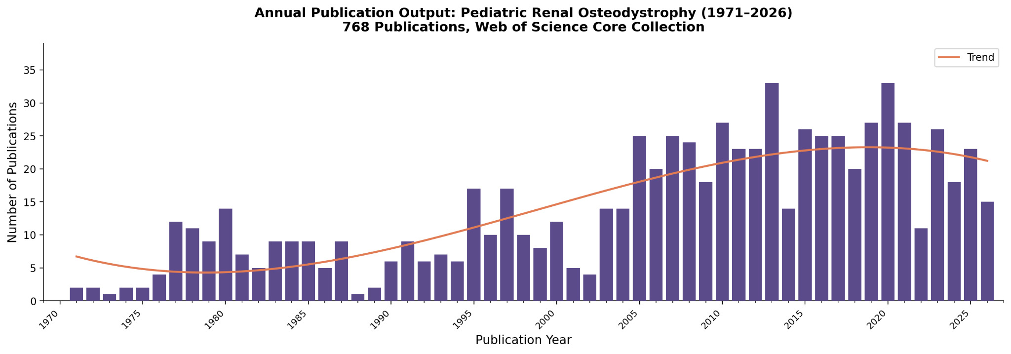
Since the first indexed publication on pediatric renal osteodystrophy in 1971, there have been 768 publications. Although activity was sporadic during the 1970s and 1980s, a marked increase occurred between 1977 and 1980, coinciding with the recognition of renal bone disease as a distinct clinical entity. There was a temporary decrease in the late 1980s/early 1990s, followed by recovery, then rapid growth after 2005. This increase may reflect growing recognition of the long-term complications of pediatric CKD, advances in biomarker research, and the 2006 KDIGO reclassification, which extended the field under the CKD-MBD umbrella. Activity peaked in 2013 and 2020 (33 papers each) and has remained fairly stable since 2019, indicating that the field has matured into an established area of research. The lower number of publications in 2026 probably reflects partial-year indexing rather than waning research interest.
.png)
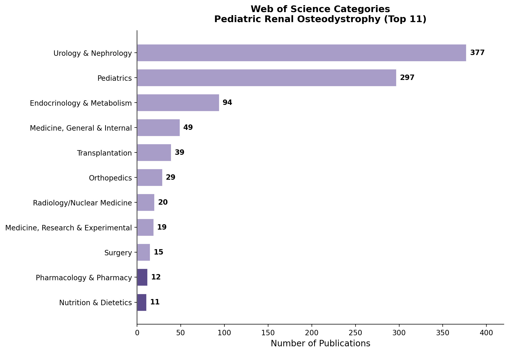
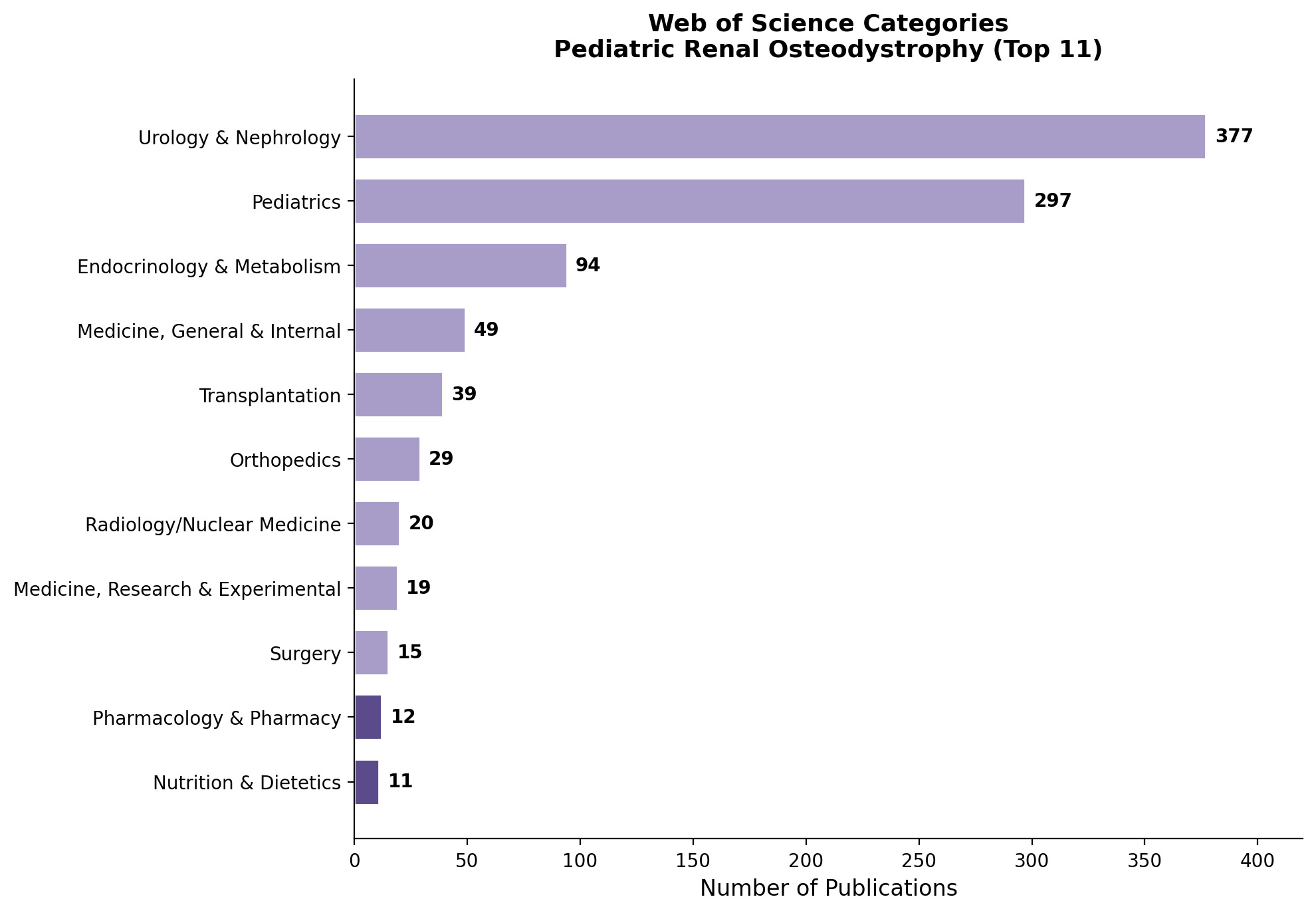
The disciplinary spread of pediatric renal osteodystrophy research reflects its inherently cross-cutting nature while remaining grounded in nephrology. Urology/Nephrology (49.1%) and Pediatrics (38.7%) dominated the discipline list, followed by Endocrinology/Metabolism (12.2%). The presence of smaller categories such as Transplantation, Orthopedics, Radiology, Pharmacology, and an extremely small number under Nutrition & Dietetics (0.3%) indicates both the broad multidisciplinary nature of pediatric renal osteodystrophy research.
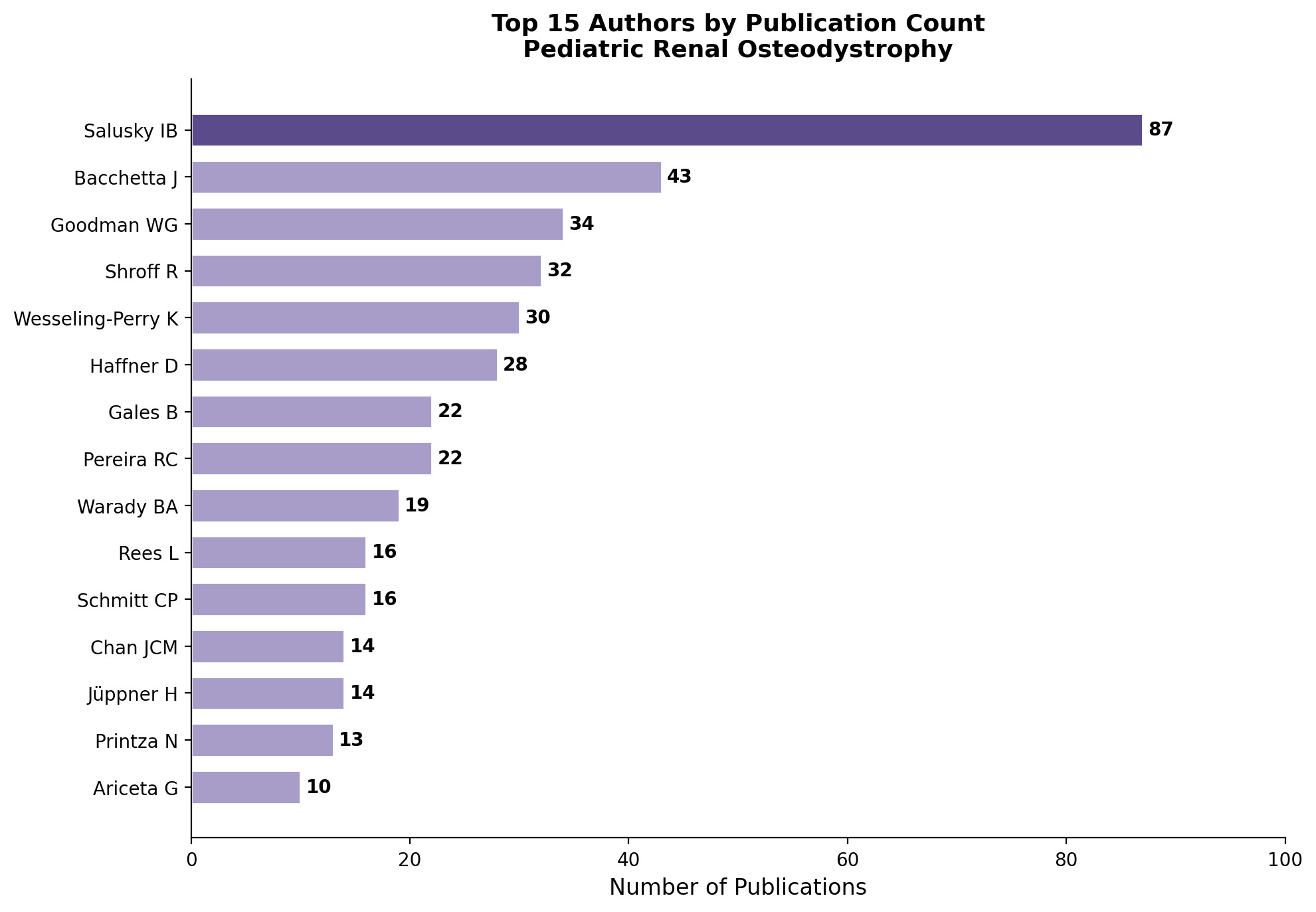
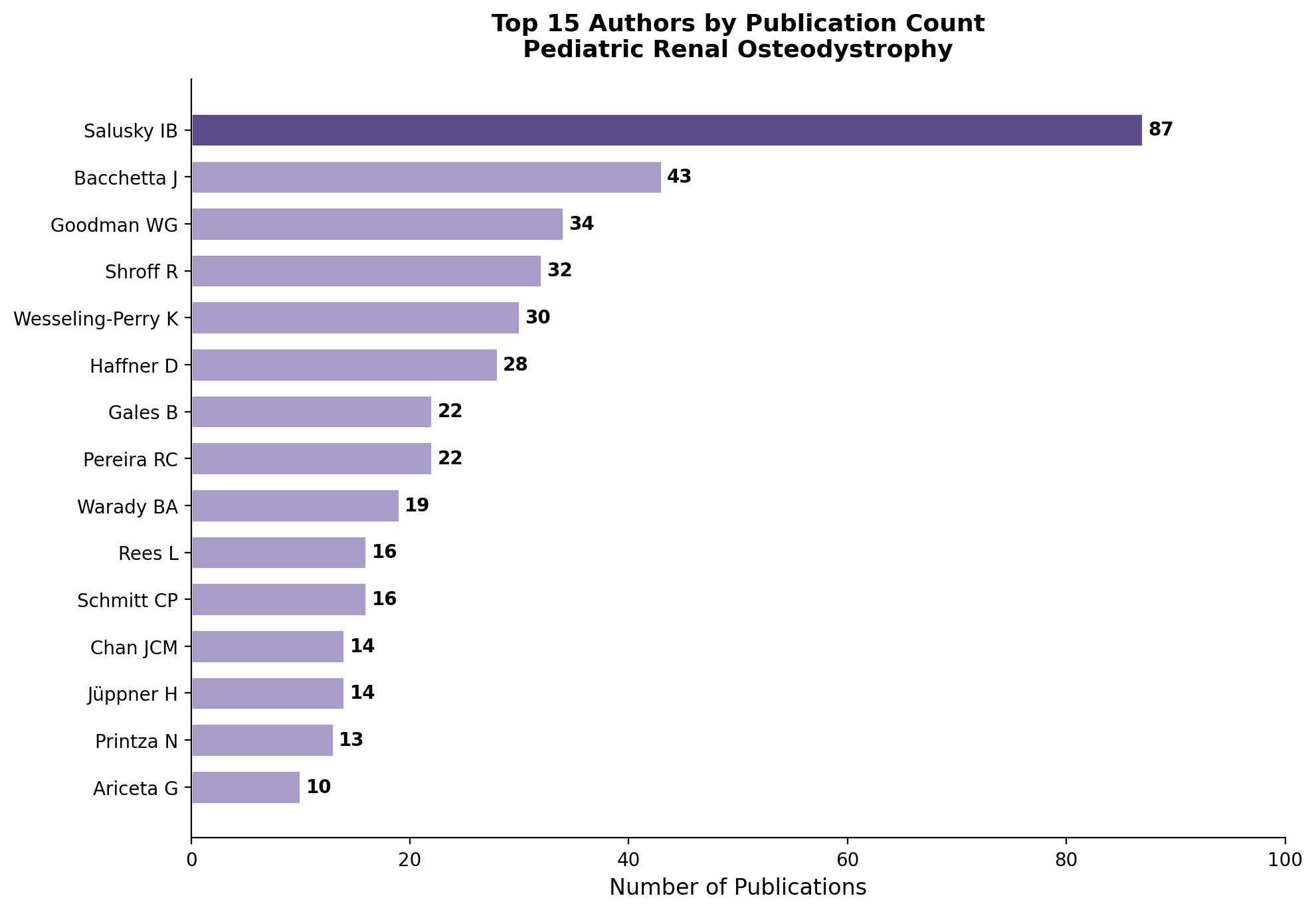
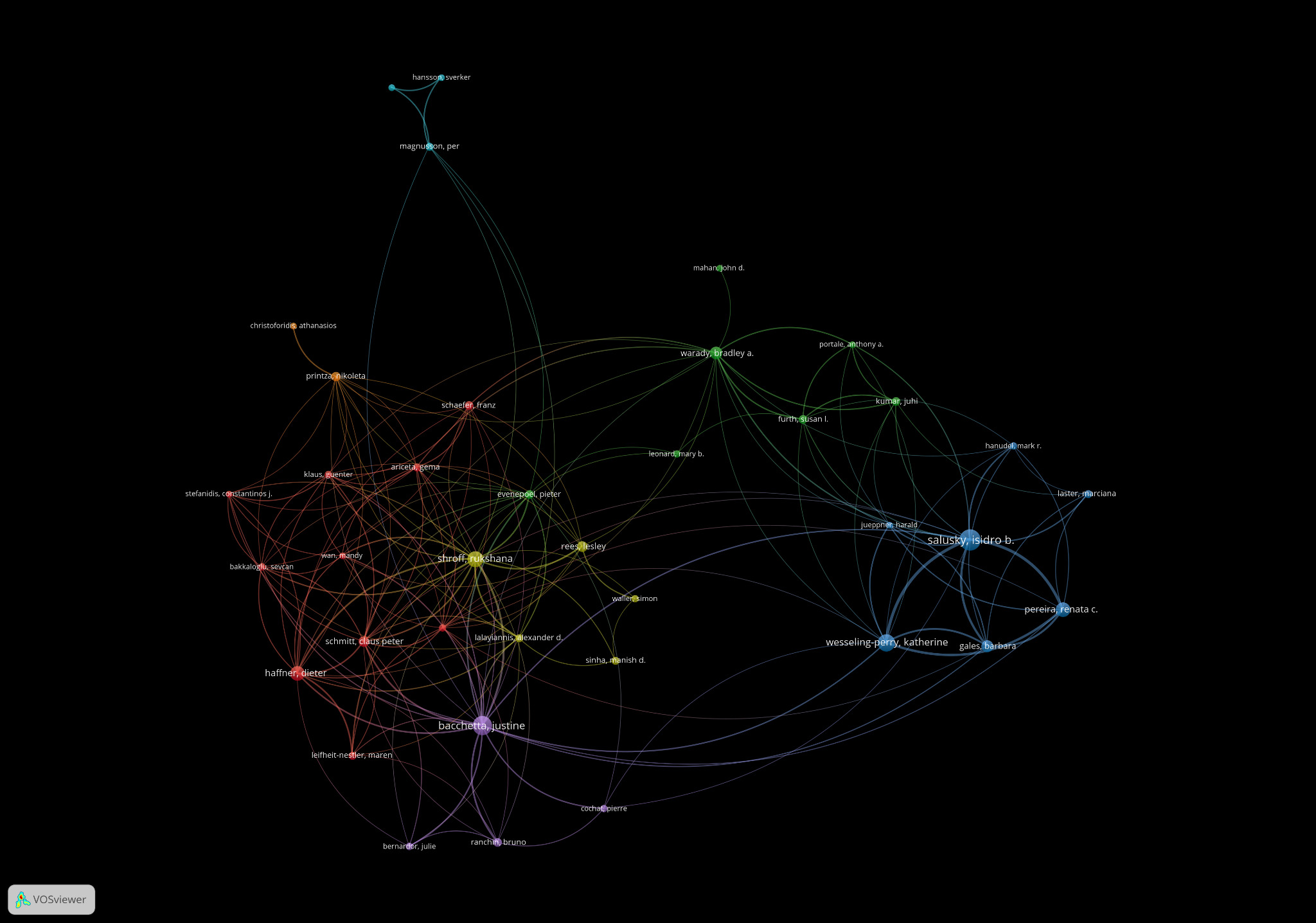
Authorship analysis shows a highly concentrated leadership structure in pediatric renal osteodystrophy. Salusky IB was the most prolific author (n = 87, 11.3%). Such extreme productivity is rare in medical subspecialties and likely reflects his central role in establishing bone histomorphometry as a gold-standard method for diagnosing ROD. Other major contributors include Bacchetta J, Goodman WG, Shroff R, Wesseling-Perry K, and Haffner D, whose institutions dominate the field.
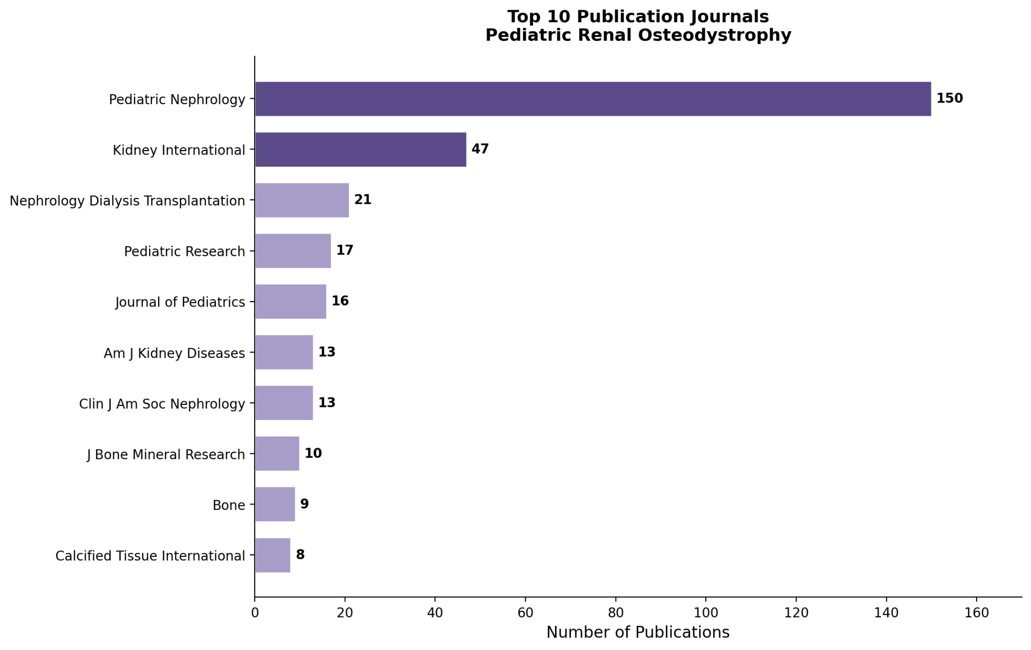
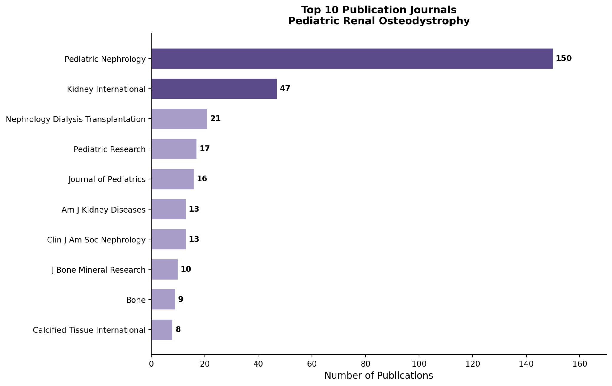
Analysis of publication venue showed an overwhelming concentration on specialist nephrology journals. Pediatric Nephrology was the top-performing journal by far (n=150), accounting for 19.5% of all papers. Kidney International came in a close second and provided representation for general nephrology journals. Journal of Bone and Mineral Research, Bone, and Calcified Tissue International also highlighted the close relationship between renal osteodystrophy and bone biology/endocrinology research communities.
.png)
Co-authorship analysis revealed several distinct collaborative communities. Salusky IB was centrally positioned between different research clusters and served as a hub for integrating multiple lines of work, reflecting his status as an intellectually central figure and leader in the community. A large North American cluster centered on UCLA investigators appears to have served as the field’s historical core, while European clusters headed up by Shroff R, Haffner D, and Bacchetta J represent complementary research traditions. While remaining linked, the network structure also suggests that pediatric ROD research remains concentrated among a relatively few leading centers and enduring collaborations.
.png)
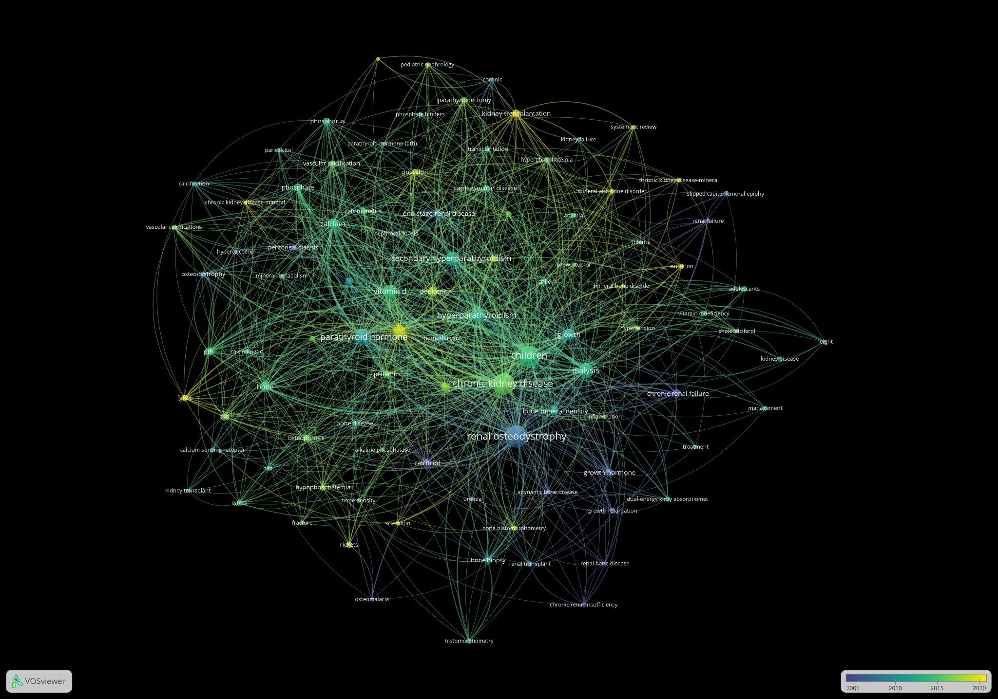
Keyword analysis yielded two prominent hubs, “Renal Osteodystrophy” and “Chronic Kidney Disease”, at the center of the network, highlighting both the historical terminology and modern CKD-MBD conceptualization. Major thematic clusters included bone histology/pathology, mineral metabolism, pharmacologic treatment, growth issues, transplantation, and nutrition. The presence of secondary hyperparathyroidism, calcitriol, phosphate binders, and adynamic bone disease terms indicated a strong emphasis on mineral metabolism and therapy, while growth terms underscored the distinctly pediatric nature of the condition. The prominence of both ‘renal osteodystrophy’ and ‘chronic kidney disease’ implies a shift away from the more familiar ROD framework and towards the broader CKD-MBD instance. The overlay network demonstrates a temporal transition from earlier themes centered on renal osteodystrophy, secondary hyperparathyroidism, and dialysis-related bone disease toward more recent interests in CKD-MBD, FGF-23 biology, and pediatric mineral metabolism.
.png)
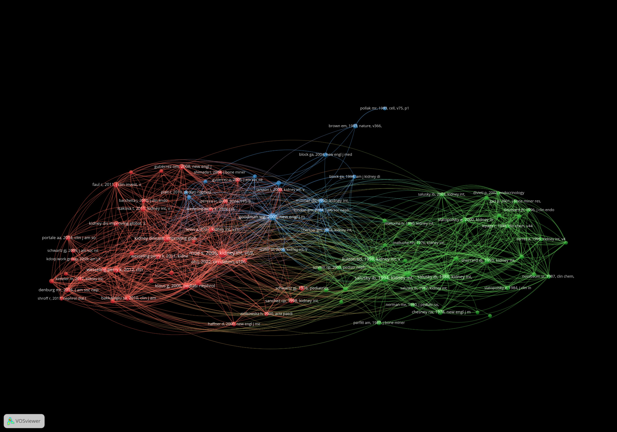
Co-citation analysis showed that the intellectual core of the field can be organized around three major historical eras. Early work was rooted in bone histology, histomorphometry, and the classification of renal osteodystrophy. Work around discoveries related to calcium-sensing receptors, vascular calcification, and cardiovascular complications of chronic kidney disease formed a second cluster. The newest cluster revolves around the CKD-MBD framework, FGF-23 biology, and guideline-driven management. Across all 3 clusters, Kidney International was the dominant journal venue, underscoring its longstanding influence on the intellectual development of pediatric renal osteodystrophy research.15,16
The most highly cited references within the retrieved literature are presented in Table 1. The most cited article was Holick’s 2004 review “Sunlight and vitamin D for bone health and prevention of autoimmune diseases, cancers, and cardiovascular diseases,” published in the American Journal of Clinical Nutrition, which garnered 1,997 citations (86.83 citations/year). Second was the 2017 KDIGO Clinical Practice Guideline Update for CKD-MBD at 1693 citations (highest citation rate of the top papers), followed by Holick’s 2009 review on Vitamin D status (#3 – 1230 citations), the 2017 KDIGO Executive Summary (#4 – 1122 citations) and Peacock’s 2008 review of calcium metabolism (#5 – 932 citations).
Together, these highly cited articles show that renal osteodystrophy research is heavily driven by broader investigations of vitamin D, calcium regulation, and CKD-Mineral and Bone Disorder (CKD-MBD) research. Notably, guidelines papers and reviews dominated the top-cited list. Several other key landmark studies focusing on calcium-sensing receptor physiology, parathyroid hormone measurement, adynamic bone disease, and renal transplantation were also highlighted, reflective of the field’s increasing focus on mineral metabolism and systemic complications of CKD. Publication years ranged from 1994 to 2020, with most papers being published after 2000, illustrating the increased attention given to CKD-MBD and associated bone disorders in recent decades.
Discussion
The field has grown steadily over the last half century. A total of 768 publications were found from 1971 to 2026, reflecting ongoing scientific interest in the skeletal complications of pediatric CKD. Early work in the late 1970s and 1980s reflected the growing clinical recognition of RBD as a separate disease, while publication output significantly increased after 2005, likely related to the 2006 KDIGO reclassification of ROD under the CKD-MBD umbrella and renewed interest in the biomarker FGF-23. Output peaked in 2013 and 2020 but has stabilized since 2019, consistent with the field maturing into one that is now well established rather than quickly emerging.17 This could reflect that most foundational questions about classifying disease and how it arises, pathophysiology, have largely been resolved, shifting the main focus toward optimizing diagnosis, monitoring, and therapeutic management.
As shown in Figure 2, pediatric renal osteodystrophy’s disciplinary profile straddles the intersection of nephrology, pediatrics, and mineral metabolism. Urology & Nephrology contributed almost half of the papers (47%), confirming that the field continues to be dominated by kidney-disease specialists, although Pediatrics ranked second (38.7%), underscoring the significance of growth/development outcomes. Endocrinology/Metabolism also played an outsized role (12.2%), highlighting the pivotal function of the parathyroid hormone–vitamin D–mineral metabolism axis in disease etiology, whereas smaller contributions from transplantation (5.1%), orthopedics (3.8%), radiology (2.6%), and pharmacology (1.6%) show its broad multiplicity. Relative sparsity of contributions from Nutrition & Dietetics (<1%) suggests that nutritional aspects of skeletal health may have remained comparatively neglected. Greater integration of nutritional research could be an important opportunity to help us understand modifiable factors in bone and growth outcomes for children with CKD. The relatively small role of nutrition research may reflect the fact that, since good growth and skeletal health are such important topics in childhood, studies of non-pharmacologic determinants of bone health are comparatively understudied.
Authorship analysis highlighted an extraordinary degree of scientific leadership in pediatric renal osteodystrophy research. Salusky IB was the most prolific author (87 publications), accounting for greater than 11% of all indexed articles, reflecting his pioneering role in establishing bone histomorphometry as a gold standard for diagnosis/classification of ROD. Co-authorship network analysis revealed that the field centers on several main clusters of collaboration. The UCLA cluster (Goodman WG, Wesseling-Perry K, Pereira RC, Gales B) dominated, while European investigators such as Shroff R, Haffner D, and Bacchetta J headed separate research streams. Collaboration between these groups occurs; however, the network suggests that much of the field’s growth took place within separate ‘regional communities’.16,18
Keyword co-occurrence analysis further illustrates the intellectual evolution of the field. Two dominant hubs, “renal osteodystrophy” and “chronic kidney disease”, organize the network and reflect the terminology transition initiated by the 2006 KDIGO reclassification—earlier studies were primarily indexed under renal osteodystrophy, while more recent literature increasingly adopts CKD-MBD terminology. Historically, ‘renal osteodystrophy’ was used as an umbrella term for the skeletal changes seen in CKD, as the original definition solely accounted for various bone abnormalities, but with increasing awareness of disturbances of mineral metabolism, vascular calcification and cardiovascular disease, the disorder transcended just bone. In response, KDIGO proposed the CKD-MBD framework to describe the systemic nature of the problem, but redefined ‘renal osteodystrophy’ as the skeletal component of the syndrome. We identified six major thematic clusters: bone histology/histomorphometry, mineral metabolism, pharmacologic management, pediatric growth, post-transplant outcomes, and nutritional risk. The distinct presence of adynamic bone disease as a keyword cluster highlights sustained attention to low-turnover bone disease resulting from excessive suppression of parathyroid hormone. Nutritional terms were relatively peripheral.17 The continued use of both terms, renal osteodystrophy and chronic kidney disease, suggests that the transition from renal osteodystrophy to CKD-MBD is incomplete, reflecting underlying tension between seeing the condition as a skeletal pathology versus a systemic consequence of chronic kidney disease.
Citation network analysis revealed a layered intellectual history shaped by several landmark discoveries. Kidney International was the dominant citation source across all major citation clusters, underscoring its longstanding role as the preeminent journal for pediatric renal osteodystrophy. The earliest cluster was centered on foundational studies by Salusky, Malluche, and Sherrard, establishing the histologic basis for the diagnosis of renal osteodystrophy. A second cluster reflected a shift toward mineral metabolism and cardiovascular complications highlighted by findings about the calcium-sensing receptor and evidence of vascular calcification in pediatric dialysis populations. The most recent cluster corresponded to the CKD-MBD era anchored by influential guideline papers and studies investigating FGF-23’s biology. The inclusion of high-impact publications from general medical journals highlights the broad clinical relevance of our field.19,20 Collectively, these citation clusters show a progression of interest: from describing bone defects to understanding their metabolic drivers to appreciating them as part of a broader web of endocrine and systemic disruptions.
We identified several important gaps. The continued coexistence of ‘renal osteodystrophy’ and ‘CKD-MBD’ as separate keyword hubs suggested that terminology standardization is still incomplete, which may hinder literature retrieval and synthesis. Nutritional and metabolic risk factors were surprisingly marginalized despite their recognized importance in childhood bone development, suggesting an area that warrants more study. Geographical representation was heavily skewed towards North America/Europe, leaving us unable to gain much insight into pediatric CKD populations elsewhere in the world. Post-transplant bone disease also appeared comparatively understudied despite its clinical importance. As with any bibliometric study, we are limited by relying on the Web of Science database, citation lag, and keyword sensitivity, but hope our results give a comprehensive overview of the field and point to some important directions for future investigation.17,21–23 Overall, these gaps in the research illustrate that these studies may provide an incomplete picture of pediatric renal osteodystrophy, especially regarding its nutritional determinants, post-transplant trajectories, and outcomes beyond those most extensively studied populations.
Further analysis of the most highly cited articles offers additional perspective on the intellectual priorities shaping pediatric renal osteodystrophy research. The prominence of vitamin D-related reviews and CKD-MBD guideline documents among the top citations underlines mineral metabolism’s centrality to the pathophysiology and treatment of pediatric renal bone disease. Highly cited papers focused on vitamin D physiology, calcium regulation, parathyroid hormone activity, and CKD-MBD reflect the field’s longstanding interest in the mechanisms behind abnormal bone turnover/mineralization. Strong citation performance for KDIGO guideline papers also indicates the profound impact of consensus recommendations guiding both clinical practice and subsequent research directions. Influential studies on kidney transplantation, adynamic bone disease, and calcium-sensing receptor biology collectively indicate that the field has broadened its scope beyond traditional descriptions of renal osteodystrophy to encompass broader issues relating to long-term skeletal outcomes and systemic consequences of chronic kidney disease. Together, the citation patterns indicate that advances in mineral physiology and CKD-MBD research have been the primary driver behind the field’s progression over the last two decades.
Conclusion
The evolution of pediatric renal osteodystrophy has important orthopedic implications, including fracture risk, skeletal deformities, impaired growth, and long-term bone health in survivors of pediatric CKD. This bibliometric analysis provides a comprehensive research focus on pediatric renal osteodystrophy, illustrating more than five decades of scientific investigation into one of the most significant complications of chronic kidney diseases in pediatric patients. Analysis of 768 publications showed an unrelenting growth in research activity. The intellectual journey of the field mirrors the same change. Initial research was around the bone histology, histomorphometry, and pathological characterization of renal osteodystrophy, while more recent work has been increasingly about FGF-23 biology, secondary hyperparathyroidism, CKD-MBD management, growth outcomes, and complications of chronic kidney disease. Co-authorship, co-citation, and keyword analyses reveal a highly interconnected research community that has shaped current understanding of CKD-related skeletal disease in children. Apart from all the progress made, there are still many gaps. Work remains unfinished towards North America/Europe, limiting the worldwide generalizability of the data. Nutrition, post-transplant bone disease, and pediatric-specific therapeutic targets appear neglected despite being important. The dual use of traditional RO terms along with CKD-MBD also indicates the struggle for a common language. As survival and life expectancy for children with chronic kidney disease (CKD) continue to improve, understanding long-term skeletal consequences of CKD will become increasingly important. To map the historical development, influential contributors, and emerging priorities within this field, we establish a foundation for future research while highlighting opportunities to advance more comprehensive, globally representative, and patient-centered approaches to pediatric CKD-related bone disease.