
Introduction
Intervertebral disc degeneration (IVDD) is one of the most prevalent musculoskeletal disorders and represents a major contributor to low back pain and disability worldwide. Epidemiological data indicate that low back pain affects a substantial proportion of the adult population and imposes a significant socioeconomic burden through healthcare costs, work absenteeism, and reduced quality of life.1 Degenerative changes of the intervertebral disc are considered a principal pathological substrate underlying chronic discogenic pain, spinal instability, and secondary deformities.2 Despite extensive research efforts, the mechanisms driving disc degeneration remain incompletely understood, limiting the development of effective disease-modifying therapies.1,2
The intervertebral disc is a highly specialized structure that relies almost entirely on diffusion for nutrient and metabolite exchange.3 In the adult spine, the nucleus pulposus (NP) and inner annulus fibrosus (AF) are essentially avascular, receiving nutrients primarily through the cartilaginous endplates (CEPs) supplied by segmental lumbar arteries.4,5 Accumulating clinical and experimental evidence suggests that impaired vascular supply, reduced tissue perfusion, and ischemic microenvironmental changes play a critical role in IVDD initiation and progression.6 Age-related atherosclerosis, lumbar artery stenosis, and diminished endplate perfusion have all been associated with disc degeneration and low back pain.7 However, the direct contribution of vascular insufficiency to disc degeneration remains difficult to isolate in human studies, underscoring the need for reliable experimental models.8
A broad range of animal models has been developed to investigate IVDD, including mechanical loading, chemical induction, genetic manipulation, and needle puncture techniques.8,9 While these approaches have provided valuable insights, most primarily induce structural damage rather than replicating the pathophysiological cascade initiated by impaired disc nutrition.6 Needle puncture models, although widely used, create an acute mechanical injury that may not adequately reflect the progressive, vascular-related degeneration observed in clinical settings.6,10,11 Similarly, previously described vascular models using limited or bilateral arterial ligation have produced inconsistent or mild degenerative changes and have often failed to demonstrate clear temporal progression across acute, subacute, and chronic stages.12,13
Given these limitations, there remains a critical gap in experimental models that can reliably reproduce progressive IVDD driven by vascular insufficiency while allowing detailed histopathological and morphometric assessment over time. A model capable of simulating unilateral, segmental hypoperfusion would more closely resemble clinically relevant conditions such as asymmetric vascular compromise and could provide a robust platform for studying disc degeneration dynamics.
Therefore, this study aimed to develop and histopathologically validate a novel unilateral three-level lumbar artery ligation model capable of reproducibly inducing progressive intervertebral disc degeneration in rats. We hypothesized that interruption of three consecutive unilateral lumbar arteries would generate sustained segmental hypoperfusion sufficient to produce sequential histopathological degeneration resembling the natural progression of human IVDD. By characterizing temporal structural alterations and quantitative morphometric changes, this study sought to establish a reproducible experimental platform for future mechanistic investigations and preclinical therapeutic studies targeting vascular-associated disc degeneration.
Materials and Methods
This experimental study was conducted in accordance with institutional guidelines and approved by the local Institutional Animal Ethics Committee (Approval No: 6/27, July 9, 2024). Fifty-two Sprague Dawley rats (26 females and 26 males), aged 8 weeks, were included. Animals were housed in sex-separated cages under controlled environmental conditions (12-hour light/dark cycle, standardized temperature and humidity) with ad libitum access to food and water. All animals were monitored daily throughout the study period.
Animals were randomly divided into four groups with equal sex distribution. One group served as the sham-operated control and underwent skin incision followed by immediate wound closure without abdominal exploration or lumbar artery ligation. This design was intended to provide a baseline control while avoiding the potential biological effects of retroperitoneal dissection and manipulation of the abdominal vasculature. The remaining three experimental groups were established to evaluate time-dependent degenerative changes following unilateral ligation of three consecutive lumbar arteries at acute (day 3), subacute (day 15), and chronic (week 8) time points. Animals were euthanized at the designated time points by intraperitoneal overdose of anesthetic agents for tissue harvesting.14 The spinal column was carefully excised after removal of paraspinal soft tissues, and intervertebral disc specimens were collected for subsequent histological and morphometric analyses.
All animals fasted for 4 hours before surgery. General anesthesia was induced via intraperitoneal administration of ketamine (50–75 mg/kg) and xylazine (5–10 mg/kg).15 When necessary, anesthesia was maintained using sevoflurane inhalation. Preoperative analgesia (paracetamol, 100 mg/kg, intraperitoneal) and antibiotic prophylaxis (cefazolin, 25 mg/kg, intramuscular) were administered, and postoperative analgesia and antibiotic treatment were continued twice daily for 3 days.16,17
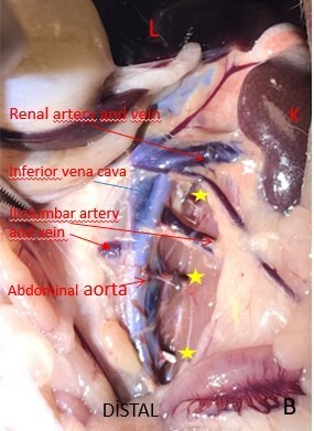
Under sterile conditions and ×5 magnification using a surgical loupe, a midline abdominal incision extending from the xiphoid process to the suprapubic region was performed. Following gentle bowel evisceration, the abdominal aorta and its lumbar segment were exposed. Careful microsurgical dissection was carried out to identify the lumbar arteries originating from the posterolateral aspect of the aorta.18
A unilateral ligation approach was deliberately selected to minimize the risk of neurological and systemic vascular complications associated with extensive spinal ischemia while still inducing sufficient segmental hypoperfusion to trigger disc degeneration. The L4–L5 intervertebral disc was selected as the primary outcome level because its nutritional supply depends predominantly on the lumbar arterial system, whereas the L5–S1 disc may receive additional perfusion from the iliolumbar and sacral arterial branches. Restricting the analysis to the L4–L5 level therefore minimized potential confounding caused by collateral vascular supply and enabled a more accurate assessment of lumbar artery ligation–induced degeneration.19
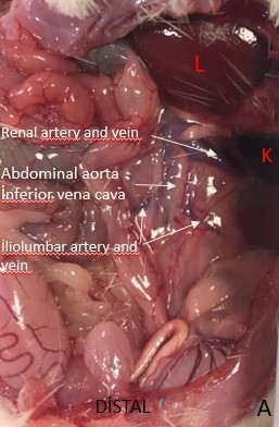
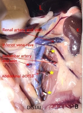
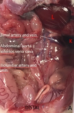
Three consecutive left lumbar arteries (L3–L4, L4–L5, and L5–S1), located proximal to the aortic bifurcation, were identified and ligated using vascular clips (LT100, Ethicon) applied with a dedicated clip applier (LX107, Ethicon). Cessation of pulsatile flow distal to the clip application was visually confirmed intraoperatively. The surgical field was irrigated with warm saline, the bowel was repositioned into the abdominal cavity, and the abdominal wall was closed in anatomical layers using absorbable sutures. Representative intraoperative images illustrating identification of the abdominal aorta, microsurgical isolation of three consecutive left lumbar arteries, and successful vascular clip application are presented in Figure 1.
Harvested specimens were fixed in 10% neutral buffered formalin for 24 hours and subsequently decalcified. Following routine paraffin embedding, serial sagittal sections (4 μm thickness) were prepared. Histological evaluation was performed using Hematoxylin–Eosin and Safranin O/Fast Green staining to assess disc architecture, cellularity, and extracellular matrix composition.4
IVDD was assessed using a standardized histopathological scoring system (score range: 0–14), evaluating NP morphology, AF organization, CEP integrity, and extracellular matrix changes. All scoring procedures were performed in a blinded manner.20
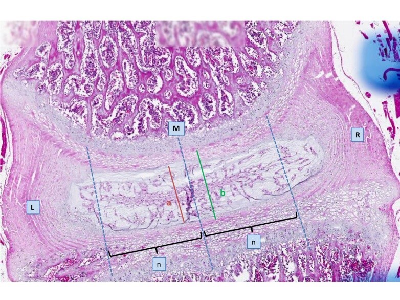
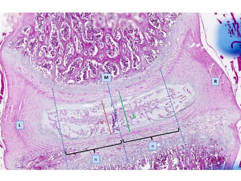
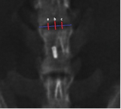
Histomorphometric measurements included intervertebral disc thickness, NP height, and AF height. As illustrated in Figure 2, thickness measurements for both NP and AF were obtained at three distinct sagittal locations for each disc specimen: at the midline and at points 750 µm lateral to the midline on both the left (L) and right (R) sides. These measurements were defined by vertical reference lines and standardized lateral distance markers (750 µm), ensuring consistency across all samples. At the chronic time point (Week 8), digital radiographs of the lumbar spine were obtained.
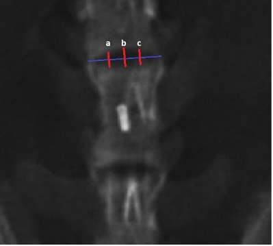
As shown in Figure 3, intervertebral disc height was measured using standardized techniques at the midline and bilateral reference points via the Picture Archiving and Communication System. Radiological measurements were subsequently compared with histomorphometric findings.21,22
Statistical Analysis
All statistical analyses were performed using SPSS software (version 23.0). Data were expressed as mean ± standard deviation, and statistical significance was set at p < 0.05.
Given the non-parametric distribution of the data, intergroup comparisons were performed using the Kruskal–Wallis test. For variables showing significant differences, post hoc pairwise comparisons were conducted using the Mann–Whitney U test with Bonferroni correction (adjusted α = 0.0083).
For repeated measurements obtained from the same animals, the Friedman test was used to evaluate within-group differences over time. When significant, pairwise comparisons were performed using the Wilcoxon signed-rank test with Bonferroni correction to control for multiple comparisons.
Effect sizes were calculated using the rank-biserial correlation coefficient (r) and eta squared (η2). A post hoc power analysis was also conducted to assess the statistical robustness of the findings.
Results
All animals tolerated the surgical procedure without major intraoperative or postoperative complications. One perioperative death occurred during the initial phase of the study because of inadvertent vascular injury during microsurgical dissection. Following refinement of the surgical technique, no additional mortality, neurological deficits, or procedure-related complications were encountered. Throughout the experimental period, all surviving animals maintained normal mobility, feeding behavior, and general health, indicating that unilateral three-level lumbar artery ligation was a reproducible and well-tolerated surgical procedure.
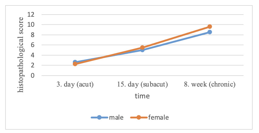
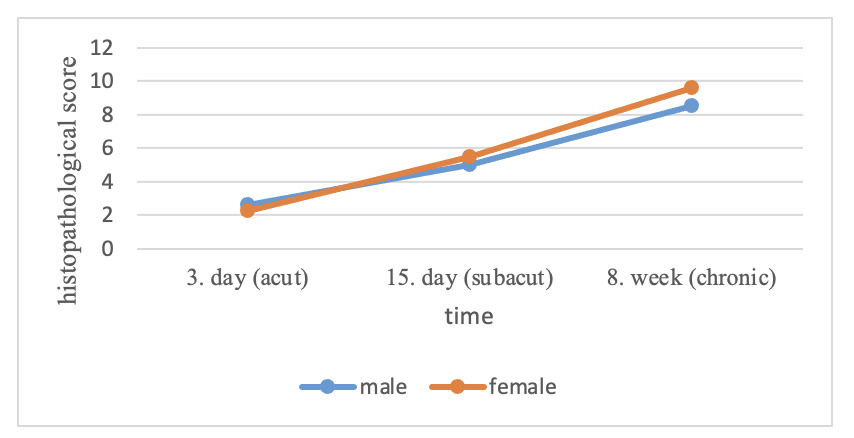
Progressive histopathological degeneration was observed following unilateral three-level lumbar artery ligation (Table 1). Sham-operated discs consistently demonstrated normal morphology and received a histopathological score of 0 throughout the study. In contrast, degeneration scores increased progressively from 2.6 ± 0.7 at Day 3 to 4.8 ± 0.6 at Day 15 and reached 8.9 ± 1.1 at Week 8, representing an approximately 3.4-fold increase in degeneration severity over the experimental period. These findings demonstrate a clear temporal progression of structural degeneration following interruption of segmental lumbar arterial blood supply.
As presented in Table 2, differences in histopathological scores among groups were evaluated using the Kruskal–Wallis test and found to be statistically significant [H(3) = 37.24, p < 0.001, η2 = 0.951]. Post hoc pairwise comparisons were performed using the Mann–Whitney U test with Bonferroni correction (adjusted α = 0.0083). All time points differed significantly from each other (all p < 0.001), with rank-biserial correlation coefficients indicating large effect sizes (r ≥ 0.90). Statistical power was calculated as 99% for all comparisons.
Histopathological scores increased progressively in both sexes throughout the study period. Although female rats demonstrated numerically higher degeneration scores at Week 8, no statistically significant sex-related differences were detected at any time point (Day 3: p = 0.39; Day 15: p = 0.34; Week 8: p = 0.056), indicating that degeneration severity was primarily associated with disease duration rather than sex (Table 3).
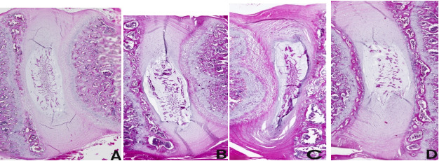
Representative histological findings are presented in Figure 4. Sham-operated discs exhibited preserved intervertebral disc architecture with a well-defined nucleus pulposus (NP), an intact annulus fibrosus (AF), and normal cartilaginous endplates, corresponding to the absence of histopathological degeneration. Following unilateral three-level lumbar artery ligation, progressive structural deterioration became evident over time, affecting both the NP and AF compartments
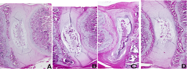
At the acute stage (Day 3), early degenerative alterations were observed, characterized by mild disorganization of the inner AF and subtle reductions in NP cellularity and matrix homogeneity, while overall disc architecture remained largely preserved (Figure 4B).
Degenerative changes progressed substantially at Day 15, with depletion of the NP matrix, transition of notochordal cells toward a chondrocyte-like phenotype, and increasing disruption of AF lamellar organization. The boundary between the NP and AF became progressively indistinct, reflecting loss of normal disc architecture (Figure 4C).
By Week 8, severe degeneration was consistently observed. The AF exhibited marked thinning and architectural disorganization, whereas the NP demonstrated profound hypocellularity, extensive extracellular matrix loss, and near-complete disruption of the NP–AF interface, consistent with advanced-stage IVDD (Figure 4D).
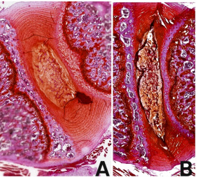
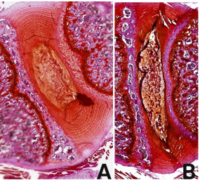
Safranin O/Fast Green staining further confirmed progressive extracellular matrix degeneration. Compared with sham controls, discs harvested at Week 8 demonstrated a marked reduction in Safranin O staining intensity within the NP, indicating substantial proteoglycan depletion and loss of matrix integrity (Figure 5).
As presented in Table 4, time-dependent changes in histomorphometric measurements were evaluated across predefined regions of the disc. Significant differences were identified in NP measurements at the midline, right, and left locations (p = 0.016, p = 0.045, and p = 0.003, respectively). Similarly, AF measurements demonstrated significant variation over time at the midline, right, and left regions (p = 0.001, p = 0.004, and p = 0.001, respectively).
These findings indicate region-specific and time-dependent structural alterations within both NP and AF components of the intervertebral disc.
Morphometric analysis demonstrated that structural alterations developed progressively following lumbar artery ligation (Table 5). During the acute (Day 3) and subacute (Day 15) stages, no significant side-to-side differences were detected in either NP or AF height (all p > 0.05). However, by Week 8, NP height was significantly reduced on the ligated side compared with the contralateral side (p = 0.001), indicating that irreversible structural collapse became evident only during the chronic stage of degeneration. AF height also demonstrated a consistent trend toward thinning, although side-to-side differences did not reach statistical significance.
Although AF height did not demonstrate a statistically significant side-to-side difference, histopathological evaluation revealed a consistent trend toward thinning in the ligated side.
These findings indicate that structural collapse of the NP becomes evident predominantly during the chronic phase of degeneration, supporting the time-dependent progression of vascular-induced disc degeneration.
Radiographic evaluation performed at Week 8 did not demonstrate significant differences in intervertebral disc height between the ligated and contralateral sides (p = 0.286). Despite advanced histopathological degeneration and measurable morphometric alterations, conventional radiography failed to detect these structural changes. These findings indicate that histopathological and morphometric analyses are substantially more sensitive than plain radiography for identifying early or moderate stages of vascular insufficiency–induced IVDD.
Discussion
The present study demonstrates that unilateral ligation of three consecutive lumbar arteries reproducibly induces progressive intervertebral disc degeneration characterized by sequential histopathological deterioration, quantitative morphometric changes, and delayed radiographic findings. Unlike mechanically induced models that create immediate structural disruption, this vascular model reproduces a gradual degenerative process that more closely resembles the temporal evolution of human IVDD. The findings further support the concept that sustained segmental hypoperfusion alone appears sufficient to initiate progressive disc degeneration in the absence of direct disc injury.
A major strength of this model is that vascular interruption alone produced progressive degeneration without introducing direct mechanical damage to the intervertebral disc. This pattern suggests that localized ischemia may initiate a cascade of secondary responses that affect the broader disc microenvironment through diffusible or paracrine mediators. Such a mechanism is supported by previous studies indicating that regression of arterial supply and disruption of anastomotic networks may precede detectable histological or radiological degeneration.23 The diffusion-dependent nature of disc nutrition further reinforces this interpretation, as the adult NP represents one of the largest avascular structures in the vertebrate body, with diffusion distances reaching up to 8 mm from the nearest blood vessel.24
The time-dependent progression observed in this model underscores the dynamic nature of IVDD. Early changes detected at the acute stage were subtle and likely reflect an initial adaptive response to reduced perfusion, whereas subacute and chronic stages exhibited progressive disruption of disc architecture and extracellular matrix depletion. This staged degeneration contrasts sharply with needle puncture models, which induce abrupt mechanical injury and fail to replicate the gradual, vascular-driven degeneration observed in clinical settings.2,9,25 Consistent with previous reports, impaired lumbar arterial blood flow has been shown to negatively affect nutrient and metabolite diffusion within the disc, thereby accelerating degenerative processes.26,27 This gradual progression closely parallels the biological evolution of human IVDD, in which vascular compromise is believed to precede overt structural degeneration. Consequently, the present model may provide a more physiologically relevant platform for investigating disease initiation than models based solely on acute mechanical injury.
Radiological disc height measurements did not reveal significant differences despite clear histopathological degeneration, a discrepancy consistently reported in both experimental and clinical studies.12 This finding highlights the limited sensitivity of conventional radiography in detecting early or moderate IVDD and underscores the importance of histological and morphometric analyses for accurate model validation. In this context, the present model demonstrates that structural and compositional degeneration may precede measurable radiographic changes, further emphasizing its relevance for studying early-stage IVDD.
Previous vascular insufficiency–based IVDD models have produced inconsistent and frequently modest degenerative changes. Imanishi et al. reported only mild and non-progressive degeneration following unilateral lumbar artery ligation, whereas Jin et al. demonstrated morphological alterations without comprehensive histopathological validation.12,13 Hou et al. combined vascular compromise with direct cartilaginous endplate injury, thereby introducing an additional mechanical component that complicates interpretation of ischemia-specific effect.6 In contrast, the present model induced progressive degeneration through isolated vascular interruption without direct injury to the intervertebral disc. The sequential histopathological deterioration observed across acute, subacute, and chronic stages therefore provides stronger experimental evidence that sustained vascular insufficiency alone is sufficient to initiate progressive IVDD.
The greater severity of degeneration observed in the present study may also be attributable to the extent of vascular interruption. Unlike previous experimental models, which generally interrupted one or two lumbar arteries, we ligated three consecutive unilateral lumbar arteries with the aim of producing sustained segmental hypoperfusion. We speculate that this approach exceeded the compensatory capacity of collateral circulation, thereby generating a more persistent ischemic environment capable of driving progressive disc degeneration. Although collateral blood flow was not directly quantified in the present study, the reproducible temporal progression of histopathological degeneration supports this interpretation.
Species-specific differences in spinal vascular anatomy should also be considered when interpreting experimental models of IVDD. In humans, lumbar arteries represent a relatively small proportion of the abdominal aortic diameter, whereas rats possess proportionally larger segmental vessels and more abundant collateral circulation.6,28–30 Consequently, direct replication of human vascular anatomy is not feasible in rodent models. Rather than reproducing anatomical proportions, the present model was designed to reproduce the biological consequences of sustained segmental hypoperfusion. The progressive histopathological degeneration observed following ligation of three consecutive lumbar arteries suggests that this degree of vascular interruption is sufficient to overcome collateral compensation and establish a reproducible ischemia-driven model of IVDD.
Importantly, the absence of significant side-to-side differences in the early stages, followed by clear divergence at the chronic stage, supports a gradual, ischemia-driven degenerative process rather than an acute injury response. In addition, the region-specific morphometric alterations observed in both NP and AF suggest that vascular insufficiency induces spatially heterogeneous degeneration within the disc.
Collectively, the present findings support the concept that vascular insufficiency is not merely associated with intervertebral disc degeneration but may represent a primary initiating factor capable of triggering progressive structural degeneration in the absence of direct mechanical injury. By reproducing sequential histopathological and morphometric changes with low surgical morbidity, the present model provides a reproducible experimental platform that more closely reflects the gradual evolution of vascular-associated IVDD than conventional mechanically induced models. This model may therefore facilitate future studies investigating the pathophysiological mechanisms underlying disc degeneration as well as the preclinical evaluation of therapeutic strategies targeting vascular compromise.
Limitations
Several limitations of this study should be acknowledged. First, although the rat model provides a controlled and reproducible platform for investigating vascular contributions to IVDD, species-specific anatomical, biomechanical, and vascular differences inevitably limit direct extrapolation to the human spine. Nevertheless, the primary objective of this model was not to replicate human anatomy precisely, but rather to reproduce the biological consequences of sustained segmental vascular insufficiency under controlled experimental conditions.
Second, the present study was designed to establish and histopathologically validate a novel vascular insufficiency–induced model of IVDD rather than to investigate the underlying molecular mechanisms. Consequently, molecular and biochemical analyses were intentionally beyond the scope of this study. Nevertheless, integration of inflammatory, hypoxia-related, and extracellular matrix remodeling markers in future investigations would further clarify the biological pathways linking vascular insufficiency to disc degeneration.
Third, imaging assessment was limited to plain radiography at the chronic time point. Although conventional radiographs confirmed the absence of substantial disc height loss despite advanced histopathological degeneration, longitudinal imaging using higher-resolution modalities such as MRI or contrast-enhanced micro-CT could provide a more comprehensive characterization of the temporal evolution of structural degeneration and vascular alterations.
Finally, although the unilateral design enabled direct within-animal comparisons and reduced inter-animal variability, it does not fully reproduce the multifactorial pathogenesis of human IVDD, in which aging, genetic susceptibility, metabolic disturbances, and mechanical loading interact with vascular insufficiency. Accordingly, this model should be regarded as a reductionist experimental platform specifically designed to investigate the contribution of vascular factors to disc degeneration rather than to replicate all aspects of the human disease.
Conclusion
Unilateral three-level lumbar artery ligation in rats provides a reproducible and biologically relevant experimental model of vascular insufficiency–induced intervertebral disc degeneration. The model consistently reproduces the sequential histopathological and morphometric changes characteristic of progressive IVDD while maintaining low surgical morbidity and enabling robust within-animal comparisons. Importantly, histopathological degeneration preceded detectable radiographic alterations, highlighting the sensitivity of this model for investigating the early stages of disc degeneration. By avoiding direct mechanical injury to the intervertebral disc, this approach more closely reflects the gradual pathophysiological evolution of vascular-associated IVDD than conventional mechanically induced models. Consequently, this model provides a robust experimental platform for investigating vascular mechanisms underlying IVDD and for the preclinical evaluation of emerging therapeutic and regenerative strategies.
Acknowledgments
This study was supported by the Health Institutes of Türkiye (TUSEB) under Grant [Call code: 2024-A4-02, Project Number: 42710].