
Introduction
The knot of Henry—an anatomical interconnection between the flexor hallucis longus (FHL) and flexor digitorum longus (FDL) tendons—has attracted increasing attention because of its relevance to tendon transfer surgery and forefoot biomechanics.1–3 Detailed anatomical investigations have further established the knot of Henry as a reliable surgical landmark that can be approached safely during plantar procedures involving the long flexor tendons.4 In the plantar foot, the FHL and FDL follow independent proximal courses before crossing at the knot of Henry.5 Previous anatomical studies have revealed that connections from the FHL to the FDL occur frequently and that there are certain patterns to their branching.6,7 Furthermore, these tendon connections have been reported to primarily contribute to the function of the second and third toes, which may extend to the fourth toe.2,8 Recent comprehensive reviews have reported that tendon connections from the FHL to the FDL are primarily distributed from the second through the fourth toes, and direct distributions to the fifth toe are extremely rare.3,9 Although this selective distribution pattern has been consistently observed in multiple anatomical studies, the reasons why the FHL’s contribution extends only minimally to the fifth toe and implications of this distribution for foot function have not been fully examined.
Previous anatomical studies have also reported that the quadratus plantae (QP) is closely associated with the tendon attachments of the FDL and FHL and is particularly involved in the control of the lateral toes.8 Few comprehensive studies have examined the possibility of the QP playing a distinctive role in the control of the fifth toe from the perspective of the division of labor within the entire long flexor tendon system.
Given these gaps, in this study, we aimed to review the existing literature on FHL–FDL tendon interconnections at the knot of Henry, focusing on the selective distribution pattern in which FHL-derived slips primarily contribute to the function of the second through fourth toes with minimal involvement in that of the fifth toe. We further examined the anatomical and biomechanical basis of this distribution, including the role of the QP and have discussed its implications for forefoot function, checkrein deformity, and tendon transfer surgery.
Methods
We conducted a narrative review of the existing literature on the FHL–FDL tendon interconnection at the knot of Henry. PRISMA guidelines were not strictly applied given the narrative design.
Literature searches were performed in PubMed and Google Scholar, targeting articles published until 2025, using the terms “knot of Henry,” “flexor hallucis longus,” “flexor digitorum longus,” “tendinous slip,” and “quadratus plantae.” Reference lists of the retrieved articles were also reviewed to identify additional sources.
Studies were included if they met any of the following criteria: (1) cadaveric anatomical studies on the FHL–FDL connection; (2) studies reporting toe-specific tendon distribution to the second through fifth toes; or (3) studies examining the functional relationship among the FHL, FDL, and QP. Studies lacking detailed anatomical descriptions, non-human studies, single-case reports, and interventional studies were excluded.
The following data were extracted from each study: sample size, population, connection pattern, toe-specific distribution frequencies, and QP involvement. The primary outcome was the presence or absence of FHL contribution to the fifth toe.
Results
Six major anatomical studies were identified for comparative review. Table 1 presents the FHL–FDL connection and toe distribution patterns identified in the major anatomical studies.1–3,6–8
In many of these anatomical studies, interconnections between the FHL and FDL were observed, with the most common pattern involving a connection from the FHL to the FDL. Anatomical variations without such connections were also reported.10 More recently, Vasudha et al. likewise confirmed a high prevalence of FHL–FDL communications while demonstrating substantial individual variation in branching configurations, further supporting the consistency of the interconnection despite morphological variability.11
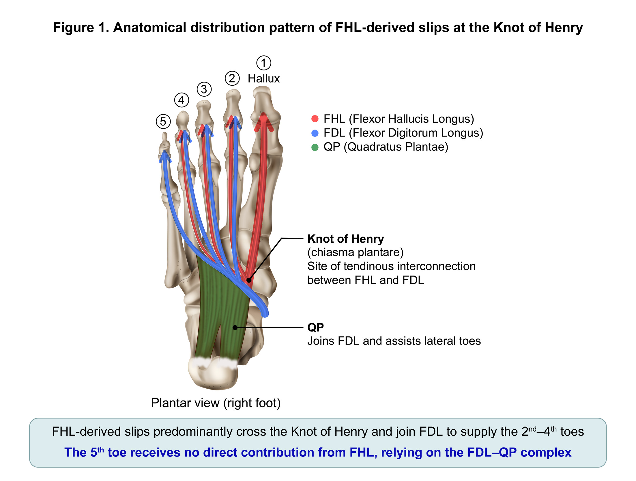
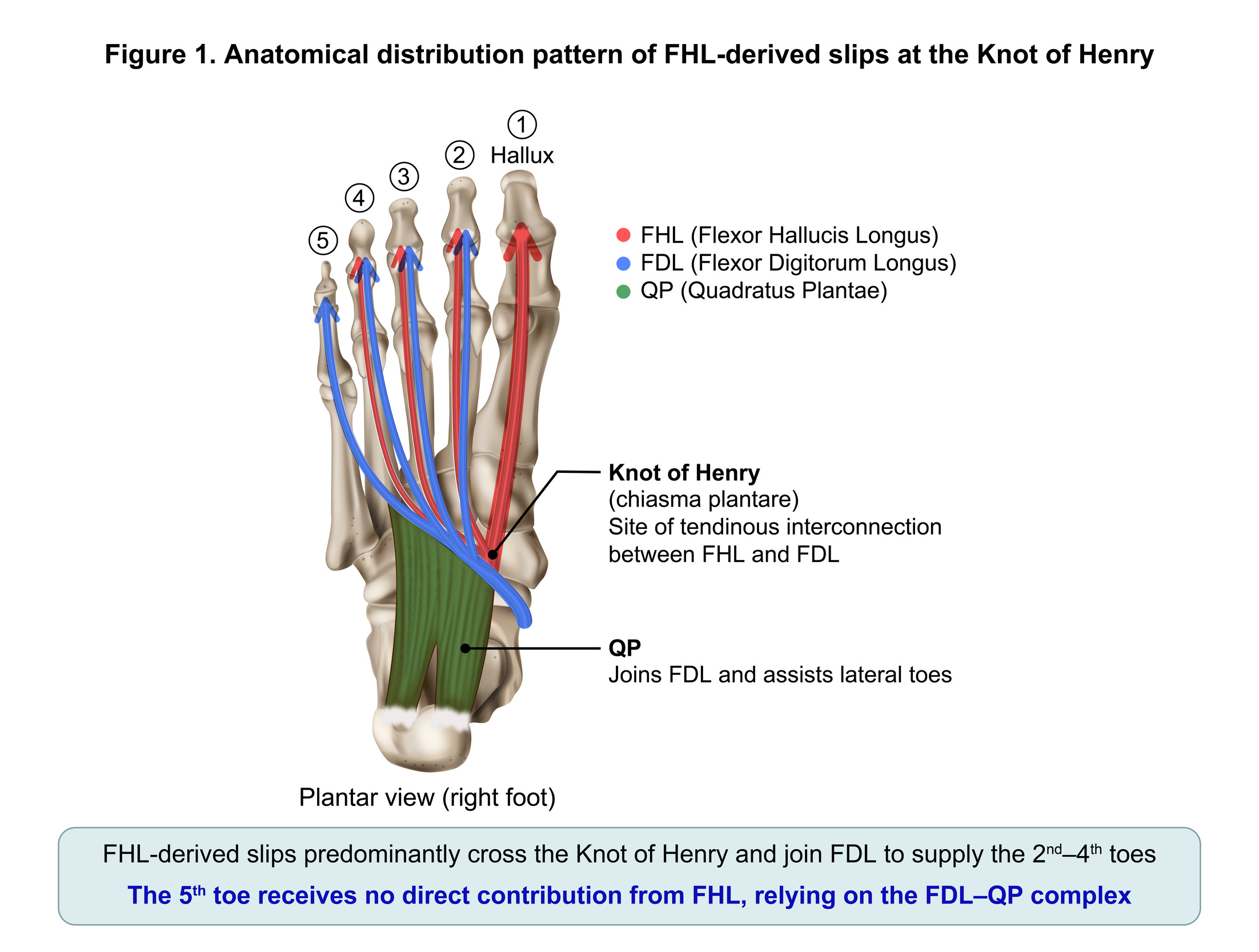
The distribution of tendon connections follows a consistent pattern from the FHL to the FDL, with connections to the second (38%–100%) and third (47%–64%) toes being frequently observed in multiple studies,2,3,7 although Hur et al. reported a slightly different distribution pattern (8% to the second toe, 64% to the third toe), and no distribution to the fifth toe was observed in any of the studies.8 Zhao et al. also reported that the FHL primarily contributes to the function of the second and third toes, extending to the fourth toe in some cases, while no contribution to the fifth toe was observed.12 On the other hand, tendon connections extending to the fourth toe were relatively rare (6%–28%).2,3,7 More recent cadaveric studies have likewise confirmed the high prevalence of FHL–FDL communications despite variation in branching patterns.11 The direct attachment of the FHL to the fifth toe was rarely reported in anatomical studies, with reports of this occurring in less than 0–1% of cases.2,3,7–9 Figure 1 illustrates the general pattern of this selective distribution of FHL-derived slips. Bai et al. also reported that they did not observe the Type D pattern extending to the fifth toe in their ultrasound evaluation of the chiasma plantare, and this distribution pattern is supported by in vivo assessments.13
Regarding the QP, several studies have reported anatomical connections with the FDL group. Hur et al. reported that the QP is attached not only to the FDL tendon but also to a slip originating from the FHL,8 which shows that the QP is a structure that is closely associated with the flexor digitorum longus system. Furthermore, Wapner et al. reported a distribution pattern in which the QP is associated with the lateral toe tendons, including those of the fourth and fifth toes.1
Observations of functional coupling between the FHL and FDL in tendon pull tests suggest that the movement of the fifth toe is primarily associated with FDL pull.6 However, this previous study did not quantify the anatomical distribution by each specific toe and is limited to suggestions based on functional observations. Similarly, in biomechanical and morphometric analyses, the functional characteristics of tendons distributed to the fifth toe were found to differ from those of tendons influenced by the FHL tendon bundle.14
Discussion
Anatomical consistency of selective distribution
The most important finding of this review was that the distribution of FHL-derived slips is consistently limited to the second through the fourth toes and is rarely observed to extend to the fifth toe. This distribution pattern has been observed in multiple postmortem studies that involved different study populations and methodologies.2,3,6–8 Furthermore, a large-scale review by Berger et al. revealed that FHL insertion into the fifth toe is extremely rare, occurring in less than 1% of cases.9
Previous studies have primarily emphasized the variability in the branching patterns of the FHL–FDL connection.2,7 However, despite this variability, the selective distribution of FHL-derived slips from the second through the fourth toes remained remarkably consistent across studies. Structural continuity between the FHL and FDL has also been demonstrated by contrast-enhanced imaging studies, further supporting the presence of a consistent tendinous interconnection at the Knot of Henry.15
Possible biomechanical basis
Such a consistent distribution pattern is unlikely to represent a random variation; rather, it appears to reflect specific anatomical and biomechanical constraints. From a biomechanical perspective, the FHL is a muscle that plays a central role in generating propulsive force in the forefoot via the hallux16,17 and is thought to be particularly involved in concentrating load on the medial forefoot during locomotion.18 The distribution of FHL-derived slips primarily from the second through the fourth toes can be interpreted to indicate that the FHL is a structure that disperses this propulsive force to the medial column and assists in the coordinated movement of adjacent toes. The FHL-derived slip that branches at the knot of Henry reaches each toe by diverging laterally from the main force transmission axis; however, mechanical efficiency may decrease as the tendon slip diverges laterally from the main force axis. This biomechanical framework provides a plausible explanation for the rarity of FHL-derived slip distribution to the fifth toe.19,20 Supporting this view, forefoot kinetic studies have consistently demonstrated that the medial column is primarily responsible for load-bearing and propulsion, while the lateral column, which includes the fifth toe, contributes relatively little to push-off forces.21–24
Role of the QP and integrated plantar flexor network
QP has traditionally been regarded as an accessory muscle of the FDL; however, comparative anatomical studies have long suggested that it represents an evolutionarily conserved component of the plantar flexor apparatus rather than merely an accessory structure.25,26 Control of the fifth toe may primarily involve the FDL and QP. Hur et al. reported that the QP is frequently attached not only to the FDL but also to the slip originating from the FHL, suggesting that the QP is anatomically integrated with the long toe flexor system.8 Variations in the morphology and insertion of the QP have also been reported, further emphasizing its close anatomical relationship with the plantar flexor system.27 Histological studies have also shown that FHL-derived slips are integrated into the FDL system via the QP, suggesting that the chiasma plantare is not a simple tendon crossover but a multilayered functional network.28,29 Pretterklieber et al. reported that while the contribution of the FHL is primarily concentrated from the first to the third toes, the QP is broadly involved in the tendons of the second to the fifth toes,29 suggesting a difference in their roles within the medial and lateral columns in this study.
Functionally, the QP is also thought to contribute to foot stability during walking as part of the intrinsic foot musculature.30,31 Sooriakumaran et al. proposed that the QP may have acquired evolutionary importance in human gait.32 In particular, the lateral column, which includes the fifth toe, is thought to be more involved in balance maintenance and load adjustment than in propulsion, and it is possible that the FDL–QP complex functions as a structure responsible for lateral column function.
Functional compartmentalization as an integrative framework
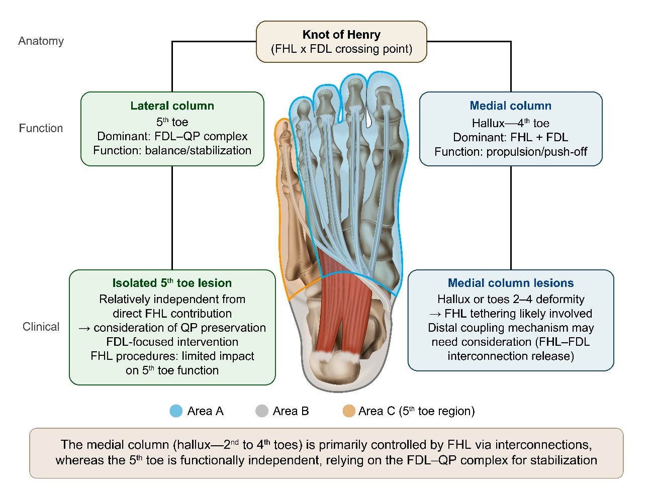
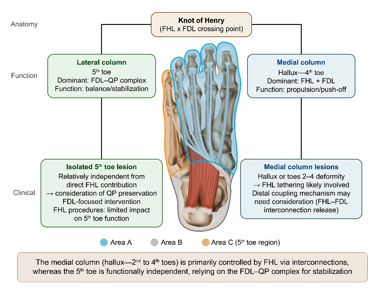
The limited contribution of the FHL to the fifth toe is unlikely to be explained by a single anatomical factor. Instead, this pattern likely reflects a combination of biomechanical characteristics of the medial forefoot and compensatory control provided by the FDL–QP complex. Specifically, the tension generated by the FHL is adapted to a propulsion mechanism centered on the medial forefoot, whereas transmission efficiency appears to decrease as force diverges laterally from the knot of Henry. Furthermore, the lateral column, which includes the fifth toe, is more heavily involved in load adjustment and balance maintenance than in propulsion, and its functional demands are likely different from those of the medial column. Additionally, as the FDL system, which includes the QP, complements the control of the lateral column, the fifth toe appears to operate through a control mechanism that is largely independent of the FHL-derived system.
These findings support the understanding that the long digital flexor system is not a single, uniform mechanism, but rather a structure with distinct functional organization between the medial and lateral columns. Figure 2 illustrates a conceptual model of functional differentiation within the long digital flexor system based on these findings.
Clinical implications
The selective distribution pattern of the FHL–FDL interconnection identified in this review provides important insights into the clinical behavior of the long flexor tendon system. Early reports described checkrein deformity as a post-traumatic consequence of FHL tendon tethering following fractures around the ankle or distal leg.33 Subsequent clinical observations suggested that FHL contracture or impaired tendon gliding may propagate to the second through fourth toes via the knot of Henry.34 Consistent with this mechanism, release of the FHL–FDL interconnection at the midfoot has been shown to improve lesser toe deformity,35,36 highlighting the clinical relevance of distal intertendinous coupling.
Recently, a pathology-oriented framework for surgical level selection proposed that deformity patterns reflect different pathological mechanisms, including proximal FHL tethering and distal intertendinous coupling.37 The present anatomical review provides an anatomical basis for this concept by demonstrating that FHL-derived slips consistently supply the second through fourth toes, whereas the fifth toe is primarily associated with the FDL–QP complex. Accordingly, deformities involving the second through fourth toes are more likely to reflect distal coupling through the knot of Henry and may therefore be suitable for procedures such as FHL–FDL interconnection release or distal FHL lengthening. In contrast, isolated fifth toe deformity may suggest a different pathological mechanism involving the FDL–QP complex rather than the FHL-derived system. Thus, the pattern of toe involvement itself may provide useful guidance in identifying the predominant tendon system and selecting an appropriate tendon-level surgical strategy.
These anatomical findings may also have implications for FHL tendon transfer procedures. When harvesting the FHL distal to the knot of Henry, surgeons should anticipate the possibility of disrupting cross-attachments to the lesser toes. Previous anatomical and clinical studies have shown that distal harvesting may require additional dissection because of these interconnections and may increase the risk of neurovascular injury.38–40 Accordingly, several surgical techniques have been proposed to facilitate safe and efficient FHL tendon harvest while minimizing injury to surrounding structures.41 These anatomical relationships should therefore be considered when selecting the harvest level and surgical approach, as they may help anticipate postoperative toe function and optimize tendon transfer procedures.
Conclusions
This study confirmed that the tendon connections from the FHL to the FDL are consistently distributed across the second through fourth toes and contribute very little to the function of the fifth toe. In particular, the limited contribution of the FHL to the function of the fifth toe can be interpreted as being the result of an interplay of multiple factors, including the attenuation of mechanical tension as it moves laterally from the knot of Henry; the low demand for propulsive function in the lateral column; and the complementary control mechanism provided by the FDL system, including the QP. These findings support the possibility that the fifth toe is a functional unit that is relatively independent of the FHL system.
The concept of “functional compartmentalization” proposed here reframes the understanding of the long flexor tendon system from being a uniform structure to being one that includes distinct medial and lateral functional units. This concept may help improve the anatomical interpretation of checkrein deformity and support tendon-level surgical planning.
Conflict of Interest
The authors declare no conflicts of interest.
Funding
This research received no specific grant from any funding agency in the public, commercial, or not-for-profit sectors.
Author Contributions
S.N: Conceptualization, Methodology, Writing – Original Draft. Y.T.: Supervision, Writing – Review & Editing. H.E., Y.O., H.K., M.N.: Data Curation, Writing – Review & Editing. Y.K.: Supervision.
Ethical statement
This study was based exclusively on previously published literature and did not involve human participants or animal experiments. Ethical approval was, therefore, not required.
Acknowledgements
AI-assisted language editing tools were used to improve grammar and readability during manuscript preparation. All scientific concepts, interpretation, and conclusions were independently developed, verified, and approved by the authors.