
Introduction
One of the possible reasons for persistent patient dissatisfaction following total knee arthroplasty (TKA) is the discrepancy between restored biomechanics and the native kinematics of the knee joint, as well as the individual lower-limb alignment of a given patient.1–3
Such a discrepancy may manifest as residual pain, a sensation of instability, or functional limitations during activities of daily living.4,5
Notably, despite substantial advances in implant technology, including improvements in implant design, the development of more anatomical component geometries, and enhanced tribological properties of bearing surface materials, the proportion of dissatisfied patients remains consistently high and has not demonstrated a clear tendency to decrease over time.6–9
In recent years, there has been growing interest in personalized alignment strategies in TKA that account for the individual anatomical and biomechanical characteristics of each patient.10–12
Hybrid alignment concepts combining elements of different philosophies – including mechanical, kinematic, and other alignment strategies – are being investigated as a means of achieving both a stable mechanical axis and optimal soft-tissue balance simultaneously.13,14
One of the most promising developments within the personalized alignment paradigm is functional alignment, interest in which has increased substantially with the introduction of robotic-assisted surgical technologies.15,16
This approach aims to preserve the patient’s constitutional anatomy, incorporates three-dimensional intraoperative component positioning, takes into account the condition and tension of soft-tissue structures, relies on dynamic joint assessment and balancing, and seeks to optimize not only tibiofemoral but also patellofemoral kinematics.17,18
This technological capability enables the practical implementation of personalized alignment principles in a consistent and reproducible manner. The primary objective of functional alignment is to achieve a balanced ligamentous envelope of the knee joint with kinematics that closely approximate physiological conditions by positioning prosthetic components according to the individual anatomy of the patient, thereby minimizing the need for aggressive soft-tissue release.19
Emerging clinical evidence suggests that this approach may potentially improve patient satisfaction and enhance early postoperative functional outcomes.20 The principles of robotic-assisted functional alignment (ra-FA) are believed to provide a high degree of accuracy and reproducibility in component positioning.21 However, despite growing interest in functional alignment, its clinical value remains a matter of debate, particularly when compared with the established “gold standard” – the mechanical alignment strategy performed using the same robotic platform.22
Although several studies have reported improvements in soft-tissue balance and alignment accuracy with robotic-assisted surgery, the evidence regarding the reproducibility of functional alignment and its translation into superior clinical outcomes remains limited and heterogeneous.23,24 Furthermore, most currently available data have been generated using different robotic systems or through comparisons between robotic-assisted and manual techniques, making it difficult to isolate the true effect of the alignment philosophy itself.25,26 Therefore, a direct comparison of functional and mechanical alignment performed within a single robotic platform is necessary to further clarify the reproducibility, radiographic accuracy, and clinical significance of functional alignment.27
The aim of this prospective randomized study was to compare the reproducibility, radiographic accuracy, soft-tissue balancing requirements, preservation of constitutional CPAK phenotypes, and early clinical outcomes of robot-assisted total knee arthroplasty performed using functional versus mechanical alignment strategies in patients with varus knee osteoarthritis.
Materials and Methods
Study Design
The study protocol was approved by the Local Ethics Committee of City Clinical Hospital №31 (Protocol № 017-25, February 2, 2025). Written informed consent was obtained from all participants.
This prospective randomized single-center comparative clinical study was conducted at the Department of Orthopaedics, City Clinical Hospital №31 named after Academician G.M. Savelyeva, Moscow, Russia, between February 2, 2025, and June 25, 2026. A total of 100 patients with primary knee osteoarthritis Kellgren-Lawrence grade III–IV scheduled for primary robotic-assisted total knee arthroplasty (ra-TKA) were included.
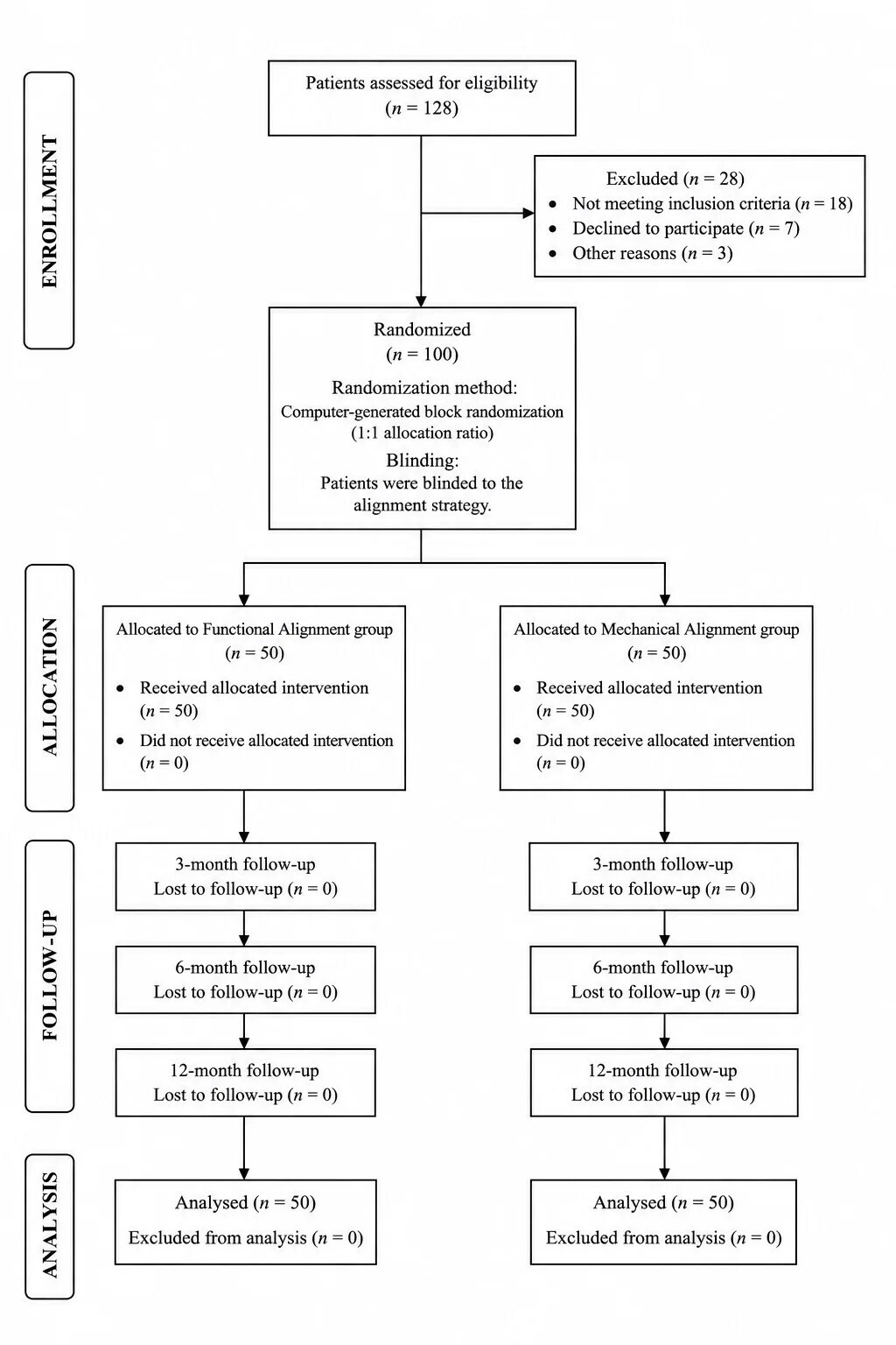
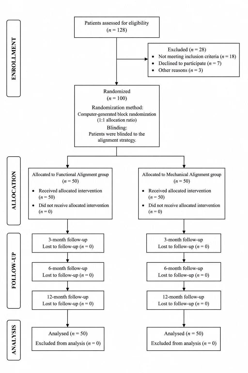
Randomization and Group Allocation Patients were randomized in a 1:1 ratio using a computer-generated block randomization sequence. Group 1 (ra-FA group, n = 50) underwent ra-TKA using the functional alignment philosophy. Group 2 (ra-MA group, n = 50) underwent ra-TKA using the mechanical alignment philosophy. Allocation concealment was achieved using sequentially numbered opaque sealed envelopes. Patients were blinded to the alignment strategy used during surgery.
Sample Size Calculation
An a priori power analysis was performed using G*Power software (version 3.1).
The primary endpoint was the difference in Forgotten Joint Score-12 (FJS-12) at 3 months postoperatively. Assuming a moderate effect size (Cohen’s d = 0.60), α = 0.05, and statistical power of 80%, the minimum required sample size was calculated as 45 patients per group.
To compensate for potential loss to follow-up, 50 patients were included in each group.
Eligibility Criteria
Inclusion Criteria were: primary knee osteoarthritis Kellgren–Lawrence grade III–IV, age > 18 years, varus deformity ≤20°, ability to provide informed consent.
Exclusion Criteria were previous surgery on the index knee, severe extra-articular deformity, collateral ligament insufficiency requiring constrained implants, body mass index >40 kg/m², ASA class IV–V, previous arthroplasty of the contralateral knee.
Patients lost to follow-up were excluded from the final analysis.
Surgical Technique
All procedures were performed by the same experienced surgical team using the CORI Surgical System (Smith & Nephew, USA) through a standard medial parapatellar approach. The Anthem Total Knee System (Smith & Nephew, USA) was implanted in all cases with preservation of the posterior cruciate ligament. Patellar resurfacing was not performed.
After osteophyte removal, anatomical landmarks were registered and a three-dimensional model of the knee was generated. In the mechanical alignment group, femoral and tibial components were positioned perpendicular to their respective mechanical axes to achieve a neutral mechanical limb alignment. In the functional alignment group, component positioning was individualized to preserve constitutional anatomy, native joint-line orientation, and soft-tissue balance while maintaining the final HKA within ±3° of neutral alignment.
For varus phenotypes, tibial positioning up to 5° varus and femoral positioning up to 2° valgus were permitted.
Soft-Tissue Release Assessment
Soft-tissue balancing procedures were recorded intraoperatively by an independent surgeon. Releases were classified as: Grade I – standard arthrotomy, osteophyte removal, and medial capsular mobilization; Grade IIa – additional release of the posteromedial capsule, deep medial collateral ligament, and/or semimembranosus insertion; Grade IIb – additional release of the superficial medial collateral ligament. The frequency and extent of soft-tissue releases were compared between groups.
Radiographic Assessment
Full-length standing radiographs of the lower extremities were obtained preoperatively and at 3 months postoperatively. The following parameters were measured: Hip–Knee–Ankle Angle (HKA), Lateral Distal Femoral Angle (LDFA), Medial Proximal Tibial Angle (MPTA), Joint Line Convergence Angle (JLCA).
Preoperative planning was performed using mediCAD® software (Hectec GmbH, Germany). To evaluate surgical accuracy, planned values were compared with intraoperative measurements recorded by the CORI system and with postoperative radiographic values. Absolute deviations from the planned alignment were calculated in degrees.
CPAK Phenotype Analysis
Constitutional coronal knee phenotypes were classified according to the Coronal Plane Alignment of the Knee (CPAK) classification.
Because only patients with varus osteoarthritis were included, preoperative phenotypes were represented by CPAK Types I, IV, and VII. For patients with unilateral osteoarthritis, the contralateral knee was used as the reference for determination of the constitutional phenotype. For patients presenting with bilateral osteoarthritis, the presumed constitutional phenotype was reconstructed using the combined assessment of overall limb alignment, joint-line orientation, and radiographic morphology.
Postoperative CPAK classification was determined using standing radiographs obtained at 3 months. The following parameters were evaluated: distribution of CPAK phenotypes before surgery, distribution of CPAK phenotypes after surgery, preservation of the constitutional CPAK phenotype, preservation of native joint-line obliquity, frequency of phenotype transformation, frequency of transition to postoperative valgus alignment.
Phenotype preservation was defined as restoration of the same CPAK category identified in the patient’s constitutional knee phenotype. Major phenotype transformation was defined as simultaneous alteration of both coronal alignment arithmetic and joint-line obliquity resulting in conversion to a different CPAK category.
Radiographic Reliability
All radiographic measurements were independently performed by two blinded orthopedic surgeons. Interobserver agreement was assessed using the intraclass correlation coefficient (ICC). An ICC greater than 0.80 was considered indicative of excellent reliability.
Clinical Assessment
Clinical evaluation was performed preoperatively and at 3, 6, and 12 months postoperatively. The following outcome measures were used: Knee Society Score (KSS), Forgotten Joint Score-12 (FJS-12), Visual Analogue Scale (VAS).
Statistical Analysis
Statistical analyses were performed using R statistical software. Continuous variables are presented as median and interquartile range – Me (Q1–Q3). Categorical variables are presented as frequencies and percentages. Between-group comparisons of continuous variables were performed using the Mann–Whitney U test. Categorical variables were compared using Pearson’s χ² test or Fisher’s exact test where appropriate. Statistical significance was defined as p < 0.05.
Results
Patient recruitment, randomization, follow-up, and analysis are summarized in the CONSORT 2010 flow diagram (Figure 1). Of the 128 patients assessed for eligibility, 100 were randomized and completed the study protocol. No losses to follow-up or exclusions from analysis occurred during the 12-month follow-up period.
A total of 100 patients were included in the final analysis: 50 patients in the Functional Alignment (FA) group and 50 patients in the Mechanical Alignment (MA) group. Baseline demographic and clinical characteristics were comparable between groups. No statistically significant differences were identified regarding age, sex distribution, body mass index, operated side, or preoperative radiographic parameters (Table 1).
Clinical Outcomes
Both alignment strategies resulted in substantial improvements in clinical and functional outcomes compared with preoperative values (Table 2).
At 3 months postoperatively, patients in the ra-FA group demonstrated significantly higher KSS Knee, KSS Function, and FJS-12 scores, as well as lower pain intensity measured by the Visual Analog Scale (VAS), compared with patients in the ra-MA group (p < 0.05). These findings reflected faster early functional recovery and a greater perception of the operated knee as a natural joint.
At 6 months, the advantage of functional alignment persisted as a trend; however, intergroup differences did not reach statistical significance (p > 0.05).
By 12 months, KSS, FJS-12, and VAS outcomes were comparable between groups (p > 0.05), indicating gradual attenuation of the early benefits associated with functional alignment as postoperative recovery progressed.
Soft-Tissue Release
Additional soft-tissue releases were required significantly less frequently in the ra-FA group than in the ra-MA group (Table 3).
Most patients treated with functional alignment required only standard surgical exposure, osteophyte removal, and limited medial soft-tissue mobilization. In contrast, grade IIa and IIb releases were performed more frequently in the mechanical alignment group to achieve symmetrical flexion and extension gaps.
The mean number of released soft-tissue structures per knee was 0.7 ± 0.5 in the ra-FA group and 1.6 ± 0.7 in the ra-MA group.
These findings indicate a more ligament-preserving nature of functional alignment.
Radiographic Outcomes
Both groups demonstrated high accuracy in reproducing the planned implant positioning (Table 4).
Postoperative HKA, LDFA, and MPTA values in the ra-MA group were closer to a neutral mechanical alignment. In contrast, greater variability of postoperative HKA, LDFA, and MPTA values was observed in the ra-FA group, reflecting intentional preservation of the patient’s constitutional anatomy and native joint-line orientation.
The absolute deviation between planned and achieved alignment parameters was minimal and comparable between groups (0.9° versus 0.8°, p > 0.05), confirming the high reproducibility of robot-assisted TKA performed using the CORI system. No complications related to malalignment, coronal instability, or early aseptic loosening were observed during the follow-up period.
CPAK Phenotype Analysis
Preoperative assessment according to the Coronal Plane Alignment of the Knee (CPAK) classification demonstrated a marked predominance of Type I phenotype in both study groups, whereas Type IV phenotype was observed considerably less frequently. No patients exhibited a Type VII phenotype (Table 5).
Substantial differences in postoperative phenotype distribution were observed between the two alignment strategies. In the mechanical alignment group, all patients were converted to CPAK Type V, reflecting restoration of a neutral mechanical axis and horizontal joint-line orientation irrespective of the native constitutional phenotype.
In contrast, functional alignment preserved the native CPAK phenotype in the majority of patients. The observed transition from Type I to Type II in a subset of patients was attributable to postoperative HKA correction toward values close to neutral alignment (178°–179°) and did not represent a change in constitutional limb morphology.
Importantly, functional alignment preserved the constitutional joint-line orientation in all cases and did not result in conversion to a valgus phenotype. All patients maintained constitutional varus limb alignment following surgery.
These findings suggest that functional alignment preserves patient-specific constitutional knee morphology substantially more effectively than mechanical alignment, while maintaining the native coronal characteristics of the lower limb.
Discussion
The results of this prospective randomized study confirm the clinical feasibility of the functional alignment concept in robotic-assisted total knee arthroplasty using the CORI robotic platform.
The present findings demonstrated a high degree of accuracy in reproducing the preoperative plan and close agreement between planned and achieved radiographic parameters, indicating excellent reproducibility of robotic-assisted TKA. The high accuracy of component positioning achieved with robotic technologies has been repeatedly demonstrated in previous studies.28,29 This is of fundamental importance for the implementation of personalized alignment strategies, as it enables surgeons to reproducibly restore individual patient anatomy, control flexion-extension gap balance, and minimize the need for aggressive intervention on soft-tissue structures.
The principal finding of the present study was the identification of differences in the dynamics of functional recovery during the early postoperative period. Patients treated with robotic-assisted functional alignment demonstrated superior KSS and FJS-12 scores at 3 months postoperatively compared with those treated using mechanical alignment. However, as follow-up duration increased, these differences gradually diminished and became statistically non-significant by 12 months. These findings suggest that functional alignment primarily influences the rate of early postoperative functional adaptation rather than the ultimate level of recovery achieved after surgery.
The most likely explanation for this observation is the substantially lower extent of soft-tissue release required in the functional alignment group. Preservation of native ligament balance and natural joint-line orientation likely facilitates earlier restoration of physiological knee kinematics. Particular attention should be paid to the ligament-preserving nature of functional alignment. In the present study, extensive soft-tissue releases were required significantly less frequently in the robotic-assisted functional alignment group, and no releases of Grade IIb or greater were performed. These findings are consistent with contemporary concepts of kinematic and functional alignment, both of which aim to maximize preservation of native soft-tissue balance.30 Similar observations were reported by Gustke et al. (2025), who demonstrated that robotic-assisted functional alignment was associated with a reduced need for ligament release and superior early postoperative functional outcomes.31 Taken together, these findings support the hypothesis that preservation of native knee biomechanics may positively influence early rehabilitation following TKA.
Despite the advantages observed during the early postoperative period, intergroup differences were no longer evident at 12 months. Comparable findings were reported by Agarwal et al. (2024), who found no significant differences in pain scores, patient-reported outcome measures (PROMs), or functional outcomes between personalized and mechanical alignment strategies.32 The authors suggested that achieving adequate soft-tissue balance may be more important than strict restoration of a neutral mechanical axis in determining patient satisfaction.
Nevertheless, the findings of the present study indicate that phenotype-oriented alignment may accelerate early recovery even in the absence of differences in long-term outcomes. Similar trends were demonstrated by Jeffrey et al., who reported higher Forgotten Joint Score and Oxford Knee Score values at 3 months following robotic-assisted TKA performed using functional alignment.27 However, the magnitude of these differences was also relatively modest. Additional insight is provided by the meta-analysis conducted by Gao et al., which demonstrated that restricted kinematic alignment was associated with superior KSS and WOMAC scores, while no consistent differences were observed for other PROMs, including FJS-12.33 The authors concluded that contemporary personalized alignment philosophies may preserve the mechanical safety of implant positioning while simultaneously providing more physiological joint biomechanics.
From a biomechanical perspective, the results of the present study further support the rationale for individualized alignment strategies in TKA. Mechanical alignment is based on the assumption that a universally neutral component orientation is optimal despite the fact that only a minority of individuals possess naturally neutral knee anatomy.34 Forced restoration of a neutral mechanical axis may alter native joint-line orientation and create asymmetric ligament tension. Functional alignment, in contrast, seeks to reproduce the patient’s pre-arthritic knee phenotype while preserving individual joint-line orientation and soft-tissue characteristics.35
CPAK PHENOTYPE ANALYSIS
An additional finding of the present study was the substantial difference between alignment strategies with respect to preservation of constitutional CPAK phenotypes. The CPAK classification has emerged as a comprehensive framework for describing native coronal knee morphology by simultaneously incorporating limb alignment and joint-line obliquity, thereby allowing characterization of constitutional knee phenotypes beyond isolated angular measurements.36
Preoperatively, the overwhelming majority of patients in both groups demonstrated a varus constitutional phenotype, predominantly CPAK Type I, which is consistent with previous observations in patients undergoing total knee arthroplasty for varus osteoarthritis.36 Following robotic-assisted mechanical alignment, all patients were converted to CPAK Type V. This finding is in agreement with the work of Corban et al. (2024), who demonstrated that mechanically aligned TKA inherently drives patients toward a standardized neutral phenotype irrespective of their pre-arthritic constitutional morphology.37
In contrast, functional alignment preserved the constitutional CPAK phenotype in the majority of patients. Importantly, the transition observed from Type I to Type II in a subset of cases was primarily attributable to correction of the HKA angle toward values approaching neutral alignment (178°-179°), while preserving constitutional varus limb orientation and native joint-line obliquity. Furthermore, no patient in the functional alignment group demonstrated conversion to a valgus phenotype or loss of constitutional joint-line orientation. These findings are consistent with the observations of van de Graaf et al. (2024), who reported that functional alignment minimizes alterations in joint-line obliquity and more closely reproduces native coronal anatomy than conventional alignment strategies.21
The clinical relevance of constitutional phenotype preservation remains an area of active investigation. However, recent evidence suggests that restoration of the native CPAK phenotype may contribute to more physiological ligament tensioning and improved patient perception of the reconstructed joint. Pangaud et al. (2024), demonstrated that restoration of the preoperative CPAK phenotype following total knee arthroplasty was associated with superior functional outcomes and patient-reported satisfaction scores.38 Although the present study was not specifically powered to establish a direct causal relationship between phenotype preservation and clinical outcomes, the superior early KSS and FJS-12 scores observed in the functional alignment group may partially support this hypothesis.
Taken together, these findings suggest that functional alignment not only restores target alignment parameters with high accuracy but also more effectively preserves patient-specific constitutional knee morphology. In contrast, mechanical alignment appears to standardize coronal alignment patterns regardless of the individual’s native phenotype. Given the increasing emphasis on personalized arthroplasty strategies, preservation of the constitutional CPAK phenotype may represent an important surrogate marker of anatomical restoration and warrants further investigation in studies with longer follow-up and larger patient populations.
Importantly, this individualized approach was not associated with an increased number of radiographic outliers or alignment-related complications in the present study. Postoperative HKA values in the functional alignment group remained within the widely accepted safe zone of ±3°, which has been associated with favorable implant survivorship.39,40 Furthermore, all LDFA and MPTA values remained within acceptable deviations from the planned targets, confirming the high accuracy of surgical plan execution.
Another important observation was the safety profile of functional alignment. No increase in perioperative complications, instability, or signs of aseptic component loosening was observed during the study period. These findings are consistent with contemporary evidence demonstrating no increased risk of early revision when moderate constitutional varus is preserved within controlled limits.33 Moreover, multicenter studies of robotic-assisted functional alignment have reported high patient satisfaction and an absence of complications related to coronal alignment when safe-zone principles are respected.41
Several limitations of the present study should be acknowledged. First, the follow-up period was limited to 12 months, precluding assessment of implant longevity and long-term survivorship. Second, the study was conducted in a single center by a single experienced robotic arthroplasty team, which may limit the generalizability of the findings to less experienced institutions. Third, despite the randomized design, the influence of additional factors–including variations in rehabilitation protocols, biological healing responses, and subjective patient perceptions of functional recovery–cannot be completely excluded. Nevertheless, the present findings demonstrate the potential of functional alignment as a contemporary personalized strategy for robotic-assisted total knee arthroplasty and provide a rationale for future investigations involving larger patient populations and longer follow-up periods.
Conclusion
Robot-assisted functional alignment using the CORI Surgical System demonstrated high radiographic accuracy, excellent reproducibility of the planned alignment strategy, and a ligament-preserving surgical profile in patients undergoing primary total knee arthroplasty for varus knee osteoarthritis.
Compared with robot-assisted mechanical alignment, functional alignment was associated with superior early clinical and patient-reported outcomes, reduced soft-tissue release requirements, and substantially greater preservation of constitutional CPAK phenotypes and native coronal knee morphology. Importantly, these advantages were achieved without compromising alignment accuracy, radiographic safety, or early implant stability.
Although differences in functional outcomes diminished during follow-up, the observed preservation of constitutional knee anatomy and earlier postoperative recovery suggest that functional alignment represents a clinically relevant personalized alignment strategy for robotic-assisted total knee arthroplasty.
Further multicenter randomized studies with larger patient populations and longer follow-up are required to determine whether preservation of constitutional knee phenotypes translates into improved long-term implant survivorship, patient satisfaction, and functional outcomes.
Acknowledgements
The authors express their sincere gratitude to the surgical and technical teams of the Department of Traumatology and Orthopaedics at the Savelyev City Clinical Hospital №31 (Moscow) for their invaluable assistance during data collection and patient follow-up.
Special thanks are extended to the Faculty of Traumatology and Orthopaedics at the RUDN University for providing academic and methodological support throughout this study.
Author contributions
Georgii Airapetov served as the principal investigator and had the leading role in the conceptualization of the study, development of the research design, and overall scientific supervision. He also contributed to the critical revision of the manuscript and approved the final version for submission.
Armen Daniliyants co-developed the study concept and design, participated in all surgical procedures, and contributed substantially to data interpretation and manuscript preparation.
Alexey Prizov conducted the literature search and contributed to manuscript drafting.
Rasul Aliev performed the statistical analysis, verified the analytical methods, and participated in the interpretation of data.
Varvara Iatsukova, Ahmat Abdulaye contributed to the review and synthesis of literature, data organization, and overall systematization of the study findings.
All authors discussed the results, reviewed the manuscript, and approved the final version for publication.
Funding
No funding was received for this study.
Ethics approval and consent to participate
The study was conducted in accordance with the principles of the Declaration of Helsinki and was approved by the Institutional Ethics Committee of the City Clinical Hospital №31 named after Academician G.M. Savelyev (Protocol № аd017-а, Date: 02.02.2025). All participants provided written informed consent prior to inclusion in the study.
Consent for publication
Not applicable.
Competing interests
The authors declare that they have no competing interests related to this study.