
INTRODUCTION
Large rotator cuff tears are a common cause of shoulder pain and functional impairment. Despite advances in arthroscopic repair techniques, the retear rate following repair remains high. Previous studies have reported retear rates of 60%–90% in patients with large rotator cuff tears.1 Therefore, in addition to achieving a mechanically stable repair, optimizing the tendon-to-bone healing environment has become a critical component of surgical treatment. Successful arthroscopic repair requires not only anatomical restoration of the torn tendon but also biological enhancement of tendon integration at the greater tuberosity. However, in large tears, poor tendon quality, fibrosis at the tendon footprint, reduced vascularity, and a chronic inflammatory microenvironment substantially impair tendon-to-bone healing, resulting in higher mechanical and biological failure rates than those observed in small- or medium-sized tears.1
Platelet-rich fibrin (PRF) provides a three-dimensional fibrin scaffold that entraps platelets within a dense fibrin network while exhibiting excellent adherence to the tendon–bone interface. This scaffold serves as a sustained delivery system for numerous growth factors, including transforming growth factor-beta (TGF-β), platelet-derived growth factor (PDGF), vascular endothelial growth factor (VEGF), and others, thereby promoting angiogenesis, fibroblast proliferation, and differentiation of tendon- and bone-forming cells to accelerate tendon-to-bone healing. However, maintaining a high local concentration of these biological factors at the repair site during arthroscopic shoulder surgery remains technically challenging. Charousset et al. (2014) and Yao et al. (2024) described arthroscopic rotator cuff repair techniques augmented with platelet-rich plasma (PRP). However, because PRP is administered in a liquid form, it tends to diffuse away from the repair site into the surrounding joint space, making it difficult to maintain an effective concentration of growth factors at the tendon–bone interface and to accurately evaluate their biological effects on tendon healing.2˒3 Weber et al. (2013) and Randelli et al. (2012) reported arthroscopic repair techniques using thrombin-activated PRF applied to the tendon footprint as either a clot or membrane.4˒5 Although these solid PRF constructs provide biological augmentation, they do not adhere directly to the tendon–bone surface and instead rely on compression between the repaired tendon and bone after fixation of the lateral-row anchors, making implantation technically demanding. The physical characteristics of platelet concentrates are largely determined by the activation method employed.6–7 To facilitate arthroscopic application, an ideal biological augmentation should remain injectable during delivery and polymerize only after reaching the tendon–bone interface. Batroxobin activation fulfills these requirements by selectively cleaving fibrinopeptide A from fibrinogen, producing liquid platelet-rich des-A fibrin monomers. Following application to the repair site together with an alkaline buffer (pH 10), these monomers undergo in situ polymerization to form an adhesive PRF matrix firmly attached to the tendon–bone interface.8˒9
In this report, we present a case of a large rotator cuff tear treated with arthroscopic suture bridge repair augmented with intraoperative platelet-rich fibrin (PRF). The report provides a detailed description of all surgical steps, including patient preparation, autologous PRF preparation, establishment of the arthroscopic portals, the suture bridge repair technique, and the method of PRF application at the tendon–bone interface.
CASE PRESENTATION
A 70-year-old male patient was admitted with left shoulder pain and an inability to actively abduct the shoulder. He had undergone conservative treatment with medication and physical rehabilitation for 1 year without clinical improvement.
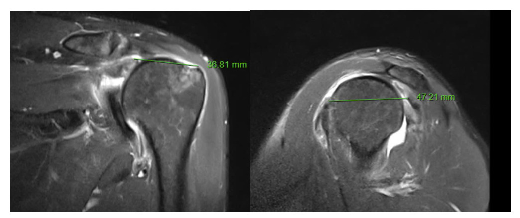
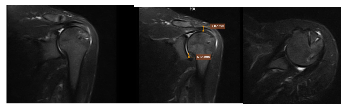
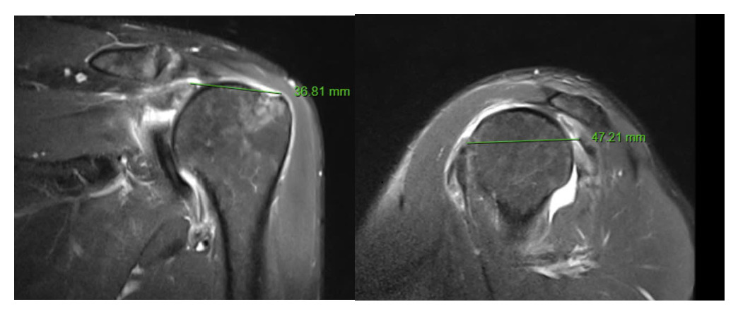
Physical examination revealed supraspinatus fossa atrophy. Passive shoulder range of motion was within normal limits. The Jobe test and drop arm test were positive. Magnetic resonance imaging demonstrated full-thickness tears of the supraspinatus and infraspinatus tendons, together with a tear involving the superior portion of the subscapularis tendon. The rotator cuff tendons showed grade 3 retraction according to the Patte classification and grade 1 fatty infiltration of the rotator cuff muscles. The tear measured 36.81 mm in the mediolateral dimension and 47.21 mm in the anteroposterior dimension.
The patient was indicated for arthroscopic rotator cuff repair using the suture bridge technique augmented with autologous platelet-rich fibrin.
The surgical procedure consisted of two steps:
Step 1. Preparation of Autologous Platelet-Rich Fibrin
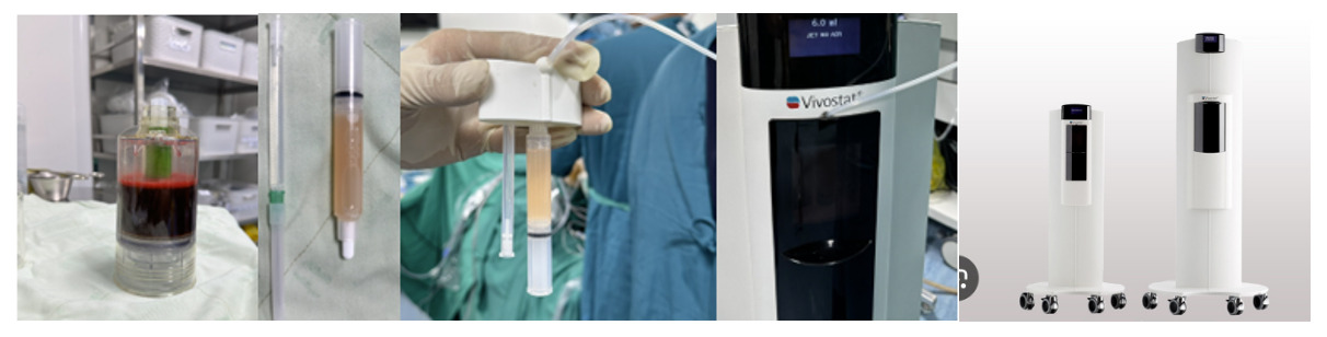
Autologous PRF is prepared by a trained hematology technician using the Vivostat® PRF preparation system.
The patient is placed in the supine position. Peripheral venous access is established and connected to the Vivostat collection set containing Obsidian ASG citrate anticoagulant and batroxobin as the activation reagent. The vertical distance between the venipuncture site and the collection kit is maintained at approximately 70 cm to provide adequate hydrostatic pressure while avoiding excessive negative pressure and air bubble formation. A total of 120 mL of autologous blood is collected. The patient-identified PRF collection cartridge is then connected to the processing kit, which is loaded into the Vivostat centrifugation chamber. The automated preparation program centrifuges the sample at 9,000 rpm for 26 minutes. At the completion of the centrifugation cycle, the processing kit is removed, inverted, and the platelet-rich des-A fibrin monomer solution is aspirated into the sterile PRF delivery syringe. The syringe containing the platelet-rich des-A fibrin monomer and the syringe containing the pH 10 alkaline buffer are subsequently mounted onto the Vivostat dual-spray applicator. Before intraoperative use, the mixing system is tested by spraying onto pH indicator paper. When the indicator changes to green, indicating appropriate mixing and alkalinity, the system is considered ready for application at the surgical site.
.png)
Step 2. Arthroscopic Double-Row Rotator Cuff Repair Augmented with Platelet-Rich Fibrin
Patient Positioning
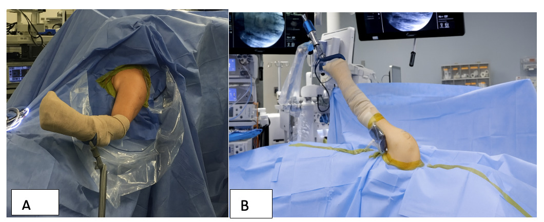
Arthroscopic repair of large rotator cuff tears can be performed with the patient in either the lateral decubitus or the beach-chair position, according to surgeon preference. In the lateral decubitus position, the patient is secured with the shoulder maintained in a neutral position and the elbow flexed to approximately 90°. Longitudinal arm traction is applied using a traction device, with the traction force limited to 7–10 kg to minimize the risk of traction-related neurovascular injury. Alternatively, in the beach-chair position, the patient is placed supine on a dedicated shoulder arthroscopy table. The operating table is then adjusted to a semi-upright (beach-chair) position, allowing optimal visualization of the glenohumeral joint and subacromial space while providing unrestricted access to the shoulder throughout the procedure.
_beach-chair_position._(b)_la.png)
Identification of Bony Landmarks and Portal Placement
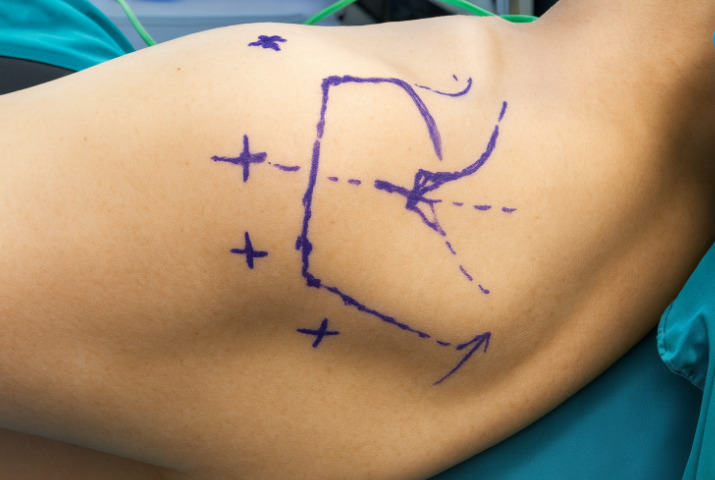
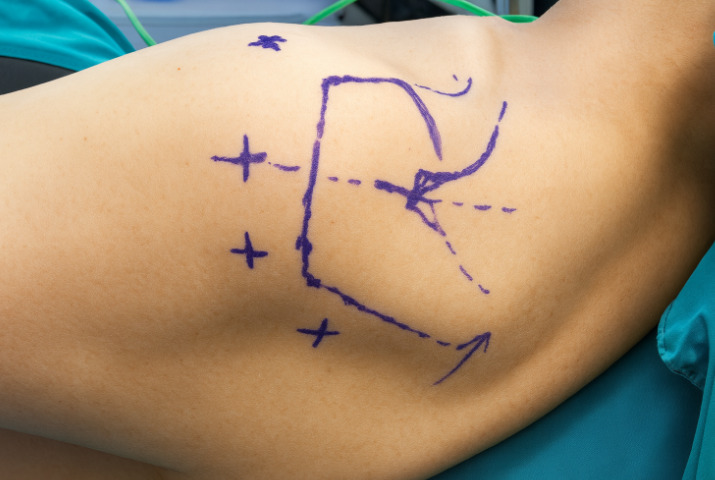
The relevant bony landmarks are identified and marked before portal placement. The standard posterior portal is established approximately 2 cm inferior and 1 cm medial to the posterolateral corner of the acromion. The lateral portal is created approximately 3–5 cm distal to the lateral border of the acromion and 1–3 cm posterior to its anterior margin. Additional accessory portals are established according to the size, configuration, and location of the rotator cuff tear. These portals are created using an outside-in technique to optimize the working trajectory and facilitate tendon mobilization, anchor placement and suture management.
Diagnostic Arthroscopy
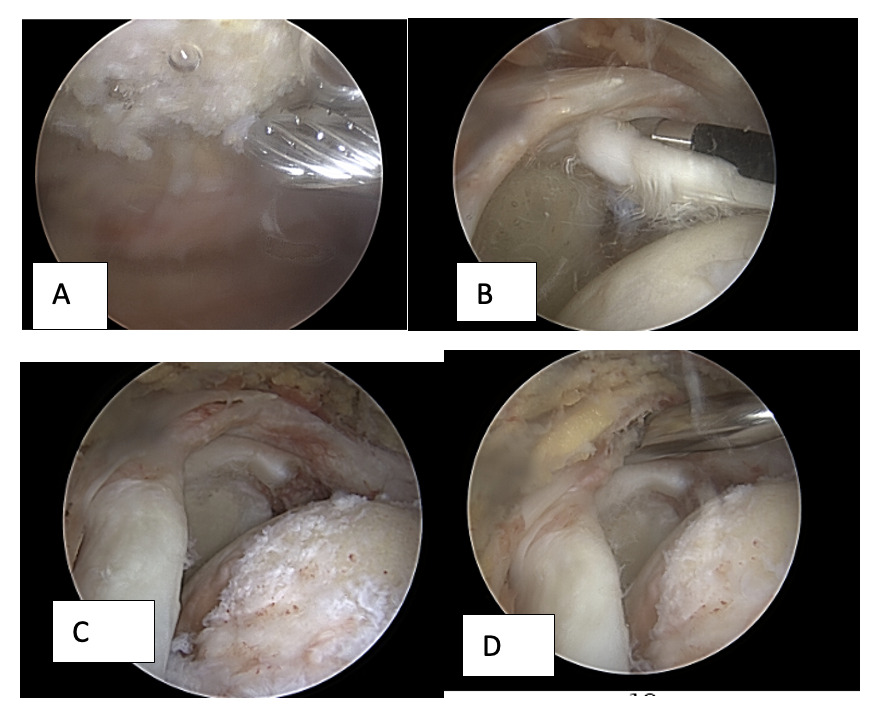
The arthroscope is introduced through the standard posterior portal to perform a systematic evaluation of the glenohumeral joint. The articular surfaces, the bare area of the humeral head, the long head of the biceps tendon, the articular side of the subscapularis, supraspinatus, and infraspinatus tendons are carefully inspected. The arthroscope is then advanced into the subacromial space to evaluate the morphology of the acromion, the degree of subacromial bursitis, the condition of the subdeltoid space, and the characteristics of the rotator cuff tear, including tear configuration, tear size, tendon retraction, and tissue quality. Accessory portals are established as required according to the location and configuration of the rotator cuff tear to facilitate subsequent surgical procedures.
_acromioplasty_performed_to_increase_the_subacromial.png)
Management of Associated Lesions
Inflamed synovial tissue and subacromial bursal tissue are debrided using a motorized shaver and radiofrequency ablation device. Acromioplasty is performed when indicated to improve the subacromial space and reduce mechanical impingement. Pathology of the long head of the biceps tendon is managed according to intraoperative findings by either tenotomy or tenodesis. Rotator cuff mobilization is then performed to achieve tension-free reduction of the tendon to its anatomical footprint.
Anterior interval slide is performed by releasing the tissue between the supraspinatus tendon and the rotator interval, including the coracohumeral ligament, through the lateral portal toward the base of the coracoid process. This release improves tendon excursion in cases of retracted tears.
Posterior interval slide consists of releasing the interval between the supraspinatus and infraspinatus tendons and may be performed alone or in combination with the anterior interval slide when additional mobilization is required. The posterior release includes careful debridement of fibrotic soft tissue adherent to the scapular spine while preserving the region adjacent to the posterior glenoid, where the suprascapular nerve traverses the spinoglenoid notch, to minimize the risk of iatrogenic nerve injury.
Preparation of the Tendon Footprint
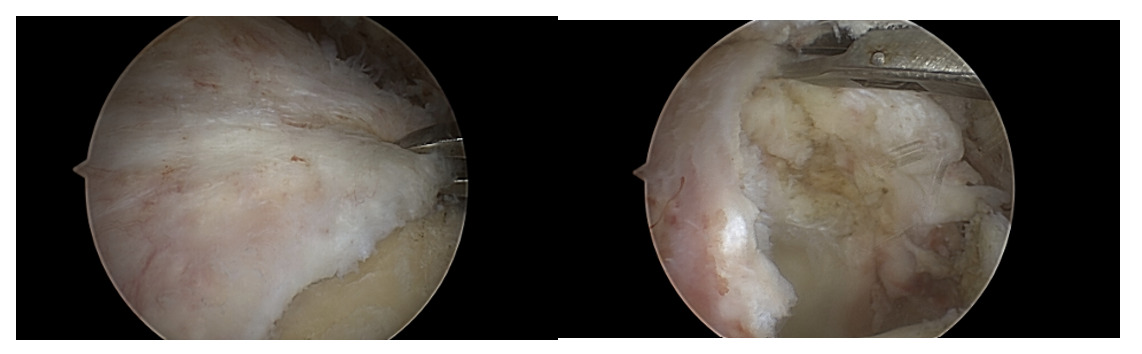
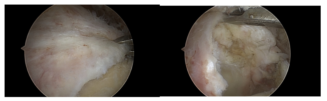
The rotator cuff footprint on the greater tuberosity is prepared by removing residual soft tissue and creating a bleeding cancellous bone surface to promote tendon-to-bone healing. Debridement and decortication are performed carefully to preserve adequate bone stock. Excessive burring should be avoided because it may compromise anchor fixation and increase the risk of anchor pullout. An arthroscopic tissue grasper is used to mobilize the torn tendon edge and assess its excursion. This maneuver facilitates identification of the optimal tendon position for suture passage and enables a tension-free reduction to the anatomical footprint prior to anchor placement.
Suture-Bridge Rotator Cuff Repair With PRF Augmentation
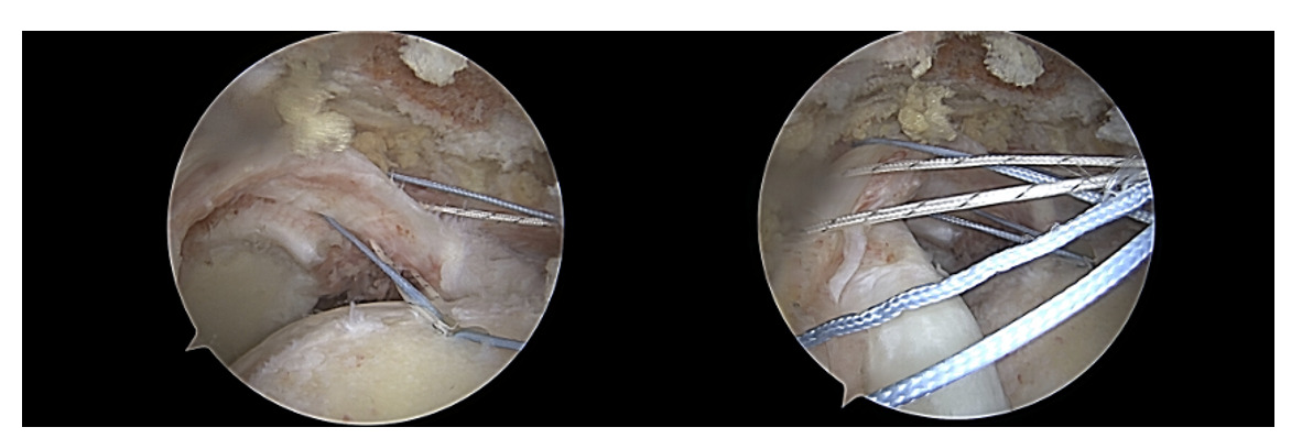
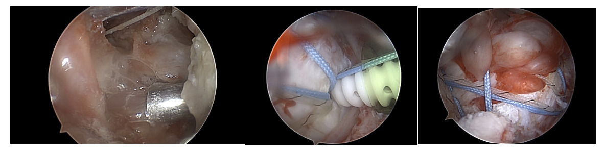
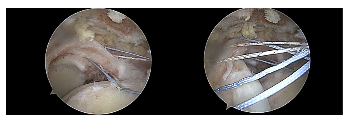
A suture-bridge technique is used for rotator cuff repair. Two double-loaded medial-row anchors are inserted approximately 2 mm lateral to the articular cartilage margin, with an inter-anchor distance of approximately 1.5–2 cm. Using an arthroscopic suture-passing device, the sutures are passed through the rotator cuff tendon at evenly spaced intervals to ensure uniform tissue purchase and balanced load distribution. Before lateral-row fixation, the arthroscopic fluid is completely evacuated from the subacromial space. The platelet-rich fibrin des-A fibrin monomer and alkaline buffer solution are then delivered onto the prepared tendon–bone interface using the dedicated applicator. After in situ polymerization of the des-A fibrin monomer into a platelet-rich fibrin matrix at the tendon footprint, lateral-row fixation is performed. Two lateral-row knotless anchors are placed along the lateral margin of the greater tuberosity. The distance between the lateral-row anchors is generally wider than that between the medial-row anchors to distribute the compressive forces evenly across the repaired tendon. The suture limbs are sequentially tensioned to achieve uniform compression of the rotator cuff tendon against the entire footprint before final fixation within the lateral-row anchors. The stability of the repair and the distribution of the PRF matrix at the tendon–bone interface are confirmed arthroscopically. The instruments are then removed, and the portal incisions are closed in the standard fashion.
_to_the_tendonbone_interface_followed_by_lateral.png)
Postoperative Rehabilitation
The shoulder was immobilized in an abduction brace for 6 weeks. During the first 6 postoperative weeks, passive forward elevation and extension of the shoulder, together with flexion and extension exercises of the elbow and wrist, were performed. Active shoulder exercises were initiated between postoperative weeks 6 and 12, including forward elevation, abduction, internal rotation, and external rotation. Resistance strengthening exercises were started 3 months after surgery.
One-Year Postoperative Evaluation
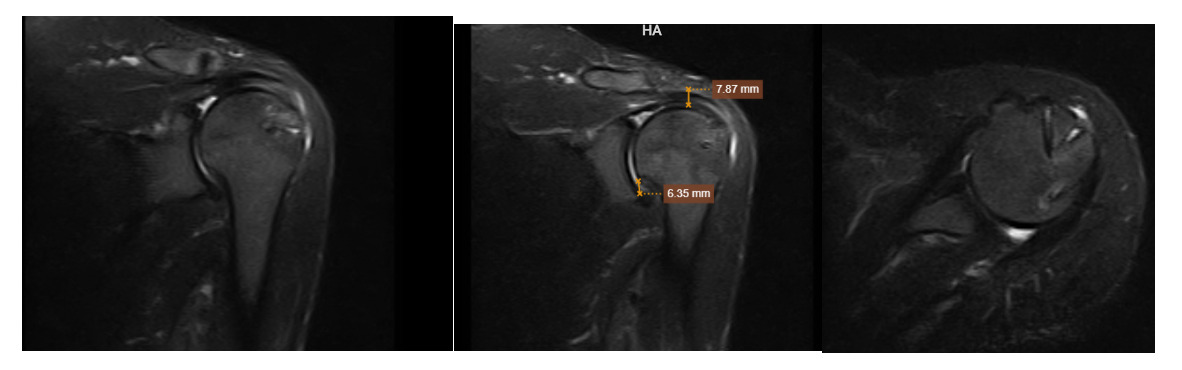
At the 1-year follow-up, the patient had no residual shoulder pain, had regained a normal range of motion, and had returned to normal daily activities. The Constant score was 98 points. Postoperative MRI demonstrated complete rotator cuff tendon healing, classified as Sugaya type I.
DISCUSSION
Arthroscopic repair of large rotator cuff tears remains one of the greatest challenges in shoulder surgery because of the persistently high retear rate despite substantial advances in surgical techniques. A systematic review by Zumstein et al. demonstrated that structural failure after repair may occur in up to 57% of large and massive tears, substantially higher than that observed in small- or medium-sized tears. This finding highlights that failure is not solely a mechanical problem but also reflects the unfavorable biological environment at the tendon–bone interface, where tendon healing occurs in the setting of compromised tissue quality and limited regenerative potential. Biological augmentation using platelet concentrates has therefore attracted increasing interest. Numerous studies evaluating platelet-rich plasma (PRP) have demonstrated its ability to enhance tendon-to-bone healing through sustained release of growth factors, promotion of angiogenesis, stimulation of fibroblast proliferation, and extracellular matrix synthesis. In a meta-analysis of seven randomized controlled trials, Yang et al. (2020) reported that PRP significantly reduced the retear rate (RR = 0.38) while improving Constant, UCLA, and VAS scores following arthroscopic rotator cuff repair. Likewise, Ryan et al. (2021) observed greater tendon thickness on postoperative MRI and more rapid pain relief during the first three postoperative months in patients receiving PRP augmentation.
Platelet-rich fibrin (PRF), a second-generation platelet concentrate, has recently gained attention because of its three-dimensional fibrin scaffold, which provides prolonged retention and gradual release of growth factors while offering superior handling characteristics during arthroscopic surgery. Nevertheless, clinical evidence supporting PRF remains limited and inconsistent. Weber et al. (2013) and Rodeo et al. (2012) reported no significant improvement in MRI healing rates when PRF was applied to the tendon footprint in patients with small- and medium-sized tears. In contrast, Villarreal-Villarreal et al. (2021) found that PRF was associated with earlier pain relief and functional recovery, although the improvement in structural healing did not reach statistical significance. More recently, Shen et al. (2024) suggested that PRF may enhance the early phases of tendon healing, while emphasizing the need for longer-term clinical studies. These conflicting findings are likely attributable to substantial heterogeneity in PRF preparation protocols, centrifugation systems, leukocyte content, delivery methods, and repair techniques.
The technique described in this Technical Note utilizes the Vivostat® PRF system, which delivers platelet-rich des-A fibrin monomer in liquid form directly onto the prepared tendon–bone interface. Upon contact with tissue and the accompanying alkaline buffer, the des-A fibrin monomer polymerizes in situ to form a stable fibrin matrix that firmly adheres to the tendon footprint. Completion of the lateral-row fixation subsequently compresses the PRF scaffold between the tendon and bone, creating a biologically favorable microenvironment capable of sustained growth factor release throughout the early healing period. From a biomechanical perspective, Barber et al. demonstrated that double-row suture-bridge repair generates greater contact pressure and a larger tendon–bone contact area than conventional repair techniques, thereby optimizing the mechanical environment for tendon healing. Integrating PRF within this compressed interface may further promote angiogenesis, cellular migration, and extracellular matrix remodeling while maintaining the stability provided by the suture-bridge construct.
Adequate tendon mobilization before repair is another critical component of this technique, particularly in large or retracted tears. Failure to sufficiently release the rotator cuff may result in excessive repair tension, which is a well-established risk factor for structural failure regardless of biological augmentation. This concept reinforces the principle that optimal mechanical fixation is a prerequisite for successful biological enhancement. PRF cannot compensate for excessive repair tension or improperly positioned anchors. From a technical standpoint, successful arthroscopic PRF application requires complete evacuation of arthroscopic fluid from the subacromial space before delivery because continuous irrigation may wash away the liquid PRF before polymerization occurs. Thorough preparation of the tendon footprint is equally important to maximize intimate contact between the PRF matrix and the tendon–bone interface.
Although high-level evidence remains limited, biological augmentation with PRF represents a rational strategy for patients with large rotator cuff tears, who constitute the subgroup at greatest risk for postoperative structural failure. The favorable biological properties of PRF, combined with encouraging preliminary clinical outcomes, support a growing emphasis on optimizing the tendon–bone healing microenvironment in addition to achieving stable mechanical fixation. Furthermore, this technique is technically straightforward, utilizes an autologous biological product, adds minimal operative complexity, and can be readily incorporated into contemporary arthroscopic rotator cuff repair. Future prospective randomized studies with larger sample sizes, standardized PRF preparation protocols, and long-term imaging follow-up are warranted to determine the true clinical value of PRF augmentation in large rotator cuff tears.
CONCLUSION
Arthroscopic repair of large rotator cuff tears augmented with platelet-rich fibrin (PRF) is a practical biological augmentation technique that enhances the healing environment at the tendon–bone interface while preserving the mechanical stability of a double-row suture-bridge repair. The use of autologous PRF provides a safe, reproducible, and cost-effective approach with minimal additional surgical complexity. This technique can be readily incorporated into routine arthroscopic practice and has the potential to improve tendon-to-bone healing and postoperative functional recovery in patients with large rotator cuff tears. Further prospective studies with larger patient cohorts and longer follow-up are required to establish its long-term clinical effectiveness and structural healing benefits.