
Introduction
Anterior shoulder instability is a frequent problem in young, active populations, with recurrence rates reported to be as high as 67% in contact athletes.1 Among the different capsulolabral injury patterns, the anterior labroligamentous periosteal sleeve avulsion (ALPSA) lesion represents a special subtype that is both diagnostically and therapeutically challenging. First described by Neviaser in 1993,2 ALPSA lesions are characterized by medial displacement of the anteroinferior labrum together with its periosteal sleeve, which heals in a non-anatomic position on the glenoid neck. This pathological displacement affects the capsulolabral tensioning mechanism, compromises the concavity–compression effect, and predisposes patients to recurrent instability.3,4
Management of ALPSA lesions ranges from open stabilization procedures to arthroscopic repair. Open repair can restore stability but it is associated with greater soft tissue dissection, postoperative stiffness, and longer rehabilitation.5 Arthroscopic techniques provide the advantages of minimally invasive access, better visualization, and lower morbidity, and are now the preferred approach for most cases of anterior instability. However, clinical studies have consistently reported higher recurrence rates following arthroscopic fixation of ALPSA lesions compared to Bankart lesions,1,6,7 underscoring both the technical difficulty and biological challenges of restoring labral anatomy in this lesion.
The double-row repair technique was developed to treat these limitations. By placing both medial and lateral anchors, the construct increases the contact area at the capsulolabral footprint and enhances biomechanical stability compared with traditional single-row repair. Cadaveric investigations have demonstrated that double-row fixation restores up to 85–90% of the native footprint, significantly greater than single-row techniques.8,9 Clinical series have also suggested reduced recurrence rates and improved shoulder function with this approach.10–13
The purpose of this study was to evaluate the clinical outcomes of arthroscopic double-row repair for anterior labroligamentous periosteal sleeve avulsion (ALPSA) lesions of the shoulder.
Methods
Study Design
A prospective case series was conducted on 50 patients (aged 18–40 years) all patients had traumatic anterior shoulder dislocation and arthroscopically confirmed ALPSA lesions. The follow-up period for each patient was 2 years, followed by remote follow up using simple shoulder score after 5 years.
Patient Selection
A total of 50 patients who met the inclusion and exclusion criteria were enrolled in the study. The inclusion criteria were as follows: Age between 18-40 years, both males and females, both athletic and non-athletic individuals, ALPSA lesion caused by a traumatic anterior shoulder dislocation. The exclusion criteria were: Failed previous labral repair, Associated rotator cuff tear, Presence of arthritic changes in the shoulder, Recurrent shoulder dislocation with significant bony defect (either of the humeral head or glenoid).
Pre-operative Evaluation
All patients underwent a standardized pre-operative evaluation consisting of detailed history taking, physical examination, validated functional scoring, and imaging assessment. Baseline history focused on variables known to influence recurrence risk and tissue quality, including the mechanism of the first dislocation, number and mechanism of recurrent instability episodes, method of reduction, requirement for anesthesia during reduction, history of epileptic seizures, and substance abuse. These data were recorded prospectively for all patients.
Clinical examination included general assessment of the cervical spine, entire upper limb, and contralateral shoulder. Generalized ligamentous laxity was evaluated using the Beighton score. Local examination of the affected shoulder assessed muscle wasting or deformity, muscle power, neurovascular status, and passive range of motion, including forward flexion, external rotation in adduction.
Shoulder stability was evaluated using standardized provocative tests to determine the presence and direction of instability, including the apprehension, relocation, as well as the anterior drawer and sulcus tests. All patients demonstrated clinical findings consistent with anterior shoulder instability.
Preoperative functional assessment was performed using validated scoring systems to allow objective baseline evaluation and postoperative comparison. Functional scores recorded preoperatively were the Instability Severity Index Score (ISIS),14 and Constant Shoulder Score (CSS).15
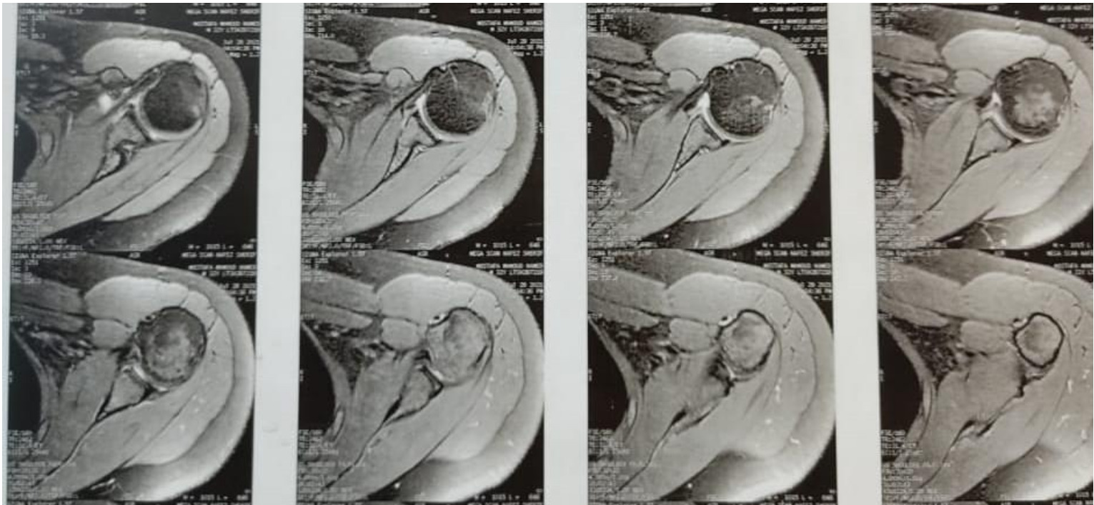
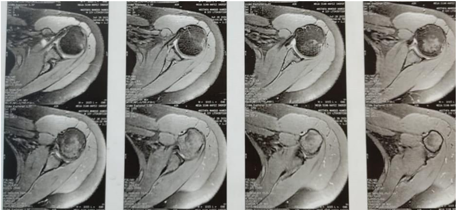
Pre-operative imaging included plain radiographs, computed tomography (CT), and magnetic resonance imaging (MRI). MRI was performed in all patients to confirm the presence of anterior labral periosteal sleeve avulsion (ALPSA) lesions (Fig 1) and to exclude associated soft-tissue pathology, including rotator cuff tears, subscapularis rupture, and biceps tendon pathology. Final confirmation of ALPSA lesions was made arthroscopically based on medialized labrum with intact periosteal sleeve.
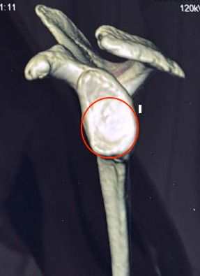
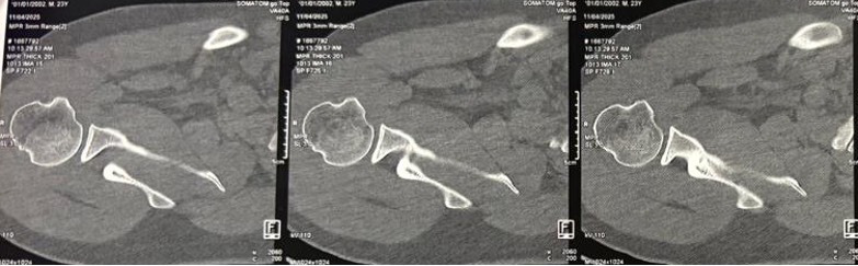
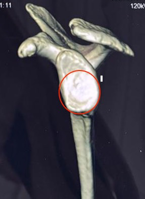
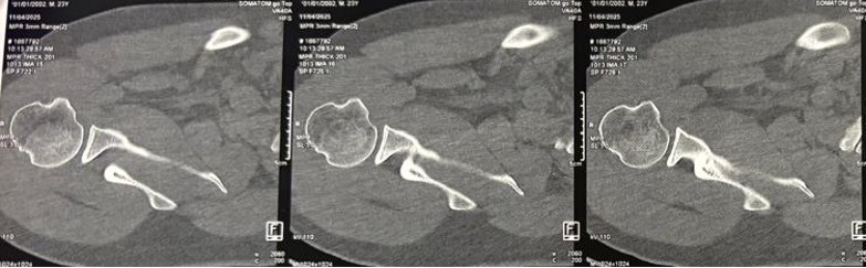
Glenoid bone loss was assessed on en-face three-dimensional CT using the glenoid width method, and glenoid bone loss greater than 20% was considered an exclusion criterion16 (Fig 2). Humeral head defects were evaluated on axial CT images (Fig 3). Intraoperative decision-making was guided by dynamic evaluation of shoulder stability, following the criteria described by Koo et al.17 and Burkhart and De Beer.18 A Hill–Sachs lesion was defined as engaging when it contacted the anterior glenoid rim with the shoulder positioned at 90° of abduction and progressive external rotation between 0° and 135°. Shoulders demonstrating an engaging Hill–Sachs lesion under these conditions were excluded from the study.
Operative Procedure
All patients underwent general anesthesia and were placed in the beach chair position. Examination under anesthesia was carried out to assess instability. Arthroscopy was performed through three standard portals: posterior (for diagnostic viewing), anterosuperior (below the biceps origin), and anteroinferior (superior to the subscapularis). The anterior labrum was mobilized, and the glenoid rim was prepared using a shaver and rasp.
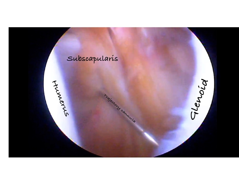
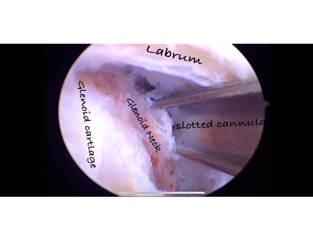
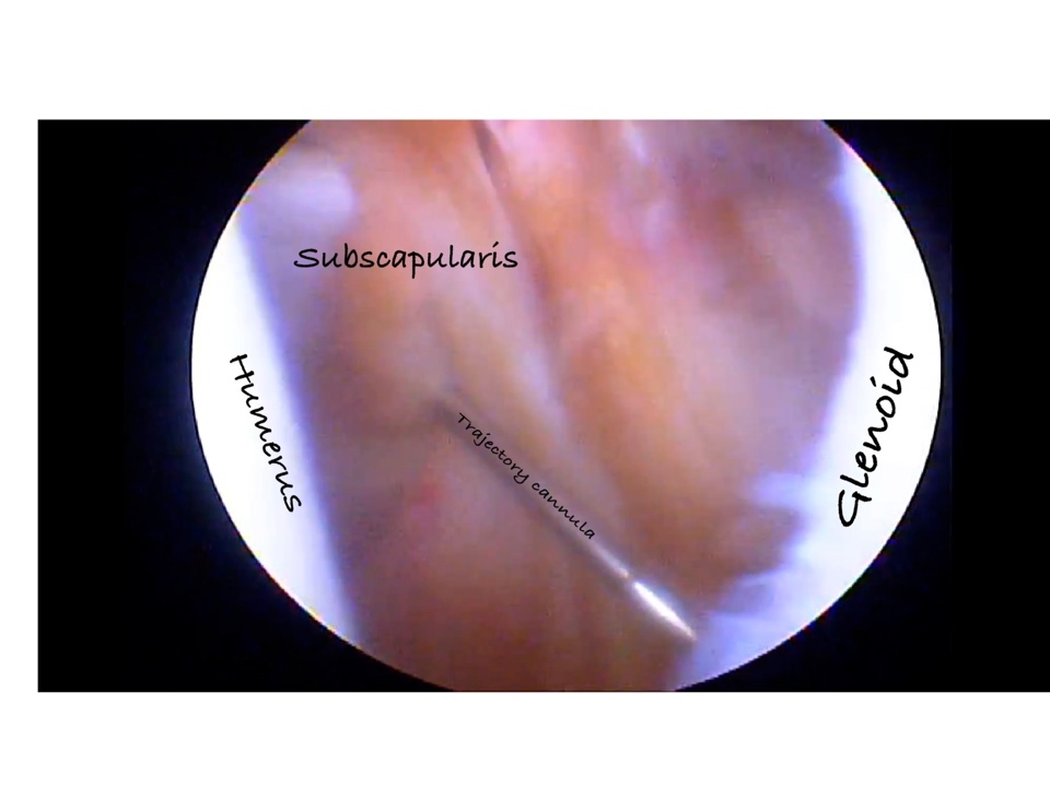
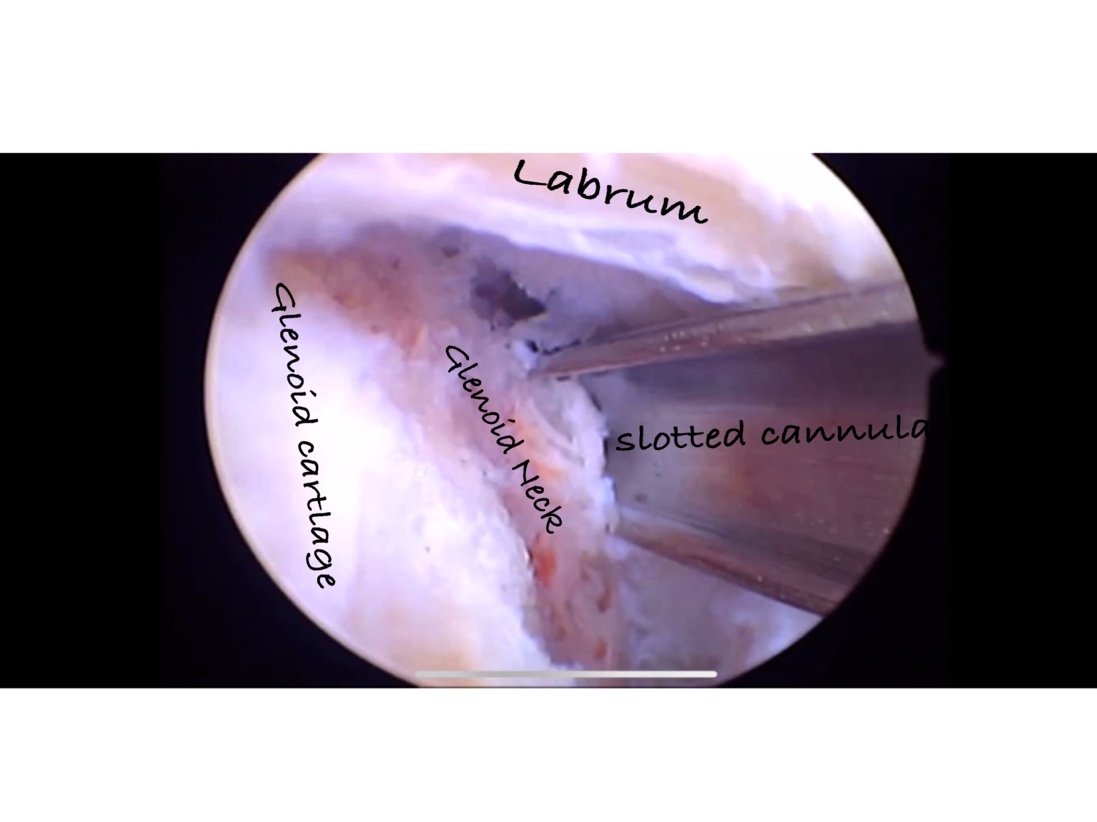
A shuttle suture was first passed through the capsulolabral tissue to facilitate its mobilization and to assist in subsequent passage of the medial-row sutures. At this stage, a trans-subscapularis portal was created by advancing a spinal needle through the subscapularis tendon to establish an end-on trajectory (Fig. 4).After shifting the arthroscope to the anterosuperior portal for optimal visualization, a medial anchor was inserted at the 4 o’clock position on the glenoid neck via the trans-subscapularis portal (Fig. 5), approximately 5 mm medial to the articular cartilage margin.
Visualization was then switched back to the posterior portal, at which sutures from the medial anchor were then passed through the mobilized capsulolabral tissue and secured using standard arthroscopic knot-tying techniques while the arm in neutral rotation, thereby reestablishing the anatomic tension and position of the labrum.
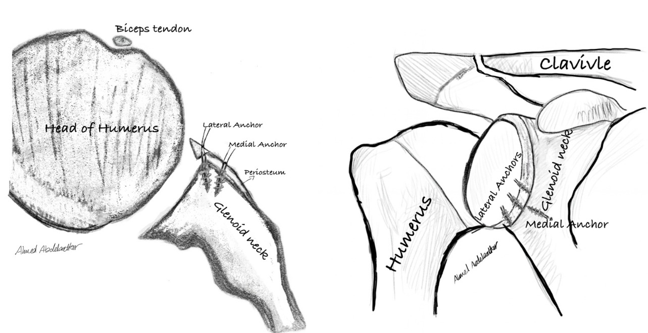
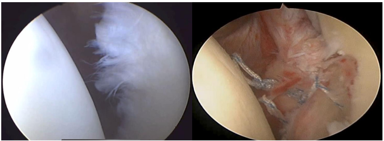
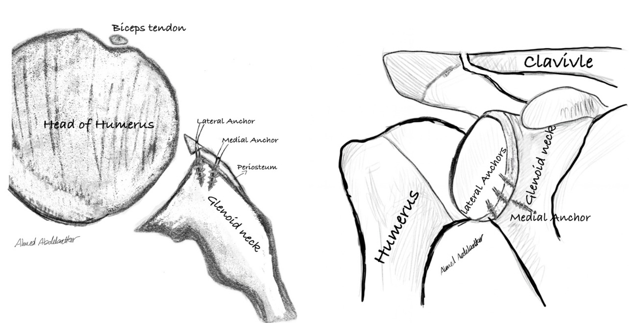
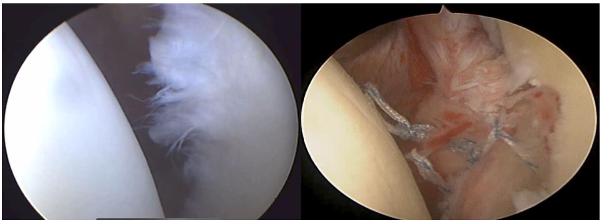
Lateral-row anchors were inserted in a conventional manner along the glenoid rim. The labrum was subsequently secured to the prepared glenoid surface using the same suture-passing technique using 3 anchors to the lateral row, thereby completing the double-row construct and restoring the capsulolabral complex to its native footprint.(Figs. 6)
In this study, one medial anchor was used in all cases. The aim of this anchor was to reduce the medially displaced periosteal sleeve and capsulolabral complex back to their anatomical position and to increase the contact area between the repaired tissue and the prepared glenoid surface. This was then supported by the lateral-row anchors to complete the repair and improve tissue compression over the footprint.
Postoperative Care and Rehabilitation
Following surgery, all patients were immobilized in a sling for 6 weeks. During this period, rehabilitation focused on protecting the surgical repair, controlling pain and swelling, and initiating gentle passive range of motion. Mobility of the accessory joints, including the hand, wrist, and elbow, was encouraged from the first postoperative day. Passive shoulder motion was permitted; however, external rotation was limited to 30° to minimize stress on the repaired capsulolabral complex.
At 6 weeks postoperatively, the sling was discontinued, and patients were allowed to progress to full, gradual passive range of motion and to begin active range of motion of the glenohumeral joint. Muscle strengthening was initiated at this stage, starting with isometric exercises and progressing to closed-chain strengthening exercises as tolerated.
Between 9 and 11 weeks postoperatively, open-chain strengthening exercises were introduced. Gradual return to unrestricted activities was typically permitted between 4 and 5 months postoperatively, depending on individual functional recovery and clinical assessment.
Follow-up Protocol
At 3, 6, 12 and 24 months postoperatively, patients were evaluated for range of motion (forward flexion, external rotation in adduction, and external rotation at 90° abduction), Constant Shoulder Score (CSS), Rowe Score, and return to previous activity levels. At 5 years, long-term functional outcomes were assessed remotely using a validated translation of the Simple Shoulder Test (SST).19
Statistical Analysis
Continuous variables were presented as mean ± standard deviation; categorical variables as frequencies and percentages. Changes over time were assessed with repeated-measures ANOVA or the Friedman test for non-parametric data. Paired comparisons were performed with Bonferroni correction or Wilcoxon signed-rank tests as appropriate. Categorical data were compared with the χ2 test or Fisher exact test. Statistical significance was set at P < .05.
Ethical Considerations
This study was approved by the Institutional Review Board of "institution "(Approval date: April 9, 2019). All procedures were performed in accordance with the ethical standards of the institutional research committee and the 1964 Helsinki Declaration and its later amendments. study enrollment.
Results
Baseline Characteristics
This prospective case series included 50 patients who met the inclusion criteria, with a mean age of 29.68 years. Of the initial 50 patients, 2 patients were lost after suture removal, leaving 48 for 2-year clinical evaluation; 4 additional patients were unreachable at 5 years, resulting in 44 patients completing SST follow-up. The dominant shoulder was affected in 86% of cases, with right-sided involvement also observed in 82% of patients. Recreational sports participation was reported in 60% of the cohort. The initial traumatic event leading to shoulder dislocation was most commonly work-related injuries (54%), followed by sports-related incidents (30%) and road traffic accidents (16%) (Table 1).
Range of Motion and Stability
Preoperative, the mean external rotation in adduction was 56.32 degrees with a standard deviation of 7.28. At three months post-operative, there was a significant decrease in external rotation in adduction with mean of 24.67 degrees (P<.001). At six months post operative, there was notable improvement, with the mean external rotation increasing to 39.25 degrees (P<.001). After one year, the mean external rotation further improved to 46.6 degrees (P<.001). At the end of 2 years, the mean external rotation further improved to 52.2 degrees (P<.001). It is noticeable that initially there was decrease in external rotation immediately after surgery, but over the course of the 2 years there was gradual and significant improvement, indicating progressive improvement in shoulder range of motion.
Constant Shoulder Score
Before surgery, patients’ constant shoulder scores ranged from 52-67 (mean 59.20 ± 4.03), while after 2 years, they ranged from 90–97 (mean 94.92 ± 1.72). The difference was highly statistically significant (p-value < 0.001), indicating a clear improvement in cases through constant shoulder score (Table 2).
Instability Severity Index Score
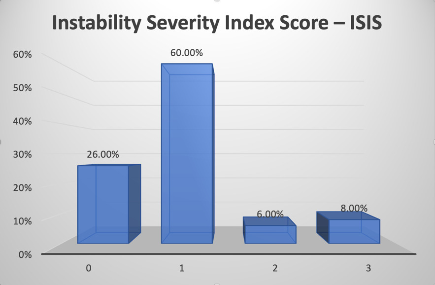
Thirteen patients (26%) had an Instability Severity Index Score (ISIS) of 0, 30 patients (60%) had a score of 1, 3 patients (6%) had a score of 2, and 4 patients (8%) had a score of 3. No patients had an ISIS of 4 or 5. Overall, according to the ISIS classification, the study cohort predominantly represented a low-risk anterior shoulder instability population (Fig. 8).
_in_the_study_cohort_(.png)
Return to Activity & Re-dislocation
Out of the 48 patients who completed the 2-year follow-up, 47 patients (97.9%) successfully returned to their previous level of activity and work. One patient decided not to resume recreational sports (tennis and football) due to fear of reinjury. No redislocations were observed during the first 2 years of follow-up.
Rowe score
At 1 year, Rowe scores ranged from 90 to 95 (mean 92.1), and at 2 years from 90 to 97 (mean 93.8), indicating excellent outcomes in terms of stability, motion, and function.
Simple Shoulder Test (5-Year Follow-up)
At 5 years, 44 patients (88%) completed remote assessment using the translated Simple Shoulder Test. Scores ranged from 9 to 12 (mean 11.36), with no cases of redislocation reported. Most patients reported excellent function; however, several noted discomfort when sleeping in certain positions, which was recorded as “No” for consistency. Although SST does not provide objective stability testing, it was chosen for feasibility in mid-term remote assessment and has been validated for Arabic-speaking populations.
Complications
One superficial portal site infection resolved after a short course of oral antibiotics.
Discussion
Historically, outcomes of ALPSA repair have been less favorable than Bankart repair, with reports of increased recurrence and failure rates after arthroscopic fixation. Nevertheless, the available evidence remains limited due to the relatively small sample sizes in most published series.1 The single-row technique remains the most widely used method for arthroscopic repair of the capsulolabral complex; however, it may not adequately restore the native anatomy. To address this limitation, several authors have introduced the double-row capsulolabral repair technique, which aims to achieve more anatomic reconstruction and biomechanical stability.8–11,20
A total of 50 patients with recurrent anterior shoulder dislocation were included in this investigation. All patients were confirmed to have ALPSA lesions and underwent arthroscopic repair using the double-row technique.
In this study, two patients (4%) were lost to follow-up after suture removal. Among the remaining cohort, 47 patients (97.9%) resumed their previous activities and occupations. One patient (2%) did not return to his prior level of physical activity, which included tennis and recreational football, due to fear of reinjury. During the first two years of follow-up, no cases of re-dislocation were reported. At the five-year follow-up, among the 44 patients who responded to remote assessment, none experienced recurrent dislocation.
In this study, the Constant Shoulder Score (CSS) showed a significant improvement following surgery. Preoperatively, CSS ranged from 52 to 67 (mean, 59.2 ± 4.03), while after 2-years follow-up it ranged from 90 to 97 (mean, 94.92 ± 1.72), indicating enhancement in shoulder stability and function.
There was an observation of significant decrease in external rotation in adduction. Preoperatively, external rotation in adduction ranged from 44° to 70° (mean, 56.3°), while at the 1-years follow-up it ranged from 38° to 59° (mean, 46.6°). This represented an average reduction of approximately 10° with the arm in adduction, which was considered clinically insignificant.But at the end of 2 years the mean external rotation in adduction further improved to 52.2 degrees that indicates good improvement.
From a biomechanical perspective, excessive humeral external rotation has been shown to place increased tension on the anterior band of the inferior glenohumeral ligament, which is a critical stabilizer of the shoulder joint. Therefore, a mild reduction in external rotation following surgical stabilization may improves the outcome by reducing stress on the repaired structures12,21
The first descriptive double-row capsulo-labral repair was performed by Lafosse et al.22 The study described a double-row technique requiring the use of 2 medial anchors and 3 lateral row anchors. This anchor fixation resulted in a W shape, so it was termed a Cassiopeia technique. The Cassiopeia technique was performed on 12 patients with an average age of 28 years. The study did not address any functional outcome results or define the length of follow-up. However, no patients had any reported intraoperative or postoperative complications. Consideration must be given to an excessive medial anterior-inferior portal for placement of medial suture anchors, as neurovascular injury becomes a greater risk when this descriptive technique is used.22
Iwaso et al23 described a similar double-row repair technique in patients with anterior shoulder dislocation sustained during athletic activity. The technique employed a V-shaped configuration, with one medial anchor placed along the glenoid neck and two lateral anchors along the glenoid rim. The procedure was performed on 28 shoulders in 25 patients, of whom 18 patients (19 shoulders) completed at least 24 months of follow-up. The majority of patients were male, with a mean age of 24.9 years. At the 24-month follow-up, all patients returned to their previous sporting activities without recurrent dislocation or subluxation. The mean limitation in external rotation was 6°, comparable to that observed following conventional single-row repair.23
Jiang et al20 described an arthroscopic suture bridge technique to create a stable and low-profile double-row repair. Six patients were included, with an average age of 30 years and follow-up ranging from 4 to 9 months. None of the patients experienced recurrent dislocation or complication.20
Moran et al12 described a double-row capsulolabral technique performed on a 30-year-old male with recurrent instability. Three months postoperatively, the patient was pain-free with negative apprehension and a painless full arc with an active range of motion.12
Wade and Reddy et al.24 described a prospective single-center, single-surgeon study evaluating arthroscopic double-row repair for Bankart lesions. The study included 49 patients, with a mean age of 29 years, who were followed clinically at 3, 6, and 12 months, with MRI assessment performed at 12 months. Their technique used medial-row anchors placed 10–15 mm medial to the glenoid articular surface, with additional medial anchors inserted according to the length of the lesion, followed by lateral-row knotless anchors on the anterior glenoid rim to complete a suture-bridge double-row construct. At one-year follow-up, the authors reported significant improvement in the Constant score from 38.37 to 88.98 and improvement in the DASH score from 52.38 to 6.80. No redislocation was seen in any of the 49 patients, and MRI showed healing with anchors in place and no evidence of retear. Although the study supports the functional benefit and stability of double-row Bankart repair, its main limitation was the short follow-up period of only one year. Compared with our study, Wade and Reddy evaluated Bankart lesions rather than ALPSA lesions and used a variable number of medial anchors according to lesion length, whereas our study used a standardized single medial anchor supported by lateral-row fixation, with longer 2-year clinical and 5-year patient-reported follow-up.24
Itoigawa et al25 investigate the clinical outcome and magnetic resonance imaging (MRI) findings after arthroscopic Bankart repair with additional double anchor footprint fixation at the 4 o’clock position on Forty-two patients (mean age 27.0 years) with average follow-up period was 19.5 ± 6.2 months. The rates of dislocation recurrence and positive apprehension test results were 2.4 %. External rotation was restricted by 3.5°. The Rowe scores at the final follow-up were 34.5 ± 1.0 points and 97.2 ± 5.7 points, respectively, representing significant improvements over the preoperative scores (p < 0.01).25
Cheng et al.26 described a retrospective case–control study investigating a new key point double-row Bankart repair and comparing it with conventional single-row Bankart repair. The study initially collected 78 patients treated between October 2010 and June 2014, and 44 patients were successfully followed up, including 24 patients treated with single-row repair and 20 patients treated with key point double-row repair. The mean follow-up was approximately 9 years. The key point double-row technique used one additional medial anchor placed at the 5 o’clock position, approximately 10 mm medial to the glenoid surface, through the 5 o’clock portal, with mattress suturing of the glenohumeral ligament–labrum complex. At final follow-up, both groups showed significant clinical improvement. In the double-row group, VAS pain improved from 5.0 to 1.8, VAS instability improved from 7.5 to 1.1, ASES improved from 62.0 to 89.5, UCLA improved from 29.5 to 33.5, and SSV improved from 59.2 to 89.5. The recurrence rate was 10% in the double-row group compared with 12.5% in the single-row group. Regarding range of motion, there was no significant limitation in forward flexion, external rotation at the side, internal rotation at the side, or external rotation at 90° abduction, while internal rotation at 90° abduction was significantly better in the double-row group. The authors concluded that key point double-row Bankart repair achieved reliable long-term outcomes, a low recurrence rate, and improved stability without excessive range-of-motion limitation.26
Strengths and Limitations
The strengths of this study include its prospective design, strict inclusion criteria, comprehensive functional evaluation with multiple scoring systems, and the inclusion of both short and intermediate follow-up. The 5-year remote evaluation using a validated translated SST is another strength, providing a practical tool for intermediate-term follow-up when in-person evaluation is not feasible.
This study has several limitations. First, it was a prospective case series without a direct comparison group; therefore, the results cannot establish superiority of double-row repair over single-row ALPSA repair. Second, the cohort predominantly represented a selected low-risk anterior shoulder instability population, as most patients had low ISIS scores and no patients had an ISIS score of 4 or 5; therefore, the favorable outcomes and absence of redislocation may not be generalizable to higher-risk patients, including those with substantial bone loss, engaging Hill-Sachs lesions, generalized ligamentous laxity, or high-demand contact athletes. Third, the sample size was relatively small and the study was performed at a single center. Fourth, the 5-year assessment was performed remotely using a patient-reported outcome measure, without objective clinical examination, range-of-motion assessment, apprehension testing, or imaging confirmation. Finally, 48 of 50 patients completed the 2-year clinical follow-up and 44 completed the 5-year remote SST assessment, creating a potential risk of follow-up bias.
Conclusion
In this selected cohort, double-row repair was associated with Acceptable functional improvement for patients with ALPSA lesions, with no redislocations observed during the two-year clinical follow-up and sustained mid-term functional outcomes. Most patients regained good shoulder function, and the mild reduction in external rotation did not interfere with daily activities. Because this study included a selected, low-risk cohort and did not incorporate a comparison group, these results should be interpreted as observational. While the outcomes are encouraging, further comparative research is needed to determine how double-row repair performs relative to single row techniques.
Conflict of Interest
No conflict to declare.
Funding Source
No funds were received.
AI Usage Declaration
Artificial intelligence tools (ChatGPT, OpenAI, San Francisco, CA, USA) were used only to improve language clarity and grammar during manuscript preparation. The authors reviewed and approved all content, and take full responsibility for the integrity and accuracy of the work.