
Introduction
Hip osteoarthritis (OA) is a common source of pain, stiffness, impaired gait, and reduced quality of life. Contemporary management is multimodal and includes education, activity modification, weight management when appropriate, exercise or physical therapy, oral or topical medications, injections, and ultimately arthroplasty for advanced disease. The American College of Rheumatology/Arthritis Foundation strongly recommends intra-articular glucocorticoid injection for symptomatic hip OA and strongly recommends imaging guidance because the joint is deep and adjacent to important neurovascular structures.1 Broader recommendations for intra-articular therapy likewise emphasize informed consent, aseptic technique, accurate placement, and individualized decisions about reinjection.2
Fluoroscopy-guided intra-articular (IA) hip injection should not be framed as disease-modifying treatment or as a substitute for appropriate surgical evaluation. Its value is narrower but clinically important: it can provide a time-limited reduction in pain and inflammation, improve participation in rehabilitation, bridge a temporary delay to definitive care, and help determine whether the hip joint is the dominant pain generator.
Diagnostic Utility and Patient Selection
Hip pain is not synonymous with intra-articular disease. Lumbar radiculopathy, spinal stenosis, sacroiliac pain, greater trochanteric pain syndrome, tendinopathy, and referred visceral pain may overlap with OA, labral pathology, or femoroacetabular impingement. In patients with concurrent hip and lumbar abnormalities, guided IA injection has demonstrated diagnostic utility for identifying the hip as the symptomatic source.3 Immediate relief during the expected anesthetic window supports an intra-articular pain generator, but lack of response should be interpreted cautiously because technical failure, severe structural disease, or mixed pain sources may confound the results.
The strongest therapeutic indication is symptomatic hip OA with concordant history, examination, and imaging when conservative measures have not provided adequate relief or are poorly tolerated. Patients should have measurable goals such as improved walking, sleep, work tolerance, or ability to participate in therapy. Diagnostic injection may also assist preoperative planning when symptoms and imaging are discordant. Suspected septic arthritis, bacteremia, infection over the needle path, uncontrolled bleeding risk, acute fracture, or an uninvestigated destructive process are reasons to defer injection.
Evidence for Therapeutic Benefit
Placebo-controlled and active-comparator trials generally support short-term benefit. Lambert and colleagues used fluoroscopic guidance and found that 40 mg triamcinolone hexacetonide improved pain, stiffness, physical function, and responder rates compared with local anesthetic and saline, with benefit maintained through three months in many participants.4 Qvistgaard et al. similarly observed improvement after corticosteroid injection in a three-arm trial comparing corticosteroid, hyaluronic acid, and saline.5 Atchia et al. reported substantial early improvements in pain and function after methylprednisolone in moderate-to-severe hip OA.6
The larger pragmatic HIT trial found that adding an ultrasound-guided injection of 40 mg triamcinolone acetonide plus lidocaine to advice and education improved average pain over six months compared with advice and education alone.7 Earlier systematic review evidence concluded that the treatment effect is largest during the first several weeks and may remain clinically meaningful for approximately eight to twelve weeks, while emphasizing small trials and heterogeneous methods.8 Thus, expectations should center on temporary symptom control rather than prevention of arthroplasty. A meaningful response may create a window for strengthening, gait retraining, or life events, even when it does not change the long-term structural course of OA.
Why Fluoroscopy?
Fluoroscopy is a well-established reference technique because it provides clear visualization of the femoral head-neck junction, continuous needle tracking, and immediate confirmation of intra-articular contrast spread if necessary. It is especially useful when body habitus, deep anatomy, prior surgery, or limited ultrasound expertise makes sonographic access less certain. Documentation of a characteristic contrast pattern reduces ambiguity about injectate location and is particularly valuable when the result will inform a diagnostic decision.
Fluoroscopy is not the only accurate option. A 2026 comparative study found significant pain and functional improvement with both fluoroscopic and ultrasound guidance and no clear difference between techniques.9 If fluoroscopy is utilized, radiation exposure should be minimized with pulsed imaging, collimation, the lowest practical magnification, avoidance of unnecessary lateral views, and appropriate shielding. In a series of 446 injections, higher body mass index increased radiation dose but not fluoroscopy time, and the absolute exposure remained low.10
Practical Fluoroscopic Technique
Before the procedure, the clinician should review relevant imaging, confirm laterality and indication, document baseline pain and function, and discuss alternatives. Consent should address variable duration of benefit, post-injection flare, bleeding, infection, allergic or contrast reaction, transient hyperglycemia or blood-pressure elevation, and any other risks deemed appropriate based off institutional guidelines. Anticoagulant and antiplatelet management should follow patient-specific bleeding and thrombosis assessment rather than a routine interruption.
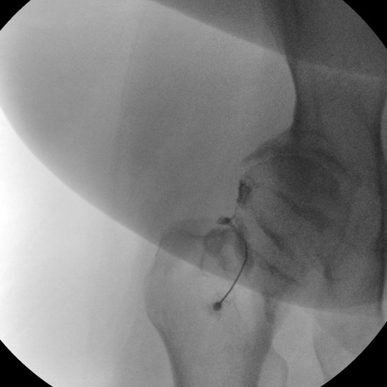
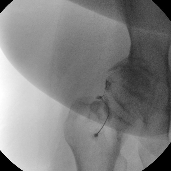
With the patient supine, the hip is prepared and draped using strict sterile technique. An anteroposterior view identifies the femoral head-neck junction while keeping the planned needle path lateral to the femoral neurovascular bundle. After local anesthesia, a spinal needle is advanced under intermittent fluoroscopy to the anterior capsular recess at the head-neck junction. A small amount of nonionic contrast should demonstrate smooth intracapsular spread without intravascular uptake. After confirmation, the therapeutic injectate is delivered without high pressure. Excessive resistance, atypical pain, or an unexpected contrast pattern should prompt reassessment rather than forceful injection. The patient is then observed briefly and given instructions regarding activity, glucose monitoring when relevant, warning signs of infection, and outcome tracking. The injection technique may be modified according to the patient’s pain distribution, body habitus, and ability to tolerate supine positioning. Physicians should individualize the approach to accommodate patient-specific circumstances. An example of an intra-articular hip injection is shown in Figure 1.
Steroid Selection and Dosing
Commonly studied hip regimens use particulate corticosteroids. Triamcinolone acetonide 40 mg is supported by the HIT trial, triamcinolone hexacetonide 40 mg was effective in the fluoroscopic placebo-controlled trial, and methylprednisolone acetate 40 mg has also demonstrated benefit.4,6,7 These agents are particulate suspensions with longer local residence than soluble dexamethasone, although direct evidence that one preparation is clinically superior in the hip is lacking. Choice should account for availability, prior response, allergy history, and institutional policy.
A practical default is 40 mg of triamcinolone acetonide or methylprednisolone acetate, combined with a small useful volume of local anesthetic for diagnostic purposes. Published protocols commonly use approximately 1-4 mL of local anesthetic, resulting in a total injectate volume of roughly 3-6 mL. The authors primarily used 40 mg of triamcinolone acetonide (1 mL) mixed with 3 mL of 0.25% bupivacaine, yielding a total injectate volume of 4 mL for intra-articular hip injections. Immediate anesthetic response can add diagnostic information, but pain relief during this window should be recorded separately from the later steroid response.
Robinson et al. reported benefit with both 40 mg and 80 mg methylprednisolone; the 80-mg cohort showed more sustained improvement in some domains at 12 weeks, suggesting a possible dose response.11 The higher dose may also increase systemic exposure. In the absence of compelling evidence that more steroid reliably produces greater net benefit, the lowest effective dose is reasonable, particularly in patients with diabetes, frailty, osteoporosis, or prior systemic reactions. Repeat injections should be based on the magnitude and duration of prior benefit, alternatives, and cumulative risk rather than a fixed schedule.
Safety, Imaging, and Arthroplasty Timing
Most adverse effects are transient and steroid related, but informed consent should be specific. Post-injection flare, facial flushing, transient hyperglycemia, blood-pressure elevation, sleep or mood disturbance, local skin change, and temporary adrenal suppression can occur; septic arthritis is rare but potentially devastating.12 Patients with diabetes should be advised that glucose may rise for several days and should have an individualized monitoring plan. Sterile technique and avoidance of injection during active infection are essential.
Reports of accelerated OA, subchondral insufficiency fracture, osteonecrosis, and rapid joint destruction have prompted closer attention to preprocedure imaging.13 A 2023 systematic review estimated a 6% incidence of rapidly progressive OA among studies capable of detecting it, but definitions varied, follow-up was inconsistent, and selection bias was substantial; causation therefore remains unresolved.14 When severe pain is acute or disproportionate to radiographic OA, clinicians should reconsider the diagnosis and obtain additional imaging when appropriate before injecting. This is particularly important when insufficiency fracture or osteonecrosis is plausible.
Injection timing matters when total hip arthroplasty is anticipated. Meta-analysis has associated IA corticosteroid injection within three months before arthroplasty with increased periprosthetic joint infection risk.15 Although the evidence is observational, a practical approach is to avoid elective IA steroid injection when arthroplasty is likely within the next three months and to coordinate with the treating surgeon. Repeated injections that provide minimal or progressively shorter benefit should trigger reassessment rather than automatic continuation.
Conclusion
The most appropriate use of fluoroscopic IA hip corticosteroid injection is targeted and goal-directed. A favorable candidate has a plausible intra-articular pain generator, inadequate relief or intolerance of conservative therapies, no urgent surgical or infectious process, and an understanding that this will provide time-limited relief. The procedure is especially useful when contrast-confirmed placement and a documented anesthetic response will resolve diagnostic uncertainty.
Fluoroscopy offers reliable access to a deep joint, clear osseous visualization, and objective confirmation of intracapsular spread. It does not guarantee therapeutic success, and ultrasound may be equally effective when performed by experienced clinicians. The injection should therefore be viewed as one component of longitudinal care, coupled with exercise, risk-factor modification, medication management, and timely surgical referral when indicated. Conservative steroid dosing, careful imaging review, explicit consent, and avoidance of poorly justified repetition preserve the procedure’s favorable clinical utility while acknowledging important evidence gaps.
Statements and Declarations
Generative AI assisted with manuscript organization, literature searches, and language editing. The authors developed the manuscript concept, supplied key references, and actively participated in writing and revision. All authors reviewed and approved the final manuscript.