
Introduction
Ceramic-on-ceramic (CoC) articulations were introduced in total hip arthroplasty (THA) to reduce bearing wear and osteolysis. Modern ceramic bearings have demonstrated excellent long-term survivorship with very low wear rate.1–3 Ceramic fracture, however, remains a potentially catastrophic complication despite improvements in manufacturing and material properties.4,5
Sandwich-type acetabular liners were developed to reduce fracture risk by interposing a polyethylene (PE) layer between the ceramic insert and metal shell. The PE interlayer was intended to absorb impact and reduce stress concentration within the ceramic. However, the design introduced an additional PE-ceramic interface and a thinner ceramic rim. Subsequent clinical reports described dissociation, rim chipping, and ceramic fracture, with fracture rates substantially exceeding those of conventional third-generation monoblock ceramic liners; most sandwich liner failures were recognized within the first several postoperative years.5–8
We report a case of ceramic-polyethylene sandwich liner fracture clinically recognized twenty-one years after revision THA, accompanied by deep extra-articular migration of ceramic fragments into the obturator internus region.
Case Presentation
Ethical Considerations
This case report was approved by the Institutional Review Board of Inje University Sanggye Paik Hospital (IRB No. 2024-12-009). Written informed consent for publication of the clinical details and accompanying images was obtained from the patient. All patient information was de-identified to protect patient confidentiality.
A 64-year-old man presented on October 17, 2022, with progressive left hip pain and audible clicking during ambulation. He denied any antecedent trauma and had no significant medical history. Primary left THA had been performed at an outside hospital in December 1998 for secondary coxarthrosis related to slipped capital femoral epiphysis. Revision THA was subsequently performed due to aseptic loosening of both components at the authors’ institution on November 7th, 2001.
The operative record of the revision THA documented implantation of a 56-mm SPH Contact cup, a 28-mm Forte third-generation alumina ceramic liner with polyethylene backing, and a size-18 revision stem (LimaCorporate S.p.A., now part of Enovis, San Daniele del Friuli, Italy). Acetabular component positioning was measured as 30.9° of abduction and 12° of anteversion. The patient remained ambulatory without any assistive devices and asymptomatic for seven years postoperatively until he was lost to follow up. Approximately 2 weeks before presentation, he developed clicking sound followed by increasing pain and progressive difficulty in walking.
On physical examination, the patient was ambulatory with mild Trendelenburg gait. Passive range-of-motion of the affected hip was limited to 90° of flexion, 5° of internal rotation, 30° of external rotation, 15° of adduction, and 35° of abduction; the range-of-motion in the well-leg was 130° of flexion, 30° of internal rotation, 40° of external rotation, 20° of adduction, and 45° of abduction. Motor strength was grade 5 bilaterally for hip flexion, knee extension, knee flexion, ankle dorsiflexion, and great-toe extension, and sensation was intact. The modified Harris Hip Score (mHHS) was 42.9. The height and weight of the patient was 170cm and 63kg, respectively, which resulted in BMI of 21.8 kg/m2. The preoperative white blood cell count was 8,260/µL, the erythrocyte sedimentation rate was 8 mm/hr, and the C-reactive protein level was 0.6 mg/dL; all values were within the institutional reference ranges.
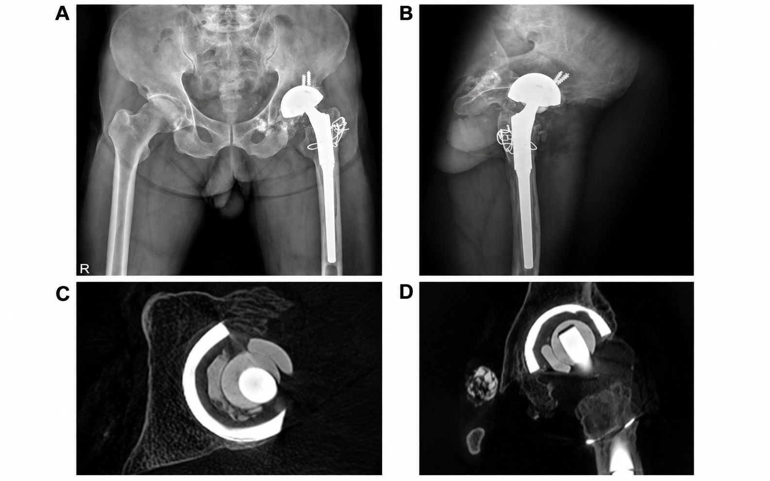
Plain radiographs demonstrated superolateral eccentric migration of the femoral head within the acetabular component and multiple radio-opaque densities located medial and inferior to the cup (Figure 1A,B). CT confirmed sandwich liner fracture and demonstrated deep extra-articular migration of multiple ceramic fragments into the obturator internus region (Figure 1C,D). Osteolysis around the acetabular component was minimal.
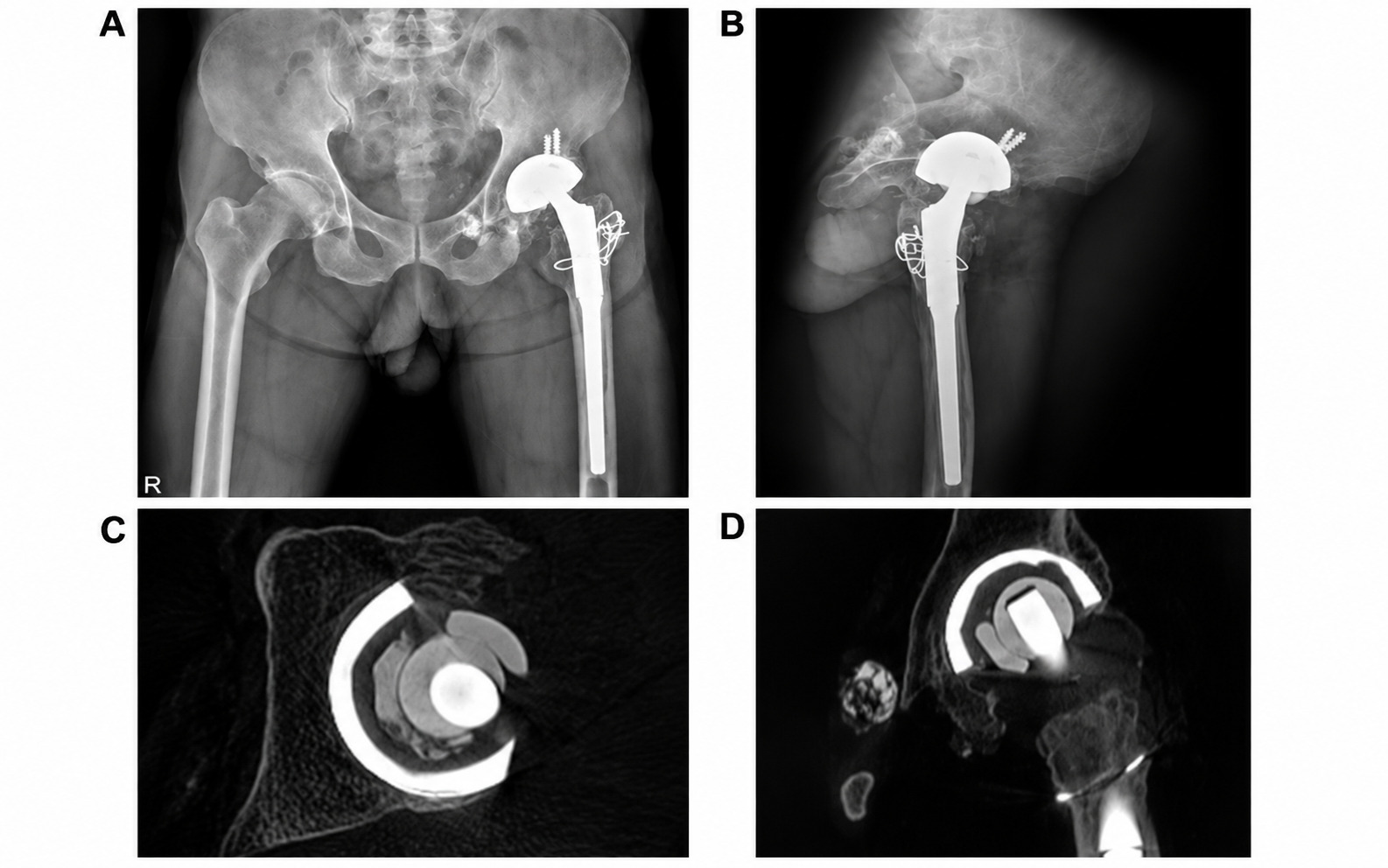
The patient was diagnosed with sandwich liner failure associated with disseminated ceramic debris. We hypothesized that the residual PE layer may have maintained partial articulation, allowing continued ambulation after ceramic disruption and delaying clinical recognition.
Management and Outcomes
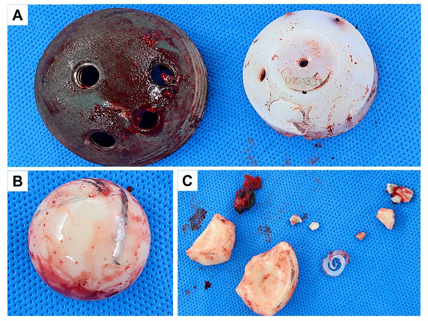
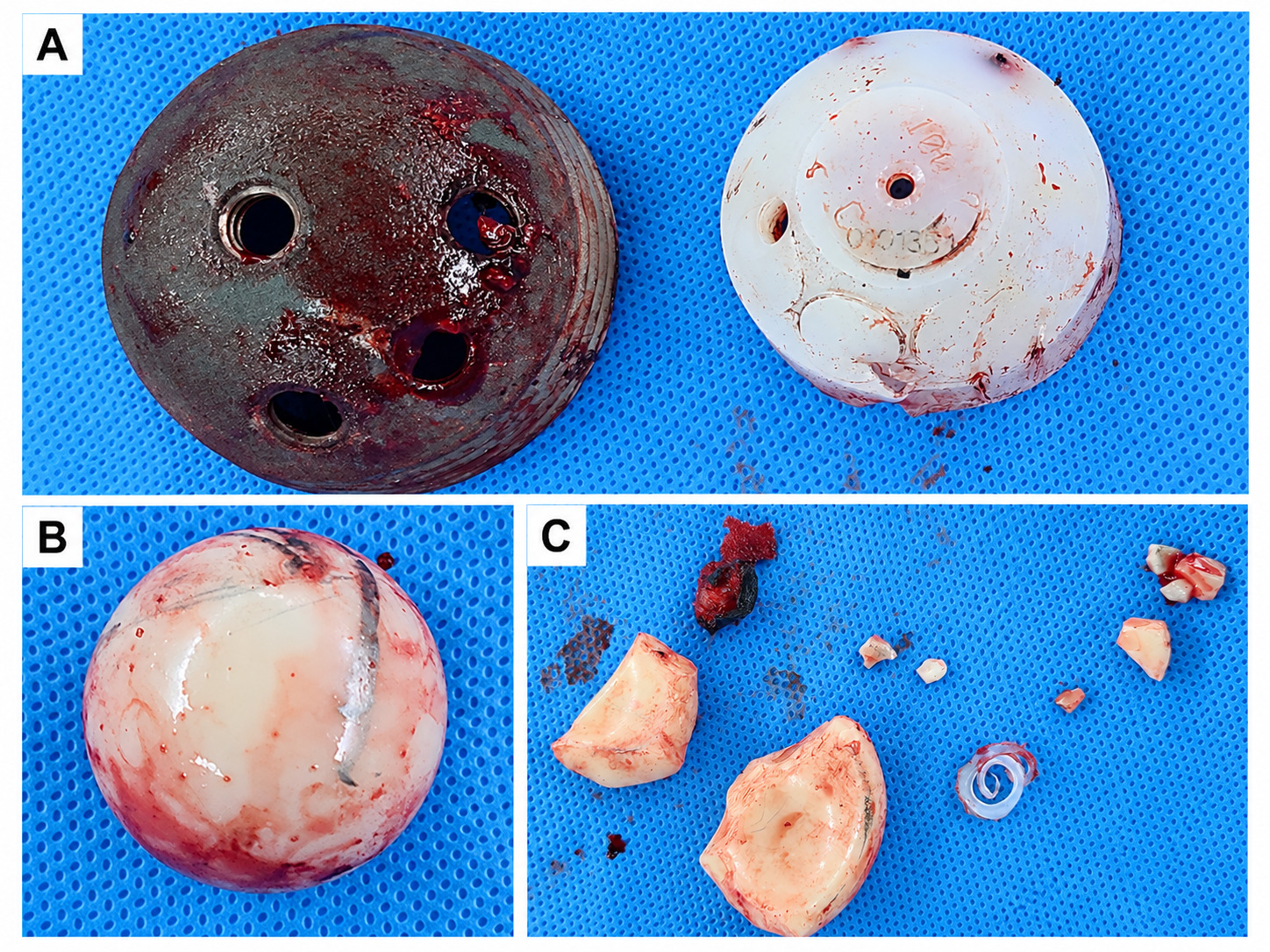
Re-revision THA was performed on October 18, 2022, through the previous incision, using a modified Hardinge approach. The fractured ceramic layer was identified (Figure 2C), and the locking mechanism for the PE backing was intact. The ceramic head showed a stripe-wear mark on its superior surface (Figure 2B). Acetabular shell, the liner and the ceramic head were removed to allow complete exchange of the failed composite bearing. Extensive synovectomy, meticulous debridement, maximal safe retrieval of ceramic fragments, and copious irrigation were performed. Most gross fragments were retrieved; however, smaller particles had migrated through the obturator internus region and could not be identified within the surgical field.
Acetabular reconstruction was performed using a cementless cup (Bencox Hybrid Cup 58-mm, Corentec Co., Ltd., Cheonan, Republic of Korea) with supplementary dome screws (6.4 × 20-mm, 6.4× 30-mm each) and a 4th generation ceramic liner (Bencox Delta 36/44, Corentec Co., Ltd., Cheonan, Republic of Korea). The femoral stem was well fixed and was retained because no loosening of the stem or trunnion damage was identified. After taper integrity had been confirmed, a 36-mm medium length BIOLOX delta ceramic head with a 12/14 taper (LimaCorporate S.p.A., now part of Enovis, San Daniele del Friuli, Italy) was implanted on the retained stem. The pre-existing cerclage wires were removed. Immediate postoperative radiographs showed satisfactory component position.
Before prophylactic antibiotics were administered, two intraoperative tissue specimens were obtained for routine bacterial culture, and both showed no growth. Frozen-section analysis demonstrated fewer than five polymorphonuclear leukocytes per high-power field. Permanent histopathologic examination showed fibrosis with foreign-body reaction and no acute inflammatory findings. Together with the normal preoperative laboratory tests, these findings did not support periprosthetic joint infection.
The postoperative course was uneventful, and the patient was allowed to full weight bearing. Follow-up CT was obtained at postoperative 6-months and showed stable residual ceramic fragments in the obturator internus region without migration or any other soft tissue abnormalities.
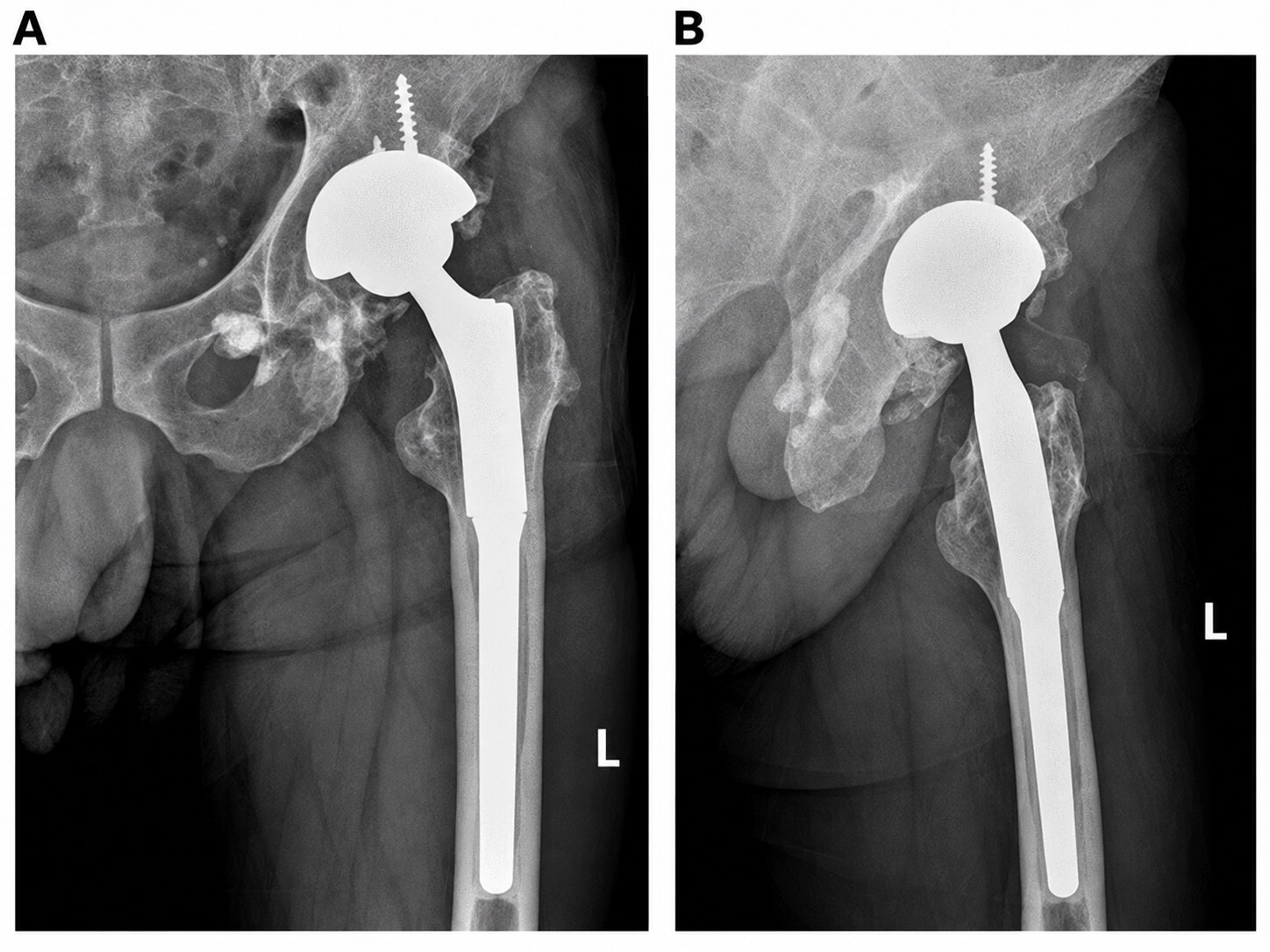
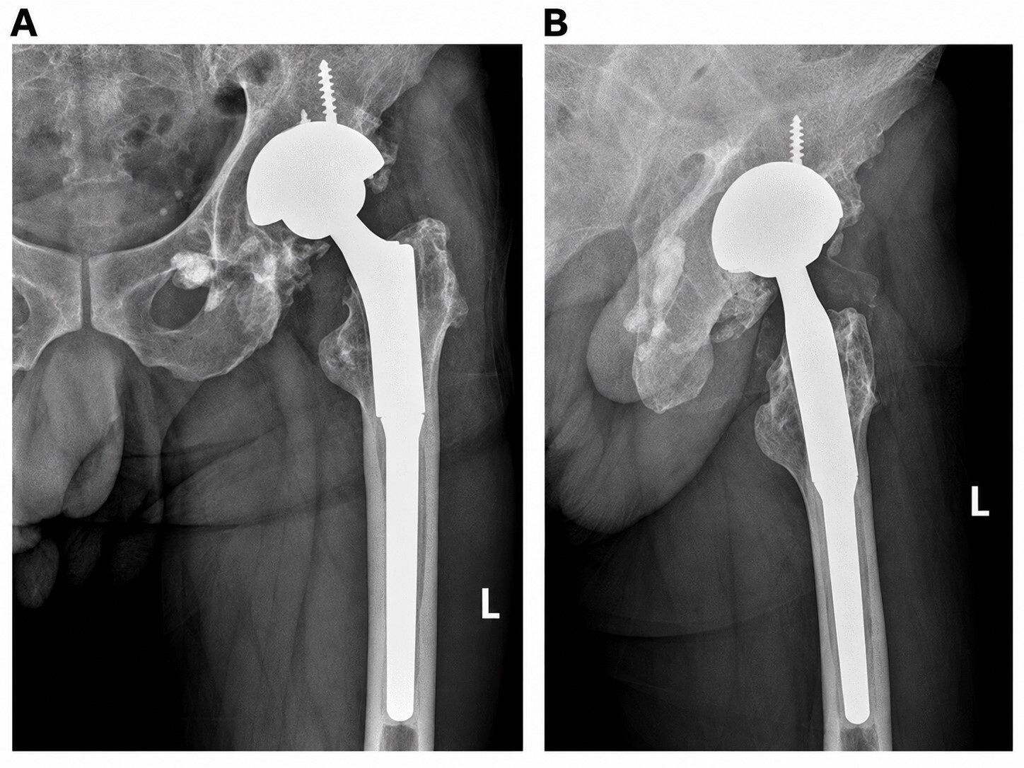
At the final follow-up on May 27, 2026, 43 months after re-revision, the patient was ambulatory independently without an assistive device, reported no meaningful limitation in level walking, and had returned to recreational hiking. The mHHS had improved from 42.9 preoperatively to 87.5. He denied squeaking, recurrent clicking, or disabling pain. Radiographs showed stable component position without dislocation, periprosthetic fracture, or radiographic loosening; the residual migrated fragments remained visible without apparent interval change (Figure 3A,B).
Discussion
The PE backing was designed to absorb impact and reduce stiffness mismatch between the metal shell and ceramic insert. However, the additional interface permitted micromotion and dissociation, while the reduced ceramic thickness increased susceptibility to rim damage and crack propagation. Clinical series reported sandwich liner fracture rates of approximately 1.6% to 6.2%,6–8 markedly higher than the 0.22% liner fracture rate reported for conventional third-generation ceramic articulation in one large retrospective review.4 Impingement and edge loading from cup malposition may further increase local stress and have been implicated in ceramic liner failure.4,9,10 Other possible patient-related factors include obesity and high activity level, neither of which was prominent in this patient. The exact mechanism of failure in this case could not be established because the retrieved components were not subjected to dedicated explant analysis. A stripe-wear mark on the ceramic head raised the possibility of abnormal bearing contact; however, stripe wear alone cannot establish the mechanism of liner fracture.11 Viste et al. found no significant difference in mean cup inclination between the fracture and nonfracture groups, whereas Hasegawa et al. reported fractures in a series using layered acetabular components, with excessive inclination implicated in some cases.7,12 The acetabular inclination of 30.9° in the present patient was at the lower end of the conventional inclination range; however, this measurement alone does not demonstrate edge loading or identify the cause of failure. Pott et al. described the “femoral neck-bite sign” as a radiographic marker of femoral-neck notching potentially associated with ceramic liner fracture or component impingement.13 This sign was not observed in our patient, although its absence does not exclude impingement.
The most distinctive feature of the present case is the very late clinical recognition of sandwich liner fracture, approximately 21 years after revision THA. The timing of the initial ceramic crack cannot be established because no serial radiographs were available during the asymptomatic interval. Traina et al. summarized sandwich liner fractures occurring at a mean of approximately 32 months and cited a series by Hwang et al. with a mean time to fracture of 25 months.5 In a multicenter study, He et al. reported five fractures among 300 hips implanted with sandwich liners, at an average follow-up of 7.3 years.6 Ten-year and longer follow-up studies have confirmed that failure may occur considerably later.7,14,15 In a case report published in 2025, Pott et al. described a sandwich liner failure recognized approximately 20 years after implantation; radiographic findings suggestive of failure had already been present four years before the reported clinical evaluation.13 In a separate 2025 series, Rodríguez et al. described fractures occurring up to approximately 17 years after implantation.15 Thus, the 21-year interval in the present case appears to be among the longest reported periods between implantation and clinical recognition of sandwich liner failure, although the actual date of fracture remains unknown.
The “delayed” component of this case refers to delayed clinical recognition of structural failure, rather than a prolonged delay in seeking medical care after symptom onset. Nevertheless, the extensive distribution of fragments into the obturator internus region suggests that liner disruption may have preceded overt clinical symptoms. Unlike a monoblock ceramic liner, a fractured sandwich liner may retain a polyethylene layer capable of maintaining some degree of articulation and permitting continued weight bearing. This residual layer may mask the severity of failure until progressive pain, clicking, head migration, or extensive fragment dissemination develops. In the present case, the polyethylene backing remained secured by an intact locking mechanism and was difficult to disengage during revision, requiring multiple extraction maneuvers, including the use of osteotomes and screws.
CT was essential for defining the extent of failure. This information was important for surgical planning because it indicated that complete removal of every fragment through a standard acetabular exposure would be neither feasible nor safe. The six-month follow-up CT showed stable retained fragments without an identifiable associated soft-tissue mass. At 43 months, radiographs showed no apparent interval change in fragment position; however, repeat cross-sectional imaging was not obtained at that time, so delayed granulomatous change cannot be fully excluded.
Revision after ceramic fracture is technically demanding because retained microscopic particles can act as third bodies and accelerate wear of the new articulation. Recommended principles include removal of grossly failed components, maximal safe retrieval of ceramic fragments, extensive synovectomy and debridement, massive irrigation, and careful assessment of the taper and retained components.16–19 Retention of the well-fixed femoral stem avoided unnecessary bone loss and operative morbidity because no macroscopic taper or stem-neck damage was identified. A contemporary fourth-generation ceramic articulation was selected to reduce the consequences of residual ceramic debris and avoid direct articulation of a metal head in a particle-contaminated joint.17–19
This report is limited by its single-case design, the absence of serial imaging during the prolonged asymptomatic period, and the inability to determine the precise timing and mechanism of the initial liner crack. Nevertheless, the well-documented implant history and CT findings show that ceramic fragments can migrate deeply into extra-articular soft tissues. Previous reports involving ceramic-bearing complications have described adverse soft-tissue reactions mimicking periprosthetic joint infection, intrapelvic granuloma requiring surgical treatment, a pelvic pseudotumor associated with a ceramic bearing, and severe pelvic bone loss with massive metallosis after delayed recognition of ceramic liner failure.20–23 The present case further illustrates the unusual pattern of intramuscular ceramic fragment migration after sandwich liner failure. At 43 months after re-revision, the patient demonstrated substantial functional improvement and stable radiographic findings after reconstruction with a modern ceramic-on-ceramic articulation.
Conclusion
Sandwich liners should not be considered exempt from surveillance simply because they have functioned for many years. Very late failure can occur, and the PE interlayer may delay clinical recognition by allowing continued ambulation after ceramic disruption. Continued long-term clinical surveillance should be considered for patients with this implant, together with patient education regarding new-onset symptoms. Surgeons should maintain a high index of suspicion even beyond two decades after implantation of the ceramic-PE composite articulation.
Acknowledgments
None
Author Contributions
Hyungtae Kim (first author): Data collection, literature review, analysis and interpretation of clinical findings, figure preparation, and writing of the original manuscript.
Ju-young Jeong (second author): Clinical data analysis, radiological image review, literature review, and manuscript review and editing.
Yerl Bo Sung (corresponding author): Direct clinical management of the patient, performance of the revision surgery, study conceptualization, supervision, interpretation of clinical findings, and critical revision of the manuscript for important intellectual content.
All authors reviewed and approved the final manuscript.
Conflicts of Interest
The authors declare no conflicts of interest.
Funding
No funding was received for this work.