
I. Introduction
Asalin Belcher, a German military surgeon, first reported femoral neck stress fractures (FNSFs) in 1905, marking them as uncommon injuries.1 Femoral neck stress fracture is caused by repetitive loading of the femoral neck that leads to either compression side (inferior-medial neck) or tension side (superior-lateral neck) stress fractures. FNSFs account for approximately 3% to 5% of all sports-related stress fractures.2 This injury has become more frequent in the civilian population because the popularity of endurance sports has increased.3 FNSFs are more common in young women, especially runners, other endurance athletes, or female military recruits, as opposed to male ones.4
We report a clinical case of a 30-year-old female patient who is a marathon runner with a healthy medical history. Two weeks before admission, she began experiencing left groin pain. The patient underwent a clinical examination, and an MRI was ordered and showed that she had femoral neck stress fractures in both the compressive side and the tension side. The patient underwent surgery for femoral neck reduction with cannulated screws. After 10 months of surgery, the femoral neck had completely healed, and the patient returned to daily activities.
II. CASE REPORT
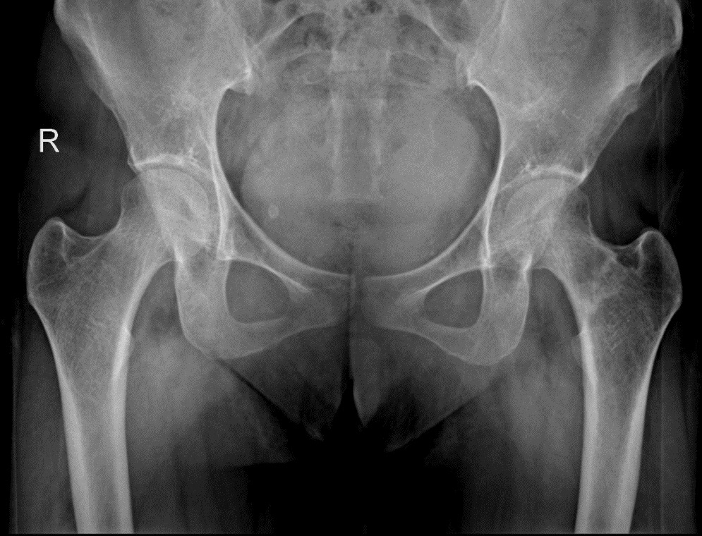
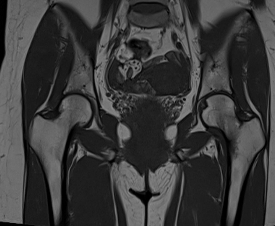
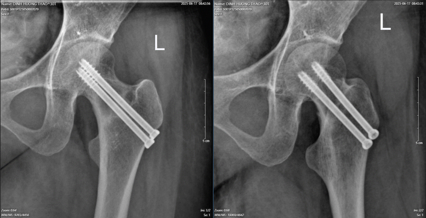
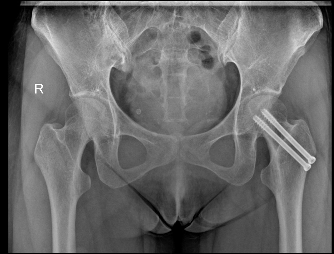
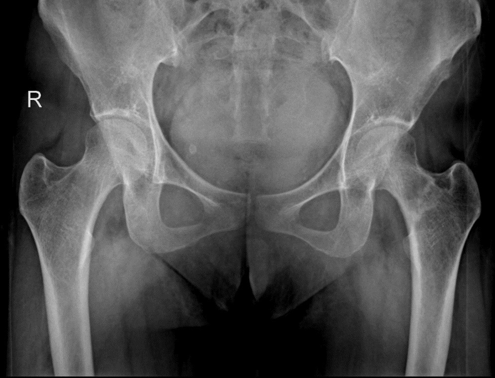
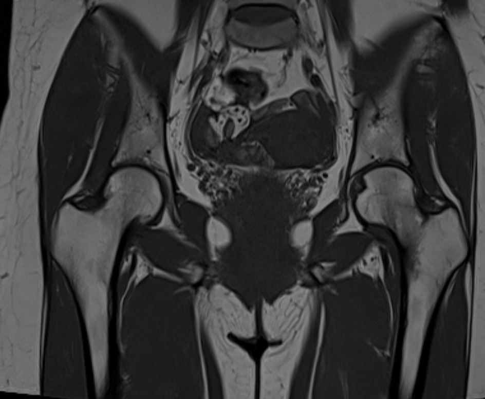
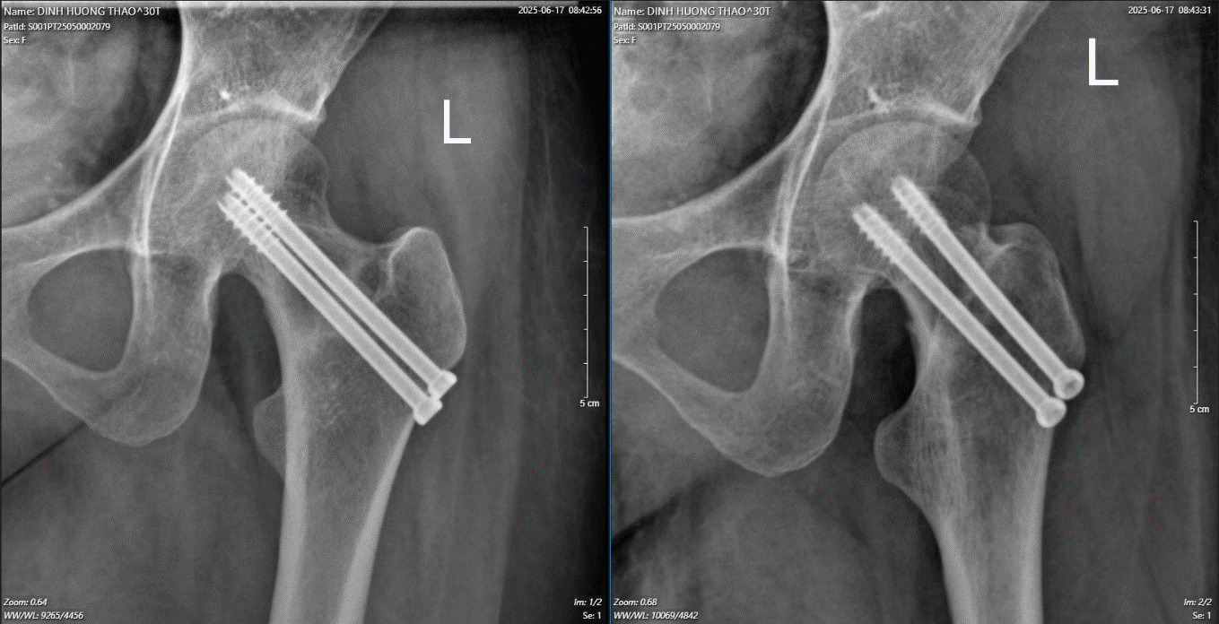
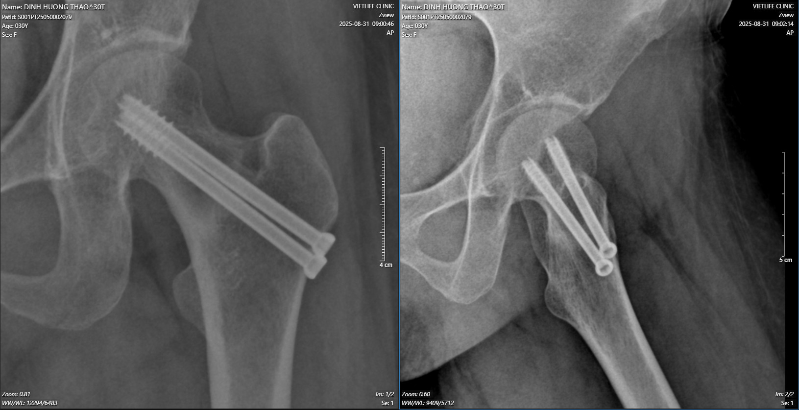
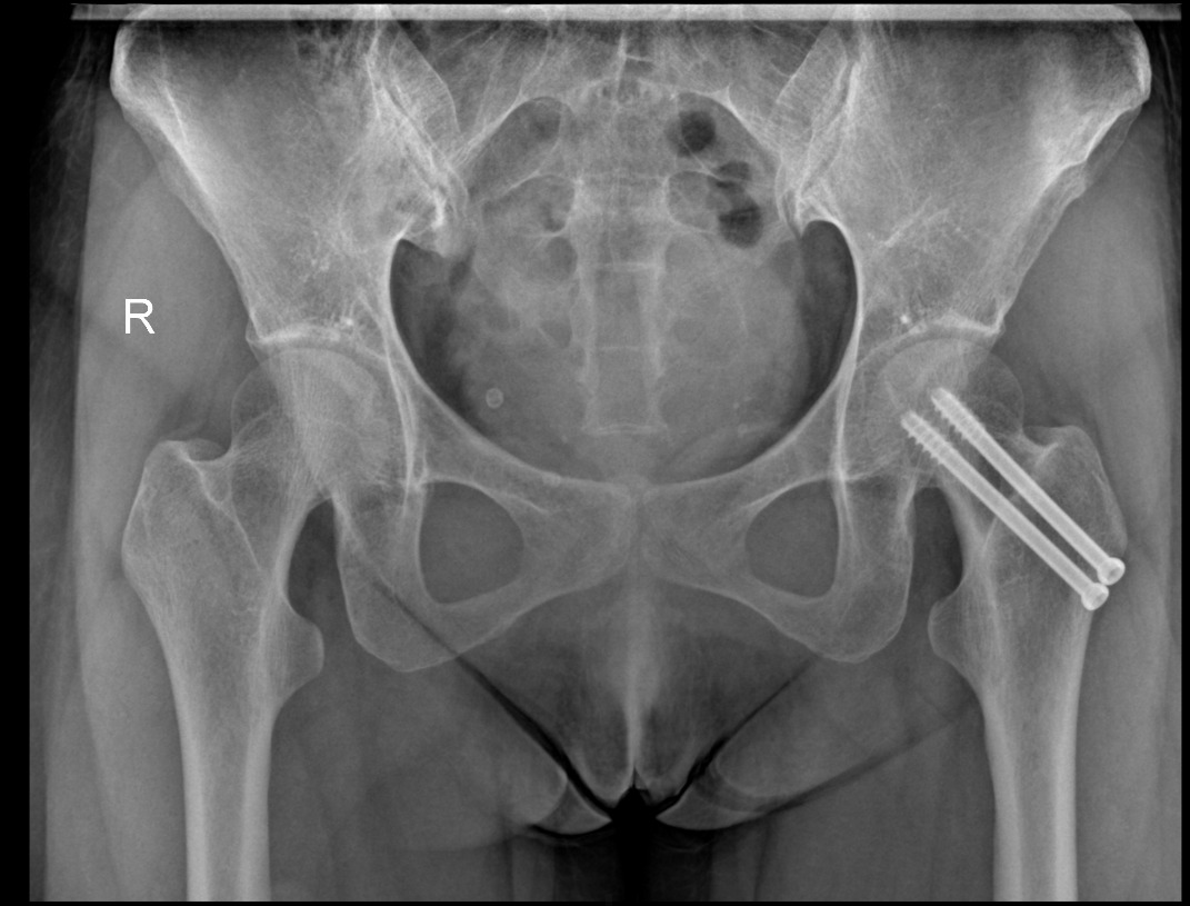
Case of a 30-year-old female patient, with a healthy medical history, who is a marathon runner, averaging 60 to 80 kilometers per week. The patient began experiencing left groin pain in April 2025, which gradually intensified over the two weeks prior to her hospital visit. She complained that she had been prepared for a marathon tournament and had been running for 200 kilometers for the last two weeks. On clinical examination we found that the patient experiences left groin pain when walking, which decreases when there is no pressure on the left leg. On the AP Pelvis X-ray, a suspected fracture line of the femoral neck was noted, and the MRI confirmed fracture lines of the femoral neck in both the compression side and the tension side. The patient was consulted and diagnosed with non-displaced femoral neck stress fracture. Surgery for fixation was indicated. The surgical incision was 2 cm long on the outer side of the upper third of the left thigh, guided under the C-arm. We drilled two tunnels to place two cannulated screws to stabilize the femoral neck fracture. Two weeks after the surgery, the patient experienced no pain in the left groin. The patient was instructed to walk with crutches after 6 weeks post-op. At 10 weeks post-op, the patient was able to walk normally and could perform light exercises. 10 months after surgery, the patient has returned to daily activities.
III. DISCUSSION
FNSFs were mostly seen in military recruits exposed to repetitive marching and running.5 Waterman et al reviewed all lower extremity stress fractures in the military from 2009 to 2012 and identified 31,758 lower extremity stress fractures, of which 9% were located within the femoral neck.5 Stress fractures can also occur in young individuals and are often a result of repetitive athletic activity. Distance runners, dancers, and military recruits are commonly affected and are at an increased risk of developing stress fractures in the hip. Our patient experienced a significant increase in running distance over 2 weeks while preparing for the marathon tournament, which we believe contributed to the development of the FNSF.
The typical presentation of a patient with an FNSF is activity-related groin pain. Patients report gradual onset hip and groin pain that worsens with repetitive weight-bearing activities and improves with rest. Patients will often have a history of a recent increase in physical activity.2 Complete displacement of an FNSF may be shown just like another displacement of a femoral neck fracture. Our patient presented with a left groin pain. Even though she was prescribed NSAIDs, the pain was not reduced, especially when she tried to weight bear.
Magnetic resonance imaging (MRI) has now become the gold-standard second-line imaging investigation for FNSFs. An MRI has been shown to have 100% sensitivity, specificity, and accuracy when investigating FNSF1, even if there’s nothing on an X-ray to show. It’s really important to diagnose this condition early to prevent complications like avascular necrosis (AVN). In a series of 42 displaced FNSFs, Lee et al6 showed that 10 patients with FNSFs developed AVN; the mean delay until surgery was 5.9 days, whereas in the other 32 that did not develop AVN, the mean delay was 1.9 days. On the other hand, Johansson et al7 noted an AVN rate of 30% in a cohort of 10 displaced FNSF in athletes who had delayed treatment by a mean of 14 weeks.
Our patient initially was taken for a pelvis X-ray in another hospital before being admitted. We barely saw any fracture line in the left femoral neck until MRI was ordered. On the MRI, the stress fracture line was on both the compression side (inferior-medial neck) and the tension side (superior-lateral neck). According to Robertson et al,1 when the forces applied exceed the elastic properties of bone and outpace the healing response of bone, micro-fractures develop and propagate at 45 degrees to the application of forces. This creates an oblique fracture line across the femoral neck, which can remain stable until it exceeds around 50% of the femoral neck width. The fracture line created with a compression fracture is more oblique than that of a tension fracture and is thus more stable and less likely to displace. There are few classifications for FNSFs, but according to the Fullerton and Snowdy (1988) classification, our patient was classified as the combination of both Type I (Compression - Side) and Type II (Tension - Side). Surgery for fixation was indicated.
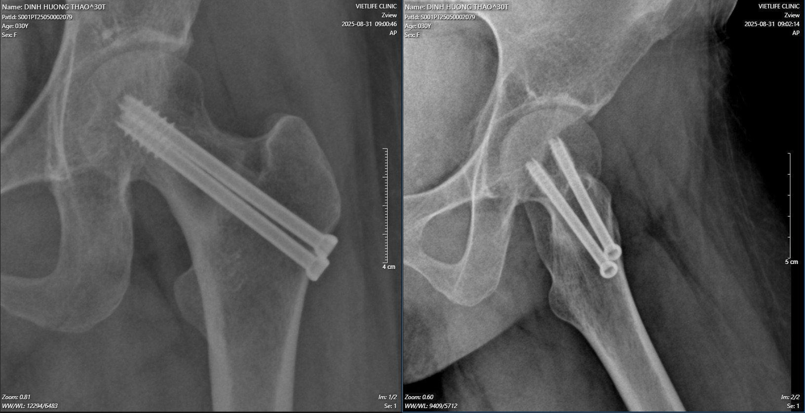
In order to prevent further displacements, we decided to have surgery for the patient. First, we chose cannulated screws instead of DHS because the patient was small (45 kilograms in weight and 158 centimeters in height—BMI was 18), and secondly, while checking on the C-arm in the operating room, there was no displacement of the femoral neck fractures. The patient was in a supine position. The surgical incision was 2 cm long on the outer side of the upper third of the left thigh, guided under the C-arm. We drilled two tunnels with K-wires to guide two cannulated screws to stabilize the femoral neck fracture. The patient was discharged the day after and then started weight-bearing after two weeks. There was no pain when she followed up, and she started to remove crunches after 6 weeks. At the 10-week follow-up, callus formation was shown in the X-ray. At 10 months after surgery, there was no pain at the left groin; the patient returned to daily activities.
IV. CONCLUSION
Femoral neck stress fracture is a rare condition. The typical clinical feature of the disease is the appearance of hip joint pain in athletes or soldiers who have undergone long-distance running during training or drills. The disease is diagnosed with clinical and paraclinical examinations. MRI is the gold standard in imaging diagnosis. Early diagnosis is important for the right treatment, preventing complications like avascular necrosis (AVN) in young patients. The results of the clinical case show that the surgery was successful, allowing the patient to return to their daily activities.