
Introduction
Revision total hip arthroplasty (THA) presents significant challenges, particularly in managing acetabular bone defects (ABDs), which are common and complex for orthopedic surgeons.1 These defects can manifest in various forms, broadly categorized as acute or chronic. Acute discontinuity often arises during acetabular component insertion and presents as a periprosthetic fracture separating the superior and inferior pelvic segments. Conversely, chronic discontinuity develops gradually as a consequence of progressive osteolysis and implant loosening over time.2 Massive ABD is characterized by a loss of pelvic continuity, frequently stemming from a persistent nonunited pelvic stress fracture associated with severe acetabular bone loss in revision THA.3 This deficiency leads to substantial acetabular bone loss and a nonunited fracture, making the simultaneous management of both issues one of the greatest challenges in revision hip surgery.
The complexity of treating massive ABD is compounded by several factors. These include the wide range of acetabular bone loss classifications, the diversity of available surgical instruments and devices, the prevalence of small case series, and the notable absence of randomized prospective studies. Furthermore, there is ongoing debate regarding the optimal type of graft material, with some surgeons favoring morselized bone grafts for their potential for rapid and predictable incorporation, whereas others advocate that structural allografts achieve stronger initial construction.4 High rates of mechanical failure and complications further underscore the difficulties associated with these reconstructions.
To standardize the assessment of ABD, several classifications have been developed. The American Academy of Orthopedic Surgeons (AAOS) categorizes bone defects into segmental (Type 1), cavitary (Type 2), combined (Type 3), pelvic discontinuities (Type 4), and fused hips (Type 5).5 The Paprosky classification system provides a comprehensive framework, distinguishing defects on the basis of the extent of bone loss and column involvement. For example, Type I involves minimal bone loss with intact columns, whereas Type III involves substantial bone loss with compromised column support, which is further subdivided into type IIIA, characterized by superolateral (up and out) migration of the hip center, and type IIIB, characterized by superomedial (up and in) migration associated with more severe bone loss and column deficiency.6 These classifications are crucial for guiding surgical planning and predicting outcomes.
Historically, ilioischial devices, such as the BS ring, have been a cornerstone in managing severe ABD. Indications for their use include significant anterior and posterior column bone loss and less than 50% remaining host bone contact, where a cementless hemispherical acetabular component would lack adequate support.7 Midterm follow-up studies have reported radiographic loosening rates ranging from 0% to 24% and failure rates between 0% and 15%.8 A notable disadvantage of this technique is the extensive debridement often needed, which can hinder healing across the defect. Since the Burch-Schneider ring does not inherently promote biological ingrowth, fatigue fracture of the metal and screw breakage are the primary causes of midterm failure. Consequently, achieving excellent purchase with multiple screws in different directions is recommended, although ischial osteolysis can compromise flange fixation.9
More recently, the Cup-Cage (C-C) construct has emerged as an alternative reconstructive strategy, particularly for massive bone loss or pathologic bone. Antiprotrusio cages, which were initially developed in the 1990s, were applied directly to the host bone or graft without an ingrowth surface, with the acetabular bearing component cemented within. While offering good initial stability, the absence of an ingrowth surface often leads to mechanical failure.10 The modern cup-cage technique, pioneered by Hanssen and Lewallen (2005), integrates a porous tantalum revision cup with screws into the remaining host bone, followed by the application of an antiprotrusio ring over the cup to increase stability and achieve biological fixation. A cemented dual-mobility cup or polyethylene liner is then inserted within the ring at the appropriate inclination and version.11 This reconstructive method is ideal when the remaining host bone cannot adequately support a hemispherical cup, or in cases of massive ABD, pathologic bone due to radiation, or primary/secondary tumors. It is particularly indicated when standard hemispherical cups are suspected to have high failure rates.12 Contraindications include rare instances of severe ilium and ischium bone loss necessitating alternative methods such as custom triflange techniques, or in cases of less severe ABD, where sufficient host bone contact with an acetabular component is achievable, rendering cup-cage reconstruction potentially unnecessary.13
Given the evolving landscape of reconstructive options for massive ABDs in revision THA, a comparative analysis of the Burch-Schneider (BS) ring and the Cup-Cage (C-C) construct is warranted. This study aimed to evaluate and describe the clinical and radiological outcomes and complication profiles of the BS ring and C-C construct in patients undergoing revision THA for massive acetabular bone defects, with each reconstruction used according to the presence or absence of pelvic discontinuity.
Patients and methods
This prospective cohort study was performed at Al-Azhar University Hospitals, Cairo, Egypt, between January 2019 and May 2023, and evaluated patients who underwent revision THA for massive ABDs. Our study followed the principals of the Helsinki declaration. The inclusion criteria were as follows: diagnosis of acetabular bone loss confirmed by both radiographic and intraoperative findings and the presence of Paprosky type 3A or 3B ABDs. The exclusion criteria were as follows: active periprosthetic joint infection, Paprosky type 1 or 2 ABDs, and severe medical comorbidities that contraindicated surgical intervention.
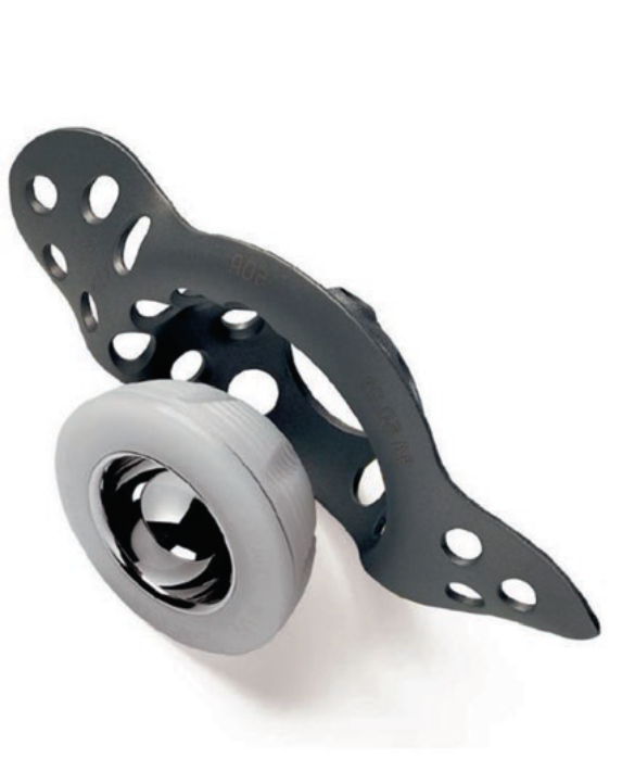
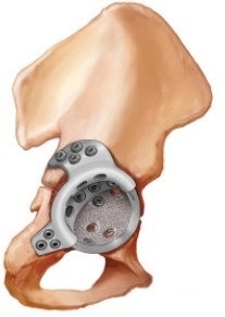
A total of 60 patients undergoing revision THA were recruited and subsequently divided into two distinct groups. Thirty patients (Group A) were managed with the use of the Burch–Schneider (BS) reinforcement ring (Figure 1)14 in the management of massive ABD. The other 30 patients (Group B) were managed by the Cup-Cage (C-C) construct (Figure 2).15 The study included a three-year follow-up period for all enrolled patients.
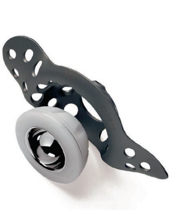
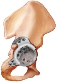
Treatment allocation was prospective but nonrandomized and was based on the pattern and severity of the acetabular defect. The reconstruction method was determined from preoperative radiographic and computed tomography (CT) assessments and confirmed intraoperatively.
The BS reinforcement ring was used for Paprosky type 3A and 3B defects without pelvic discontinuity, whereas the C-C construct was used for Paprosky type 3B defects with pelvic discontinuity, where pelvic stability required both initial mechanical stability and subsequent biological fixation.
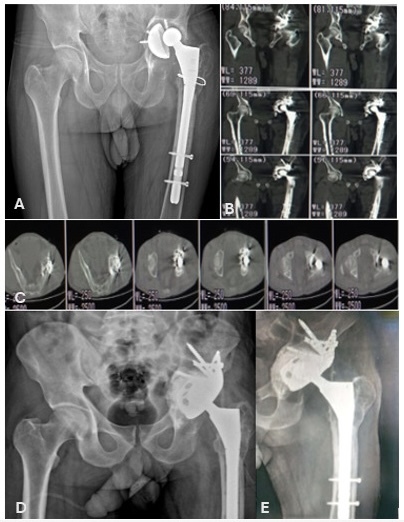
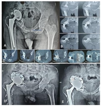
A complete medical history and general examination were obtained from every patient at the time of recruitment. Massive ABDs were identified through specific radiographic features observable on anteroposterior (AP) pelvis radiographs (Figure 3A & Figure 4A). These included a fracture line traversing the ilioischial line, medial migration of the inferior hemipelvis, or asymmetry of the obturator ring. Computed tomography (CT) scans (Figure 3B, C & Figure 4B, C) were additionally utilized to provide more precise visualization and localization of periacetabular bone loss, particularly in the coronal view, to assess the extent of column disruption.
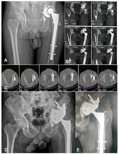
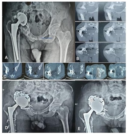
Preoperative laboratory assessments included inflammatory markers specifically the erythrocyte sedimentation rate (ESR) and C-reactive protein (CRP) level. Values were considered elevated when ESR exceeded 30 mm/h or the CRP level exceeded 1 mg/dL.16 and joint aspiration was prompted to analyze fluid cultures, differential cell counts, and cell counts. Patients with suspected prosthetic joint infection were excluded in accordance with established diagnostic criteria.
The functional outcomes were evaluated via the Harris hip score (HHS)17 both preoperatively and three-year post-operatively. The standardized observation period of 36 months for all patients allowed for a consistent midterm assessment of both clinical and radiological outcomes and patient satisfaction.
The study protocol was reviewed and approved by the Institutional Ethical Committee of Al-Azhar University Hospitals, Cairo, Egypt with Institutional Review Board (IRB) registration number Pat_3Med.Research.0000003. All procedures adhered strictly to national and institutional research ethics guidelines. Prior to enrollment, informed written consent was obtained from all participants following a thorough explanation of the study objectives, procedures, and methodology. Confidentiality and individual privacy were maintained throughout the study, and all collected information was used exclusively for the current investigation.
Surgical technique
All surgical procedures were carried out with the patient placed in the lateral decubitus position, with the affected side positioned superiorly, under either general or spinal anesthesia. To ensure adequate exposure and manipulation during revision and fixation, the operative field was extended from the iliac crest to the ipsilateral knee. Strict aseptic precautions, including meticulous skin preparation and sterile draping, were maintained throughout the procedure.
The surgeon was positioned posteriorly, while the first assistant stood anteriorly to facilitate retraction and instrumentation. A lateral approach was consistently employed, offering optimal visualization of both the acetabulum and proximal femur. Through this approach, any previously implanted spacer or failed acetabular cup was carefully removed. Thorough debridement of fibrotic and membranous tissue was performed, followed by copious irrigation with saline to minimize bacterial contamination and remove debris. The acetabular defect was then intraoperatively assessed and classified according to the Paprosky classification system, allowing for a tailored reconstruction strategy on the basis of the individual defect type. The acetabular cavity was prepared by progressive reaming until viable cancellous bone was encountered, ensuring a bleeding host bed conducive to optimal graft incorporation.
Group A: Burch-Schneider reinforcement cage
In Group A, acetabular reconstruction utilized a BS reinforcement cage. Initially, either structural allografts or morselized cancellous grafts were meticulously packed into the acetabular defect to restore deficient bone stock and provide structural support for cage construction. The reinforcement ring was then inserted and fixed via multiple screws into the ischium and ilium. Following confirmation of stable cage fixation, a cemented polyethylene cup was implanted within the ring. The inclination, alignment, and cup anteversion were verified under direct visualization.
Group B: Cup-Cage Construct
In Group B, reconstruction was performed via a cup-cage construct. This involved the initial implantation of a trabecular titanium revision shell, which was press-fitted into the acetabulum to achieve primary stability. In instances of significant bone loss, structural allografts or modular titanium augments were employed to fill defects and provide additional support to the construct. A Cup-Cage was subsequently positioned over the revision cup and secured to the ilium and ischium with screws, thereby enhancing mechanical stability. Finally, a cemented polyethylene liner was inserted into the cup within the cage to restore joint congruity. This dual-implant technique aims to provide both biological fixation via the porous shell and robust mechanical reinforcement through the cage, making it suitable for massive acetabular defects.
Following implant placement, meticulous hemostasis was achieved via electrocautery. Suction drains were strategically placed in both the deep and superficial layers to prevent hematoma formation. The wound was closed in layers via absorbable sutures for deep tissues and nonabsorbable sutures or staples for the skin. After sterile dressing was applied, the limb was positioned in slight abduction. Postoperatively, patients were closely monitored for hemodynamic stability and received antibiotics and thromboprophylaxis in accordance with institutional protocols. Early physiotherapy and mobilization were initiated, tailored to the specific type of reconstruction and the quality of the patient’s bone.
Postoperative follow up
Standard anteroposterior (AP) X-rays (Figure 3D & Figure 4D) were obtained within the first twenty-four hours postsurgery and subsequently at 6 weeks, 3 months, 6 months, 9 months, and one year. For patients with longer follow-up durations, annual radiographs (Figure 3E & Figure 4E) were taken via the same X-ray equipment to monitor component stability, graft incorporation, and restoration of the hip center.
Postoperative weight-bearing ability was determined according to the characteristics and expected biological behavior of each reconstruction. Patients treated with the Burch–Schneider reinforcement cage were allowed immediate weight bearing as tolerated because the construct provided immediate mechanical stability through rigid screw fixation of the reinforcement cage and cemented fixation of the polyethylene cup within the cage.
In contrast, patients treated with the cup-cage construct were maintained on protected weight bearing for approximately 4–6 weeks. This period was selected to allow initial biological fixation and osseointegration of the porous trabecular titanium revision shell before progressive loading. Weight bearing was subsequently advanced according to the patient’s clinical and radiological progress and the stability of the reconstruction.
Statistical analysis
GraphPad Prism 6 was used for statistical analysis. Continuous variables are summarized as the means ± standard deviations (SDs) or medians with interquartile ranges (IQRs), depending on the data distribution, whereas categorical variables are reported as counts and percentages. Normality was evaluated via the Shapiro–Wilk test. Comparisons of parametric data were performed via either paired or independent Student’s t-tests as appropriate, whereas nonparametric data were analyzed via the Mann–Whitney U test. Categorical variables were compared via the chi-square test or Fisher’s exact test when needed. Statistical significance was defined as a p-value < 0.05."
Results
Sixty patients with massive ABDs classified as Paprosky type IIIA or IIIB undergoing revision THA were prospectively enrolled and equally allocated into two groups. Group A (30 patients) was treated via a Burch–Schneider (BS) reinforcement ring, whereas Group B (30 patients) underwent reconstruction via a cup–cage (C-C) construct.
Demographic analysis revealed no statistically significant differences between the two groups in terms of age or sex. The mean age was 64.8 ± 7.9 years in Group A and 65.5 ± 8.3 years in Group B (p = 0.78). The sex distributions were comparable (Group A: 18 females and 12 males; Group B: 16 females and 14 males; p = 0.64). The distribution of Paprosky defect types was comparable between the groups, with Group A having 16 (53.3%) Type IIIA defects and 14 (46.7%) Type IIIB defects, whereas Group B had 17 (56.7%) and 13 (43.3%) defects. The preoperative Harris hip score (HHS) was 41.5 ± 6.1 in Group A and 40.7 ± 6.4 in Group B. The mean follow-up duration was 36 months. The detailed demographic and clinical characteristics are presented in (Table 1).
At 36 months, both reconstruction groups demonstrated significant improvement in HHS. In Group A, the mean HHS increased from 41.5 ± 6.1 preoperatively to 78.4 ± 7.1 (p < 0.001). In Group B, the mean HHS increased from 40.7 ± 6.4 to 83.6 ± 6.0 (p < 0.001). The postoperative HHS was numerically higher in the C-C group; however, the between-group difference was not statistically significant (p = 0.07) (Table 2). These findings describe outcomes observed with each reconstruction strategy in its respective clinical setting and do not establish the superiority of either technique.
All the components were radiographically stable at early follow-up. At 3 years, stable fixation without migration or radiolucency was observed in 24 patients (80%) in Group A and 27 patients (90%) in Group B with no statistically significant difference between the groups (p = 0.472). Radiolucent lines involving ≥2 zones were detected in 6 patients (20%) in Group A and 3 patients (10%) in Group B (p = 0.472). Component migration >2 mm occurred in 2 patients (6.7%) in Group A, with no cases reported in Group B (p = 0.492). Restoration of the anatomic hip center was achieved in 21 patients (70%) in Group A compared with 27 (90%) in Group B; however, this difference was not statistically significant (p = 0.104). The mean time to radiographic bone graft incorporation was 8.0 ± 1.1 months in Group A and 7.7 ± 1.0 months in Group B, with no statistically significant difference between the groups (p = 0.274) (Table 3).
The overall complication rate was 30% in Group A and 20% in Group B, with no statistically significant difference in the overall complication rate (p = 0.552). Dislocation occurred in 3 patients (10%) in each group (p = 1.000). Aseptic loosening was observed in 3 patients (10%) in Group A, whereas no cases were reported in Group B this difference was not statistically significant (p = 0.237). Deep infection occurred in 3 patients (10%) in Group A and 2 patients (6.7%) in Group B (p = 1.000). All five patients in both groups had early postoperative infections and were managed successfully with surgical debridement and targeted antibiotic therapy. Transient sciatic nerve palsy occurred in 1 patient (3.3%) in Group B and in none of the patients in Group A (p = 1.000); the deficit resolved spontaneously within 3 months. No cases of cage fracture, implant breakage, or catastrophic mechanical failure were observed in either group during the follow-up period (Table 4).
Discussion
The present study evaluated the midterm clinical and radiological outcomes of two reconstruction strategies used for massive acetabular bone defects during revision of total hip arthroplasty. Both the Burch–Schneider (BS) reinforcement ring and the cup-cage (C-C) construct were associated with substantial improvement in the postoperative Harris Hip Scale score (HHS) at 36 months. Our results align with a growing body of literature supporting the efficacy of both techniques in addressing complex acetabular revision surgery.
For example, Perticarini et al. (2025) evaluated 57 cases of acetabular revision using cages and cemented cups and reported an 87% implant survival rate and an average HHS of 89.4 at a minimum 60-month follow-up. Their study, which included Paprosky IIB, IIIA, and IIIB defects, highlighted the success of cemented cups in conjunction with titanium acetabular cages for pelvic discontinuity and severe bone loss.18 Similarly, Cochonat et al. (2025) reported acceptable midterm survival (83.1% at five years) and good functional outcomes (mean HHS of 82.7) in 59 patients treated with a cemented modular dual mobility cup combined with an acetabular reinforcement construct over a 10-year period.19 These studies underscore the general effectiveness of reinforcement strategies in complex revision THA.
In the present study, both groups demonstrated significant improvement in HHS at 36 months. Several radiological outcomes also numerically favored the C-C group, including stable fixation (90% vs. 80%), radiolucent lines ≥ 2 zones (10% vs. 20%), and restoration of the anatomic hip center (90% vs. 70%). The mean time to bone graft incorporation was also comparable between the groups (7.7 ± 1.0 vs. 8.0 ± 1.1 months, p = 0.274). These findings suggest that both techniques can provide satisfactory mid-term clinical and radiological outcomes within their respective indications. These findings are broadly consistent with the work of Abolghasemian et al., who reported a higher 7-year survival rate for cup-cage constructs than for traditional cages alone (87.2% vs. 49.9%), with the difference attributed to reduced migration and loosening.20
The proposed advantages of the cup-cage construct may be related to its combination of porous biological fixation with mechanical reinforcement. However, the present study did not demonstrate statistically significant between-group differences in stable fixation, component migration, graft incorporation, or other principal radiological parameters. Therefore, these theoretical biomechanical advantages should be interpreted cautiously in the context of the present findings.11
Marti et al. (2025), in their analysis of 200 hips treated with the BS reinforcement ring, identified pelvic discontinuity as an important predictor of failure.14 This observation is relevant to the present study because the two reconstruction techniques were used according to different defect patterns, with the BS ring used for Paprosky IIIA and IIIB defects without pelvic discontinuity and the C-C construct used for Paprosky IIIB defects with pelvic discontinuity.
The occurrence of transient sciatic nerve palsy exclusively in the cup-cage group (3.3%) warrants attention. This could be attributed to the more extensive surgical manipulation or the bulk of the construct in some cases, although these resolved spontaneously. The absence of implant/cage fracture or catastrophic mechanical failure in either group throughout the follow-up period is reassuring and speaks to the durability of both reconstructive options when appropriately applied. Our findings indicate that both techniques yield significant functional improvements and can provide acceptable midterm outcomes when selected according to the underlying acetabular defect.
Importantly, the present comparison was nonrandomized, and the reconstruction technique was determined by the defect pattern and the presence or absence of pelvic discontinuity. The interpretation of the present findings requires consideration of the indication-based treatment allocation. The BS ring was used in defects without pelvic discontinuity, whereas the C-C construct was used in cases involving pelvic discontinuity. Therefore, differences in outcomes cannot be attributed solely to the reconstruction technique, as the underlying defect characteristics may have influenced both treatment selection and postoperative results. Consequently, selection bias and confounding by indication cannot be excluded. In addition, the groups differed in terms of grafting/augmentation strategies and postoperative weight-bearing protocols, which may have influenced functional recovery and radiological outcomes. These differences should therefore be considered when interpreting the comparative findings.
The strengths of our study include its prospective comparative design, standardized surgical techniques, and consistent three-year follow-up period, providing valuable midterm data. However, the findings should be interpreted in light of the study’s limitations, including its single-center design and relatively small sample size (n = 60), which may limit the generalizability of the results. The absence of randomization, while common in complex surgical studies, introduces potential selection bias. Furthermore, current evidence in this field, as noted in our initial literature review, is largely based on small, prospective case series with a scarcity of randomized controlled trials, making definitive conclusions challenging. The inherent limitations of widely used classification systems such as Paprosky and AAOS in fully capturing the three-dimensional nature of severe defects also present a challenge in precise defect characterization. Another limitation is the postoperative weight-bearing differed between groups: BS-ring patients were allowed immediate weight bearing, whereas cup-cage patients required protection. This difference may have affected recovery and complications, confounding the independent effect of reconstruction technique.
Our findings support the use of both the BS ring and the C-C construct for appropriately selected patients with massive acetabular bone defects. The choice of reconstruction should be individualized according to the extent and type of bone loss, particularly the presence of pelvic discontinuity, the quality of the remaining host bone, and the requirements for mechanical and biological stability. Future investigations should aim to include larger multicenter randomized controlled trials to validate these findings to overcome the limitations of single center studies and small sample sizes.
Conclusion
Both the BS ring and the C-C construct were associated with improved clinical outcomes in appropriately selected patients with massive acetabular bone defects. The C-C construct may be useful in the management of pelvic discontinuity, whereas the BS ring remains an option when pelvic continuity is preserved. However, owing to the nonrandomized, indication-based treatment allocation and the limited sample size, these findings should not be interpreted as evidence of the superiority of one reconstruction technique over the other.
Ethical approval
This article does not contain any studies of animals.
Informed consent was obtained from all individual participants included in the study according to the rules of the hospital research ethical committee.
The study obtained approval from the Institutional Review Board (IRB) under registration number Pat_3Med.Research.0000003, ensuring ethical compliance and participant protection.
All procedures performed in our study were in accordance with the ethical standards of the institutional research committee and with the 1964 Helsinki declaration and its later amendments or comparable ethical standards.
Competing interests
There are no conflicts of interest in the manuscript, including financial, consultant, institutional and other relationships that might lead to bias or a conflict of interest. Authors had no disclosing of any financial ties to the subject of this research.
Funding
No benefits in any form have been received or will be received from a commercial party related directly or indirectly to the subject of this article.
Data availability
The datasets used and analysed during the current study are available from the corresponding author upon request
Acknowledgment
The authors appreciate all the staff members in the Department of orthopaedic surgery, Al-Azhar University, Cairo. Egypt.
Author contributions
Sameh A. Abdulall, Study design, Participation in writing the manuscript. Mohamed A. A. Ibrahim, Participation in writing and revising the manuscript, revising statistical analysis, Grammer and Language editing and follow up of patients, corresponding author. Ahmed M. Khairy, Surgical technique, participation in writing the manuscript, statistical analysis and follow up of patients. Yasser A. Ghonim, Elsayed S. Elbanna, Surgical technique, participation in writing the manuscript, statistical analysis. Mahmoud G. Abdelazim and Abdelhamid E. Hendy, Participation in revising the manuscript and revising statistical analysis. Mahmoud M. Hassaan, Study design, Participation in writing the manuscript. Lotfy Shwitter: participation in revising the manuscript, revising statistical analysis, final approval for submission.