
INTRODUCTION
The medial and lateral menisci distribute load, improve joint congruity, absorb shock, and contribute to knee stability. Their circumferential collagen architecture converts axial compression into hoop tension and limits focal articular cartilage stress.1 Congenital meniscal variants occur more often in the lateral compartment. Discoid morphology is the best-recognized variant, whereas true hypoplasia, partial deficiency, and absence are exceptionally uncommon.2–7
Ramp lesions involve the peripheral meniscocapsular and/or meniscotibial attachments of the posterior horn of the medial meniscus. In anterior cruciate ligament (ACL)-injured knees, reported prevalence varies substantially because of differences in lesion definition, chronicity, imaging technique, and arthroscopic exploration. These lesions may be missed on routine magnetic resonance imaging (MRI) and through standard anterior arthroscopic portals.8–10
Persistent non-visualization of only the posterior horn of the lateral meniscus is particularly unusual. In a patient without previous meniscal surgery, the principal diagnostic possibilities include a developmental deficiency, severe post-traumatic or degenerative tissue loss, and a retracted or scarred remnant that is not readily visible on MRI. We report an adult with this finding in association with chronic ACL insufficiency and a medial meniscal ramp lesion, and we discuss the diagnostic and management implications.
CASE PRESENTATION
Clinical History and Examination
A 41-year-old man with obesity (body mass index (BMI), 37.44 kg/m2; height, 182 cm) presented with sudden severe medial joint-line pain and recurrent instability of the right knee. Eight years earlier, he had injured the knee while playing football. Examination and MRI at that time demonstrated a complete ACL tear; reconstruction was recommended but declined.
He subsequently experienced intermittent anterior knee pain, giving-way, and popping during knee extension and stair climbing. Symptoms worsened with prolonged walking and improved with rest. His medical history included appendectomy and nephrolithiasis. He reported no previous knee operation or known drug allergy.
He walked with an antalgic gait. Medial thigh muscle wasting was present. Range of motion was full, with joint-line tenderness. Lachman, pivot-shift, anterior drawer, and McMurray tests were positive. Varus and valgus stress tests were negative, and distal neurovascular findings were normal.
Imaging Findings
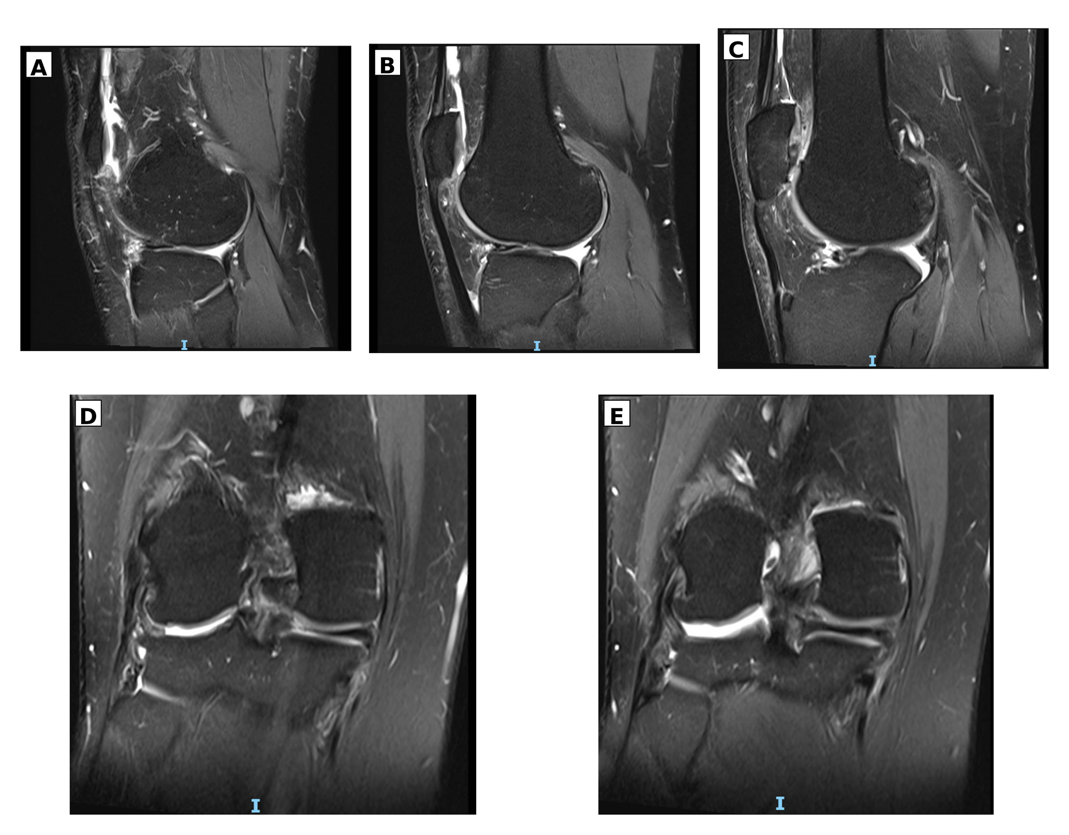
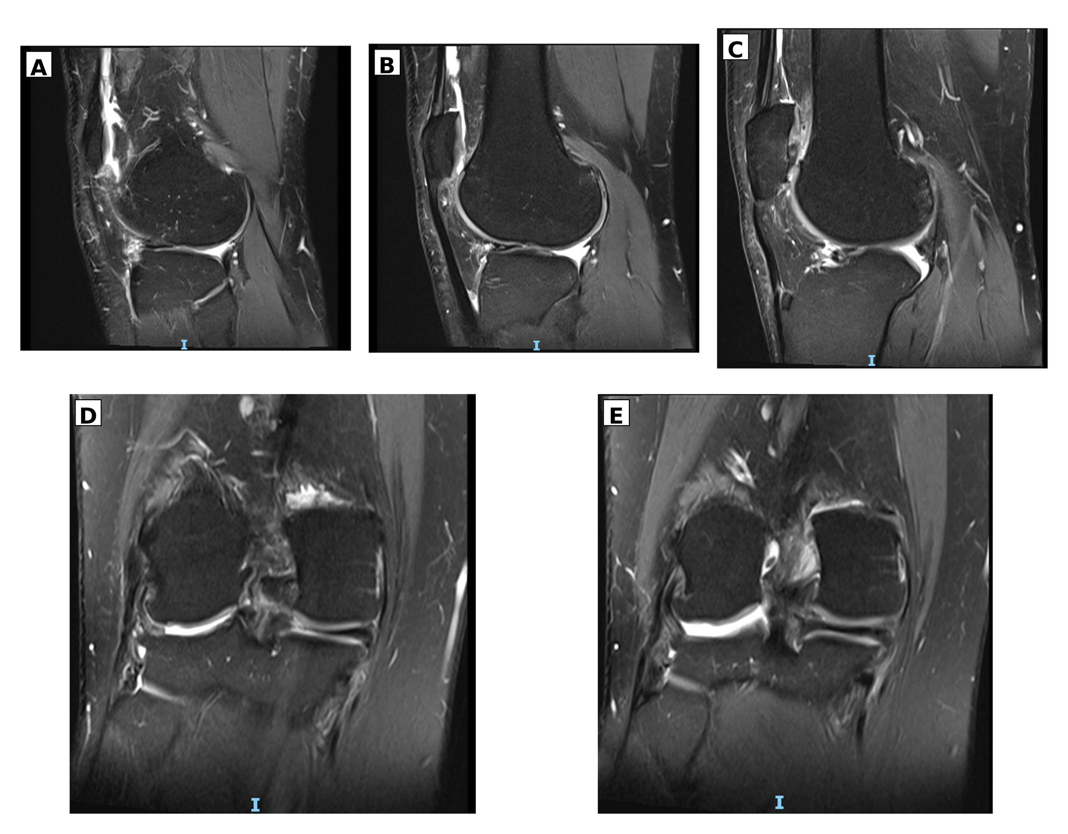
MRI showed chronic discontinuity of the ACL and a vertical tear at the meniscocapsular junction of the medial meniscus posterior horn, consistent with a ramp lesion. On the available sagittal and coronal images, the posterior horn of the lateral meniscus was not visualized, and it remained non-visualized across the available MRI sequences; the segment was therefore interpreted as apparently absent. The patient denied previous meniscal surgery, and no arthroscopy portal scars or hardware were identified. Additional findings included full-thickness cartilage fissuring in the lateral and patellofemoral compartments, marginal osteophytes, reactive subchondral change, patellar tendinopathy, and mild joint effusion. The posterior cruciate and collateral ligaments were intact (Figure 1).
MANAGEMENT AND OUTCOMES
Clinical Management and Current Outcome
At the time of manuscript preparation, diagnostic arthroscopy and ACL reconstruction had not been performed. Consequently, the lateral meniscal finding had not been confirmed surgically, the stability of the ramp lesion had not been assessed by probing, and no postoperative or longitudinal outcome data were available. The patient had presented for surgical assessment; no subsequent operative or follow-up data were available at the time of reporting.
Diagnostic Interpretation
Published reports of meniscal hypoplasia or absence are limited and heterogeneous. They include partial lateral meniscal deficiency, complete absence of both menisci and cruciate ligaments, isolated medial meniscal absence, bilateral hypoplasia of both menisci, and arthroscopically confirmed complete absence of the lateral meniscus.3–7 Selected reports are summarized in Table 1. The present case differs because the abnormality appears confined to the lateral posterior horn and coexists with chronic ACL insufficiency and a medial ramp lesion.
The imaging appearance cannot establish a congenital origin. The absence of operative scars and hardware supports the history of no previous surgery but does not exclude tissue loss from the original injury, repeated instability, or degeneration. MRI also cannot reliably exclude a markedly attenuated, scarred, displaced, or retracted remnant. The term apparent absence is therefore more appropriate than proven agenesis. Arthroscopy with systematic inspection of the posteromedial and posterolateral compartments would provide the most direct confirmation if the result would alter treatment. Normal variability at the posteromedial meniscocapsular junction may also mimic a ramp lesion on MRI, reinforcing the need for context-specific interpretation and arthroscopic correlation.11
Relationship to Anterior Cruciate Ligament Insufficiency and Ramp Lesions
Ramp lesions can increase anterior and rotational laxity in an ACL-deficient knee. Cadaveric studies have shown that disruption of the meniscocapsular and meniscotibial attachments increases tibial translation and rotation, and that isolated ACL reconstruction may not fully correct rotational instability unless unstable ramp-related tissue is addressed.12,13 Meniscal deficiency also increases contact stress and is associated with progressive cartilage degeneration.14
Posterior lateral meniscal pathology is well recognized in ACL-injured knees. Arthroscopic series have documented lateral posterior root avulsions, and combined medial ramp and lateral posterior root lesions have been reported in a substantial proportion of primary and revision ACL reconstructions.15,16 Ramp-like peripheral tears of the lateral meniscus have also been described, suggesting that posterolateral meniscocapsular injury may be underrecognized.17 These observations support an acquired mechanical explanation for the present finding, although they do not prove that the entire posterior horn can disappear through this mechanism.
Management Implications
If surgery is undertaken, arthroscopic inspection should determine whether lateral meniscal tissue is truly absent, severely attenuated, or displaced and whether any residual tissue is repairable. The medial ramp lesion should be probed for stability. Evidence generally supports repair of unstable lesions, whereas short-term studies have not shown a consistent advantage to routine repair of small stable lesions during ACL reconstruction.8,12,18,19 However, long-term observational data suggest that some stable lesions left untreated may subsequently fail.20 Management should therefore be individualized according to symptoms, instability, cartilage status, chronicity, and arthroscopic findings rather than the imaging label alone.
Limitations
This report describes one patient and cannot establish prevalence, cause, or prognosis. No diagnostic arthroscopy, biopsy, genetic assessment, or previous MRI images were available. The apparent absence is therefore based on current imaging and patient history, and a severely attenuated remnant cannot be excluded. No treatment or longitudinal outcome data were available. The literature concerning the exact combination of lateral posterior horn deficiency, a medial ramp lesion, and chronic ACL insufficiency is also extremely limited.
CONCLUSION
Persistent non-visualization of the lateral meniscus posterior horn in a knee without documented surgery is unusual. In this patient, it occurred with chronic ACL insufficiency, a medial meniscal ramp lesion, and degenerative joint changes. MRI defined the abnormalities but could not distinguish congenital deficiency from severe acquired tissue loss. Careful history, compartment-by-compartment imaging review, and arthroscopic confirmation when clinically indicated are required before an absent-appearing meniscal segment is labeled congenital. Future reports should document arthroscopic findings and longitudinal clinical and imaging outcomes.
Acknowledgments
The Researchers would like to thank the Deanship of Graduate Studies and Scientific Research at Qassim University (https://www.qu.edu.sa) for financial support (2026-QU-APC).
Authors’ contributions
All Authors made equal contributions to the manuscript
Ethical Considerations
This report was conducted in accordance with the Declaration of Helsinki. Written informed consent was obtained from the patient for publication of anonymized clinical information and MRI images. Direct identifiers were removed, and clinical information was handled in accordance with institutional confidentiality requirements.
Conflict of interest
The authors declare no relevant financial or non-financial competing interests.
Funding
The authors received no funding for this work.
Permission to publish
Written informed consent was obtained from the patient for publication of the clinical details and images. The signed permission form should be uploaded as a supplementary file.
Data availability
Data are available from the corresponding author on reasonable request, subject to privacy and ethical restrictions.
Reporting guideline
This case report was prepared in accordance with the CARE guidelines.