
1. Introduction
Osteochondral lesions of the talus (OLT) are focal lesions involving the articular cartilage and subchondral bone of the talus. They are frequently associated with ankle trauma and may lead to persistent pain, restricted motion, and mechanical symptoms. Although OLTs are relatively uncommon in the general population, an incidence of approximately 27 cases per 100,000 person-years has been reported in an active-duty military population.1 In a systematic review of 2,087 OLTs, 78% of lesions were associated with a history of trauma, highlighting the substantial contribution of ankle injury to the development of these lesions.2 Clinical assessment is based on the history and physical examination, while magnetic resonance imaging (MRI) is important for characterizing lesion location, cartilage integrity, and subchondral bone involvement.3,4
Treatment is determined by lesion stage, stability, size, subchondral bone involvement, and response to conservative management. Conservative treatment may be considered for selected stable lesions, whereas surgery is generally considered for persistent symptomatic lesions, unstable fragments, or failed nonoperative treatment. Microfracture is a marrow stimulation technique intended to promote repair tissue formation within the osteochondral defect. The procedure penetrates the subchondral plate to recruit marrow-derived progenitor cells and other marrow elements into the defect, promoting formation of reparative fibrocartilaginous tissue. However, this repair tissue differs biologically and mechanically from native hyaline cartilage. Biologic augmentation using platelet-derived products or scaffolds has increasingly been investigated as a means of improving the local repair environment.4–6
Platelet-rich fibrin (PRF) is a platelet-containing fibrin matrix that can provide a three-dimensional scaffold and sustained release of biologically active mediators. Its fibrin network may provide a temporary three-dimensional matrix for cell retention and tissue repair, while platelet-derived mediators may contribute to the local regenerative environment through sustained release of biologically active factors. Recent clinical studies of PRF in OLT have reported improvements in pain and functional outcomes, although the available evidence remains limited and is largely based on small observational cohorts. In particular, the incremental benefit of PRF over microfracture alone remains uncertain.7,8
Therefore, this study aimed to describe the clinical and imaging characteristics of patients with OLT treated with arthroscopic microfracture combined with autologous PRF and to evaluate early clinical and MRI outcomes following surgery.
2. Materials and Methods
2.1. Ethical Considerations
The study protocol was reviewed and approved by the Thesis Proposal Review Committee of Hanoi Medical University before study initiation.
All study variables and outcome measures were collected accurately and systematically in accordance with the study protocol.
Participation was entirely voluntary and the study was conducted solely for the purpose of improving the quality of diagnosis and treatment. Eligible patients were provided with detailed information about the study, and those who agreed to participate provided written informed consent. All patient information and clinical data were coded and kept strictly confidential.
2.2. Study Design and Participants
This prospective observational case series included 18 patients with OLT who underwent ankle arthroscopy with microfracture and autologous PRF application at Viet Duc University Hospital between January 2025 and January 2026.
Inclusion criteria Age ≥18 years. Failure of conservative treatment for at least 3-6 months before surgery. OLT diagnosed clinically and by imaging and classified as Hepple grade III or IV.9 Lesion size of 0.5-1.5 cm2 and subchondral bone defect <15 mm. Complete medical records and agreement to participate in the study.
Exclusion criteria Patients were excluded if they had extensive cartilage lesions or grade III or higher ankle osteoarthritis, uncontrolled systemic disease, current anticoagulant or antiplatelet therapy, previous surgery involving the affected ankle region, local ankle infection, or untreated ankle instability.
2.3. Surgical Technique
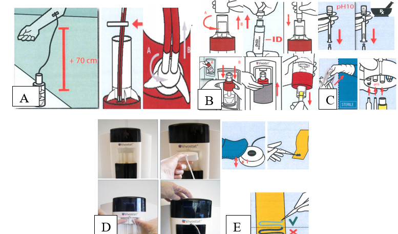
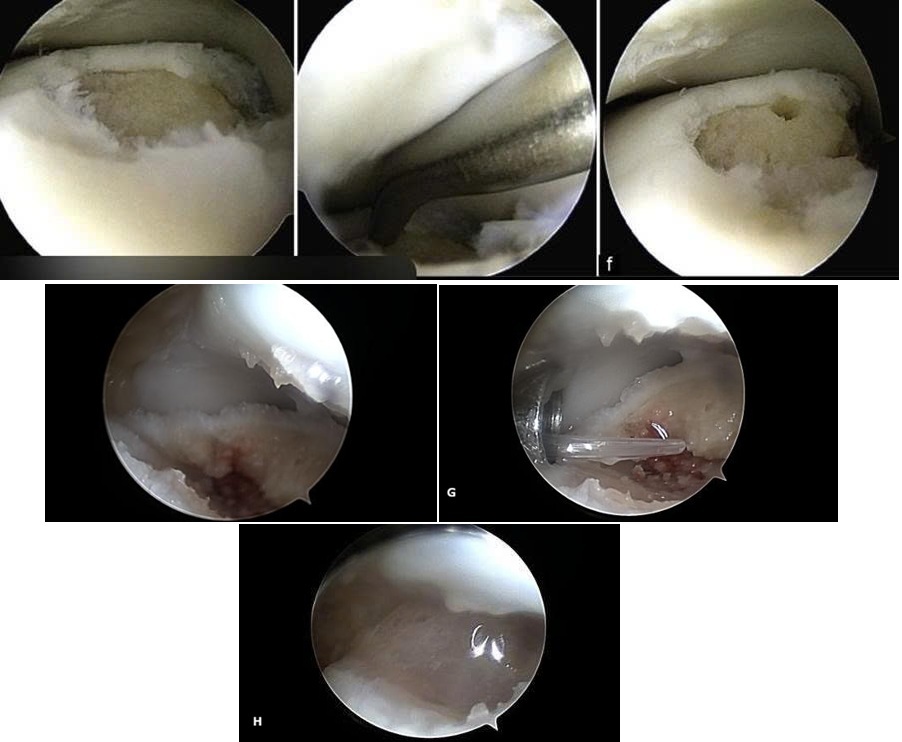
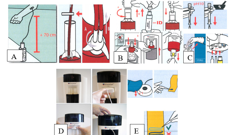
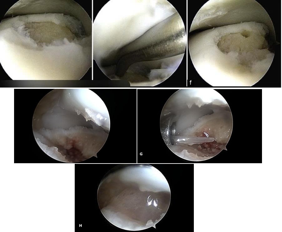
Patients were placed in the supine position with a thigh tourniquet, and the foot was supported in a functional position. Spinal anesthesia was administered. Standard anteromedial and anterolateral ankle portals were established. Arthroscopic assessment included identification and treatment of unstable cartilage, scar tissue, inflamed synovium, fibrous bands, and loose osteochondral fragments. The OLT was identified and debrided to stable margins, followed by microfracture of the subchondral bone using a microfracture awl. Multiple perforations were created evenly throughout the osteochondral defect, while preserving the intervening subchondral bone. The awl was advanced sufficiently to penetrate the subchondral plate, and bleeding from the perforations was confirmed as evidence of access to the underlying marrow space. Autologous platelet-rich fibrin (PRF) was prepared from 120 mL of whole blood using the Vivostat System (Vivostat A/S, Denmark). According to the manufacturer’s protocol, the collected blood was centrifuged at 9000 rpm for 26 minutes. Type I PRF was subsequently collected and transferred to the spraying/mixing system. The preparation process of autologous PRF using the Vivostat System is illustrated in Figure 1. The final volume of PRF obtained after processing 120 mL of whole blood was approximately 5-6 mL. After completion of the microfracture procedure, the joint fluid and residual saline were evacuated. Approximately 5-6 mL of the prepared autologous PRF was then applied directly to the microfractured osteochondral defect using the Vivostat spraying/mixing system, allowing the PRF to cover the treated lesion as a biological scaffold. The intraoperative application of autologous PRF to the microfractured osteochondral lesion is shown in Figure 2.
2.4. Postoperative Care and Outcome Assessment
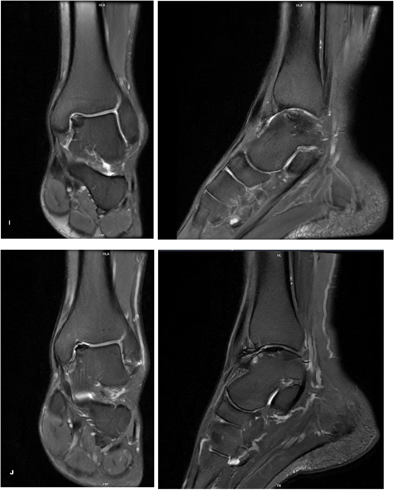
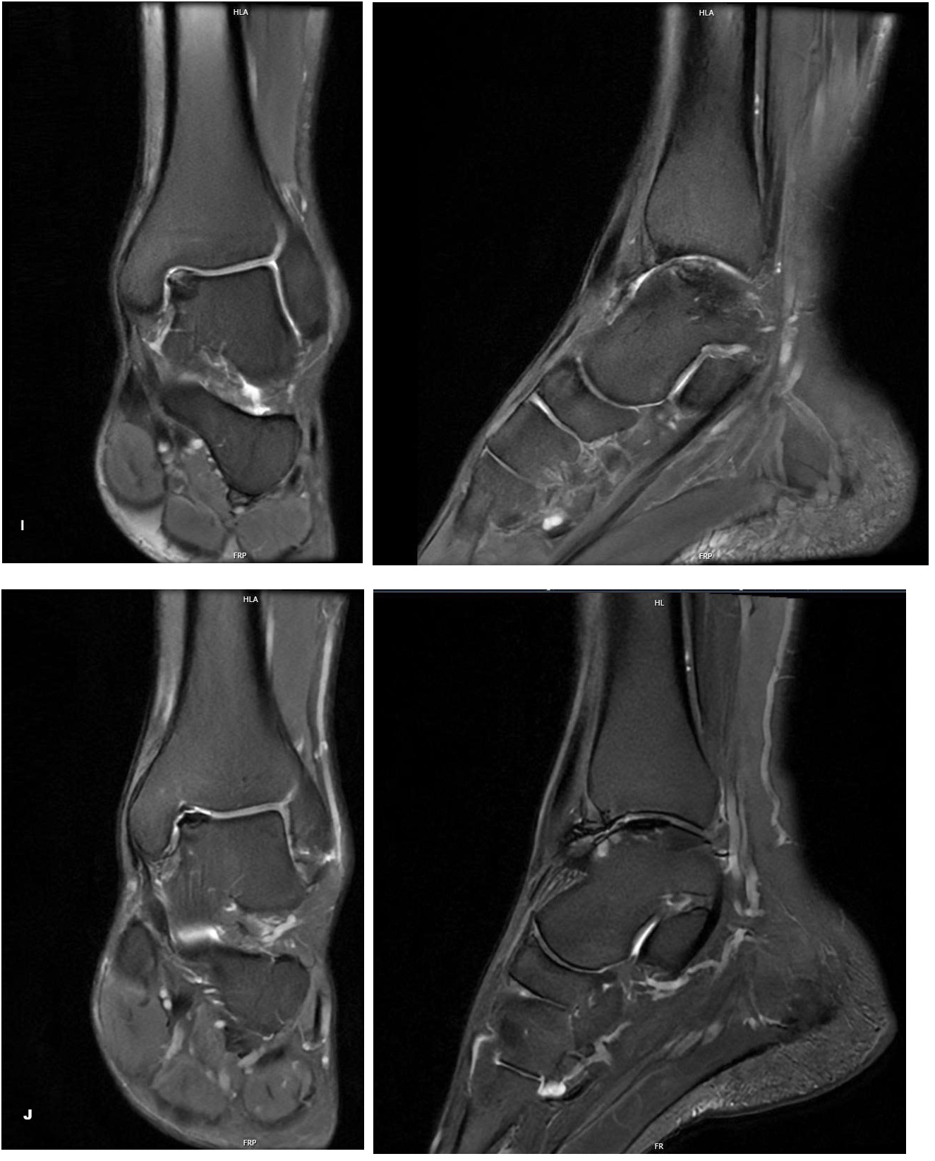
The ankle was immobilized in a functional position with a cast after surgery. During the first 2-6 postoperative weeks, patients performed active range-of-motion exercises without weight bearing. After 6 weeks, progressive weight bearing and ankle strengthening were initiated. Clinical assessments were performed preoperatively and during follow-up. AOFAS and VAS were assessed at 6 months after surgery. Preoperative MRI was graded using the Hepple classification, whereas postoperative MRI at 6 months was evaluated using the MOCART 2.0 ankle score. Postoperative MRI examinations were performed on a 3.0-T Siemens MRI system using sagittal, coronal, and axial proton-density fat-suppressed sequences, together with T1-weighted and T2-weighted sequences. MRI images were interpreted by a radiologist. The MOCART 2.0 ankle score comprises seven structural features: degree of defect fill, integration into the adjacent cartilage, cartilage surface, cartilage signal intensity, bone defect, bone marrow edema, and subchondral cysts. The total score ranges from 0 to 100 points. Representative preoperative and postoperative MRI findings demonstrating the osteochondral lesion and the postoperative appearance of the repair tissue are shown in Figure 3. These imaging findings illustrate the structural features evaluated when assessing cartilage repair using the MOCART 2.0 ankle score. Because there is currently no universally established classification defining specific severity categories for the overall MOCART 2.0 score, the score was treated as a continuous descriptive measure in this study. The AOFAS score was categorized as excellent (90-100), good (80-89), fair (70-79), or poor (<70). Pain intensity was assessed using a 0-10 VAS. The MOCART 2.0 ankle score ranges from 0 to 100 and evaluates structural features of cartilage repair tissue, including defect fill, integration, surface, repair-tissue signal, bony changes, marrow edema-like signal, and subchondral cysts.10
2.5. Statistical Analysis
Data were initially collected and entered using Microsoft Excel and subsequently analyzed using SPSS version 20.0 (IBM Corp., Armonk, NY, USA). Quantitative variables, including age, VAS scores, and AOFAS scores, were expressed as mean ± standard deviation (SD) or median with interquartile range (IQR), depending on the distribution of the data. For comparisons of preoperative and postoperative measurements, a paired t-test was used for normally distributed variables, whereas the Wilcoxon signed-rank test was used for non-normally distributed variables. Categorical variables, including sex, affected side, and lesion location, were presented as frequencies and percentages. Comparisons between groups were performed using the chi-square test or Fisher’s exact test, as appropriate, based on the expected cell frequencies. A two-sided p-value of <0.05 was considered statistically significant.
3. Results
3.1. Patient Characteristics
Eighteen patients were included. Mean age was 37.67 ± 13.75 years (range, 18-60), with an equal male-to-female ratio. Patients aged 20-40 years accounted for 66.67% of the cohort. A history of ankle trauma was present in 77.78% of patients. The mean interval from symptom onset to surgery was 11.89 ± 6.01 months (range, 6-26). Right-ankle lesions accounted for 61.11% and left-ankle lesions for 38.89%.
3.2. Clinical and Imaging Characteristics
All patients reported ankle pain. Mean preoperative VAS score was 5.22 ± 0.82 (range, 4-7). All 18 patients had a preoperative AOFAS score below 70.
On MRI, 88.89% of lesions were anteromedial and 11.11% were anterolateral; no central lesions were reported. According to the Hepple classification, 77.78% were grade III and 22.22% were grade IV. Synovitis was reported in 55.6% of patients.
3.3. Operative Findings and Complications
Mean operative time was 25.0 ± 7.8 minutes (range, 20-45). Intraoperative findings included ankle synovitis in 55.56% of patients, three patients with a loose osteochondral fragment causing mechanical locking, and two patients with anterior ankle impingement/space narrowing in the nontraumatic OLT subgroup. No vascular or nerve injury, wound infection, or ankle joint infection was reported. All patients had uncomplicated initial wound healing.
3.4. Clinical Outcomes
Mean follow-up was 11.82 ± 3.62 months (range, 6-19). Four of 18 patients (22.22%) continued to report pain after surgery; two experienced mild pain after prolonged walking and two had persistent mild-to-moderate ankle pain. One patient (5.56%) developed postoperative restriction of ankle motion, which the source manuscript associated with rehabilitation and prolonged immobilization.
Mean AOFAS score improved from 53.22 ± 3.80 preoperatively to 82.83 ± 4.24 at 6 months (p<0.001). Mean VAS score decreased from 5.22 ± 0.82 to 1.83 ± 1.07 (p<0.001). Sixteen patients (88.89%) reported a return to normal activities of daily living during follow-up. Return to sport was not systematically assessed in the present study and was therefore not included as an outcome measure.
3.5. MRI Assessment Using the MOCART 2.0 Ankle Score
Figure 3 presents a representative case demonstrating the preoperative osteochondral lesion and the postoperative appearance of the repaired cartilage tissue on MRI.
Mean postoperative MOCART 2.0 score was 76.0 ± 8.5 (range, 60-91).
4. Discussion
4.1. Clinical and Imaging Characteristics
The mean age of the patients in this study was 37.67 ± 13.75 years, and two-thirds were aged 20-40 years. This profile is consistent with the clinical setting in which symptomatic OLT is frequently encountered among relatively young, active individuals. A history of ankle trauma was reported in 77.78% of patients, supporting the importance of trauma as a major etiologic context in this surgical cohort. However, because of the small sample size and study design, this proportion should not be interpreted as an estimate of the prevalence of traumatic OLT in the general population.3,4
Anteromedial lesions accounted for 88.89% of cases in the present cohort. Previous reviews have shown that OLTs are commonly located along the medial talar rim, although the reported distribution varies according to the classification system and study population.4 The high proportion of anteromedial lesions in the present series may reflect the characteristics of patients selected for arthroscopic treatment.
All patients reported ankle pain, and all had poor preoperative AOFAS scores. These findings indicate substantial symptomatic and functional impairment at the time of surgery. Interpretation should nevertheless account for concomitant pathology, including synovitis and degenerative changes described in some patients.
4.2. Surgical Technique and Rationale for PRF Augmentation
In this series, microfracture was performed after debridement of the osteochondral defect, followed by application of autologous PRF. Microfracture creates controlled openings in the subchondral plate, allowing marrow-derived cells and biologically active blood components to enter the defect and contribute to reparative tissue formation. The resulting tissue is predominantly fibrocartilaginous rather than native hyaline cartilage. Current operative recommendations emphasize tailoring portal selection and access to lesion location and morphology.5,6 PRF may complement the marrow stimulation response by providing a three-dimensional fibrin matrix that can retain cells and biologically active mediators at the defect site, while platelet-derived factors may support the local reparative environment. However, the present study did not include a microfracture-only control group and therefore cannot determine whether PRF provides an incremental benefit beyond marrow stimulation. Previous studies of platelet-derived products used as adjuncts to microfracture have reported improvements in pain and functional outcomes, but differences in product composition, application technique, lesion characteristics, and study design limit direct comparison.11
4.3. Clinical Outcomes and Complications
AOFAS improved from 53.22 ± 3.80 to 82.83 ± 4.24, while VAS decreased from 5.22 ± 0.82 to 1.83 ± 1.07 at 6 months, with both changes reaching statistical significance. These findings are directionally consistent with previous clinical reports of PRF-augmented treatment for OLT. Dimitrov et al. reported an increase in mean AOFAS from 52.8 to 84.9 in 15 patients with high-grade OLT treated arthroscopically with PRF.7 More recently, Baumfeld et al. reported improvement in AOFAS from 54.1 to 82.6 and a reduction in VAS pain from 7.58 to 2.69 in 36 patients treated with an autologous PRF scaffold.8
The studies differ in design, sample size, follow-up duration, and PRF preparation. The Baumfeld study was a retrospective case series with substantially longer follow-up, whereas the present series included only 18 patients with a mean follow-up of 11.82 months. Therefore, the current findings should be interpreted as early evidence of feasibility and clinical improvement rather than evidence that PRF is superior to microfracture alone.
No vascular or nerve injury or infection was reported in the present cohort. Nevertheless, the small sample size limits the ability to detect uncommon complications. In the 36-patient PRF series by Baumfeld et al., the overall complication rate was approximately 11%, including transient paresthesia, superficial wound infection, and one reoperation.8
4.4. MRI Findings and MOCART 2.0
The mean postoperative MOCART 2.0 ankle score was 76.0 ± 8.5, with 50.0% of patients scoring 60-79 and 44.4% scoring 80-89. The MOCART 2.0 ankle score was specifically developed for MRI-based assessment of cartilage repair tissue in the ankle and evaluates seven structural domains, with a total score ranging from 0 to 100.10 In the present study, the score provides a descriptive assessment of postoperative repair tissue. Because no preoperative MOCART score and no control group were available, the findings cannot establish that PRF increased cartilage regeneration.
4.5. Limitations
This study has several limitations. First, the sample size was small and the study was conducted at a single center, limiting generalizability. Second, there was no microfracture-only control group, preventing assessment of the incremental effect of PRF. Third, follow-up was relatively short and heterogeneous (6-19 months), limiting assessment of mid- and long-term outcomes. Fourth, return to sport was not systematically assessed, and the return-to-daily-activities measure was based on reported functional recovery rather than a validated activity scale. Finally, postoperative MOCART 2.0 was assessed without a preoperative score, preventing quantification of structural change over time.
5. Conclusion
Among 18 patients with Hepple grade III-IV OLT treated with arthroscopic microfracture combined with autologous PRF, AOFAS and VAS scores improved significantly at 6 months. The mean postoperative MOCART 2.0 ankle score was 76.0 ± 8.5. These early findings support the feasibility of this approach and its association with improvement in pain and ankle function. Larger controlled studies with longer follow-up are required to determine whether PRF provides additional benefit over microfracture alone.