
1. Introduction
Lumbar disc herniation (LDH) is a common degenerative spinal disease that causes low back pain and radiating lower extremity neurological symptoms. Traditionally, surgical intervention has been considered the primary option for relieving nerve compression and salvaging neurological function in cases of sequestered or massive prolapsed LDH.1 However, with in-depth research into the biological characteristics of intervertebral discs in recent years, abundant clinical evidence has confirmed that some prolapsed or sequestered discs possess the potential for spontaneous resorption, providing a solid theoretical basis for non-surgical treatment.2,3
Extracorporeal shock wave therapy (ESWT), a non-invasive physical therapy, was initially primarily used for extracorporeal lithotripsy in urinary calculi.4 In recent years, due to its high-energy mechanical pulse effects, which can stimulate the reconstruction of targeted tissue microcirculation, promote tissue regeneration, and reduce muscle tension, it has been widely applied in the rehabilitation of musculoskeletal disorders.5
This report discusses a typical case of a patient with acute prolapsed LDH who experienced significant symptomatic improvement and facilitated resorption of the herniated mass following systematic, staged conservative treatment combined with ESWT. This aims to provide novel insights for the clinical management of such conditions.
2. Case Presentation
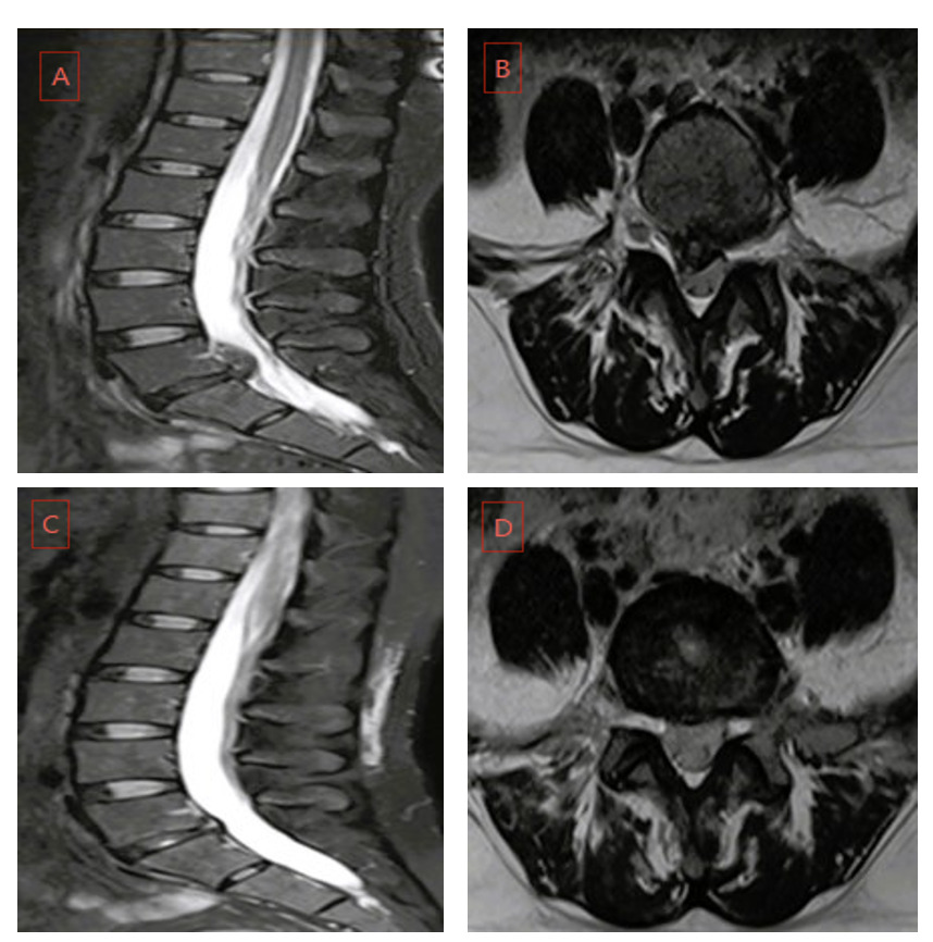
A 36-year-old female patient presented with “radiating numbness and pain in the right lower extremity for 1 week following a severe sneeze.” Lumbar magnetic resonance imaging (MRI) revealed a massive right posterolateral prolapse of the L5/S1 intervertebral disc. She had a history of recurrent low back pain, which was generally relieved by rest. Significant radiating pain and numbness were observed in the right buttock, posterior thigh, lateral calf, heel, and lateral malleolus. The straight leg raise test was positive and Braggard’s test was positive. The patient strongly refused surgical intervention and requested conservative management. During the acute phase, intravenous infusion of mannitol injection combined with dexamethasone sodium phosphate injection was administered for rapid dehydration and non-specific anti-inflammatory therapy. This aimed to alleviate acute nerve root edema and the infiltration of inflammatory algogenic substances. Following initial pain relief, the remission phase began, where the patient was instructed to maintain strict bed rest for one month. During this period, she was guided to perform static “Dead Bug” core stabilization exercises daily to activate the deep transversus abdominis and multifidus muscles, rebuilding active spinal stability. Concurrently, she received oral mecobalamin for neurotrophic support and eperisone for muscle spasm relief. Entering the rehabilitation phase, ESWT was introduced as the core physical intervention. The patient was placed in a prone position, and pain points and palpable trigger points were localized targeting the lumbosacral erector spinae muscles, the right groin area along the sciatic nerve pathway, and the posterior calf muscle belly. The treatment frequency was once every two days, while wrist-ankle acupuncture was concomitantly applied to consolidate the analgesic effect. Fig.1.
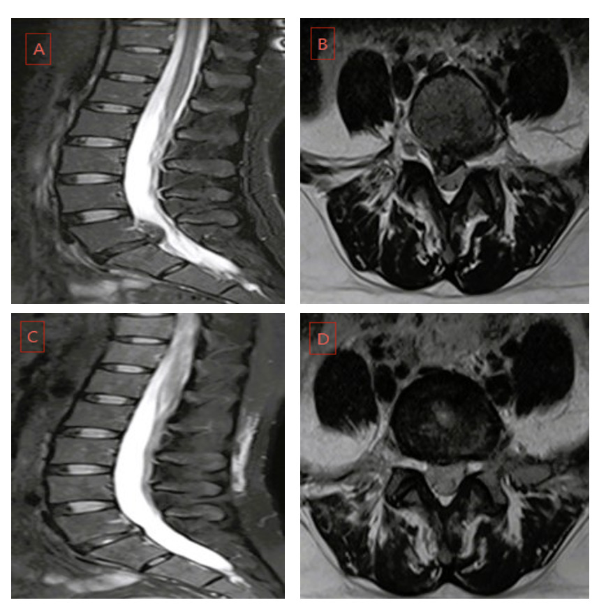
Treatment Outcomes: Upon completing a systematic rehabilitation cycle, the patient’s right lower extremity radiating pain and numbness almost entirely resolved, with only a mild tingling sensation remaining when touching the local skin of the right lateral malleolus, Fig. 1.Her daily activities were unrestricted, indicating a highly satisfactory clinical outcome.
3. Discussion
The successful intervention in this case not only demonstrates the definitive efficacy of a staged, systematic conservative treatment for massive prolapsed LDH, but also highlights the potential clinical value of ESWT in assisting and accelerating the process of disc resorption.
Previous meta-analyses have indicated that approximately two-thirds of LDH patients may experience varying degrees of spontaneous resorption of the herniated mass following systematic conservative management.6 This “spontaneous resorption” is not driven by a single mechanism but is the result of intertwined pathophysiological processes, including immune recognition, neovascularization, enzymatic degradation, and physical dehydration.
Initially, there is a breakdown of immune privilege and activation of the inflammatory response. Under physiological conditions, the intervertebral disc is an avascular tissue. The nucleus pulposus is tightly enclosed by the annulus fibrosus and cartilaginous endplates, maintaining a state of immune privilege.7 When disc prolapse or sequestration occurs, the nucleus pulposus enters the epidural space of the spinal canal through tears in the annulus fibrosus and posterior longitudinal ligament. The originally sealed nucleus pulposus matrix is directly exposed to the epidural blood circulation, recognized by the immune system as a “foreign antigen,” and rapidly triggers autoimmune and aseptic inflammatory responses. Locally released chemokines attract monocytes from the systemic circulation, which aggregate and differentiate into macrophages in situ. Subsequently, neovascularization occurs.8 Within the inflammatory microenvironment of the epidural space, local tissue cells secrete large amounts of pro-angiogenic factors, such as Vascular Endothelial Growth Factor (VEGF).9 These factors induce the rapid expansion of capillary networks on the surface of the herniation and surrounding granulation tissue, growing inward into the originally avascular nucleus pulposus, thus establishing crucial channels for material transport. Finally, enzymatic degradation and phagocytosis take place. The locally aggregated macrophages and neovascular endothelial cells secrete abundant matrix metalloproteinases. These enzymes can specifically degrade collagen fibers and proteoglycans within the nucleus pulposus, breaking down large molecular matrices into soluble small-molecule peptides, which are ultimately phagocytized by immune cells and cleared through the newly formed capillary network and systemic circulation.10,11
In this case, the treatment protocol was designed following a sequential principle of early dehydration and analgesia, mid-term bed rest and core activation, and late-stage physical promotion of resorption. Early strict bed rest and pharmacological dehydration effectively reduced the water content of the herniated mass and decreased its physical volume, directly relieving mechanical compression on the nerve roots and the dural sac. This not only alleviated severe radicular pain but also bought a crucial time window for subsequent rehabilitation training and physical therapy. The introduction of ESWT during the rehabilitation phase acted as a catalyst for the resorption process at the biological level. First, it promotes angiogenesis. The mechanical stress of shock waves stimulates endothelial cells in the target area, which can specifically upregulate the expression of angiogenic factors like VEGF.12 This accelerates the infiltration of granulation tissue and neovascular capillary networks into the nucleus pulposus, breaking the anatomical barrier of its avascularity. Second, it accelerates immune cell recruitment and degradation. The acoustic shear forces of shock waves can induce the local release of chemokines, shortening the body’s immune recognition cycle for the foreign nucleus pulposus. This attracts more macrophages to gather at the lesion, thereby increasing the local concentration of proteases such as MMPs, which significantly accelerates the enzymatic degradation, breakdown, and phagocytic clearance of nucleus pulposus fragments.
Limitations
Although this case demonstrates the ideal efficacy of systematic conservative treatment combined with ESWT for prolapsed LDH, its evidence level is limited as a single case report. Due to the lack of continuous pre- and post-treatment imaging to directly confirm the volume reduction ratio of the herniation, and the persisting confounding factors such as individual differences and the natural temporality of spontaneous resorption, future multi-center, large-sample randomized controlled trials are required to further elucidate the dose-response relationship and standardize clinical protocols of ESWT in promoting resorption.
4. Conclusion
Prolapsed lumbar disc herniation holds clear potential for spontaneous resorption. For patients in the acute flare-up phase, smoothly navigating the acute period through early strict bed rest, pharmacological dehydration, and core muscle activation, followed by the timely introduction of ESWT during the rehabilitation phase, can effectively harness its biological effects of promoting micro-neovascularization and accelerating immune phagocytosis. Consequently, this safely and non-invasively accelerates the resorption process of the herniated mass. This sequential therapy provides a novel and highly valuable clinical strategy to spare patients with prolapsed LDH from surgical trauma.
Ethics approval
The present study was approved by the institutional review board of our institution before data collection and analysis.
Consent to publish
We confirm that patient provided written informed consent for the use of the anonymized clinical data and images in this publication.
Competing interests
All authors certify that they have no affiliations with or involvement in any organization or entity with any financial interest or non-financial interest in the subject matter or materials discussed in this manuscript.
Authors’ contributions
Drafting the article: Luolong Tao and Shengtao Zhang. Acquisition of data: Ming Cheng. Conception and design: Bin Zhang. Study supervision: Fuchong Lai
Acknowledgements
We would like to extend our heartfelt thanks to the patient who participated in this study. Her cooperation and understanding have been the foundation of this research.
Availability of data
The datasets used during the current study are available from the corresponding author on reasonable request.
Funding
No funding was received for this study.