
Introduction
Delbet Type 1b fracture-dislocations of the femoral neck are a rare but severe form of pediatric hip injury, often resulting from high-energy trauma.1 Avascular necrosis (AVN) of the femoral neck is a frequent and devastating complication, occurring due to the disruption of blood supply to the proximal femur.2 The incidence of AVN following such injuries is nearly 100%, making it a significant concern for both clinicians and patients.1 Along with AVN, long-term complications of proximal femur fractures in pediatric patients include pain, coxa vara, and nonunion.3
Another study reported that after 8 months of internal fixation for a Delbet-type 2 fracture of the femoral neck in a 9-year-old girl, AVN of the femoral neck developed.1 We present a case of a 13-year-old male who sustained a Delbet Type 1b fracture-dislocation of his left femoral neck and posterior left acetabulum following a scooter versus car accident. Remarkably, the patient showed no signs of AVN at a two-year follow-up, defying the almost certain prognosis commonly associated with these injuries.4 The case presented in this report is notable for its absence of avascular necrosis in a 13-year-old male patient, two years after sustaining a traumatic epiphyseal separation and dislocation, as well as a posterior wall fracture of the left acetabulum. There is an extremely limited number of pediatric Delbet Type 1b fracture-dislocations reported with such an outcome.
Case Report
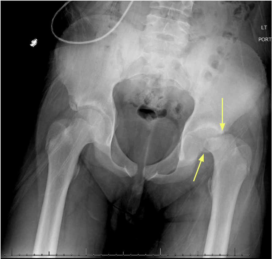
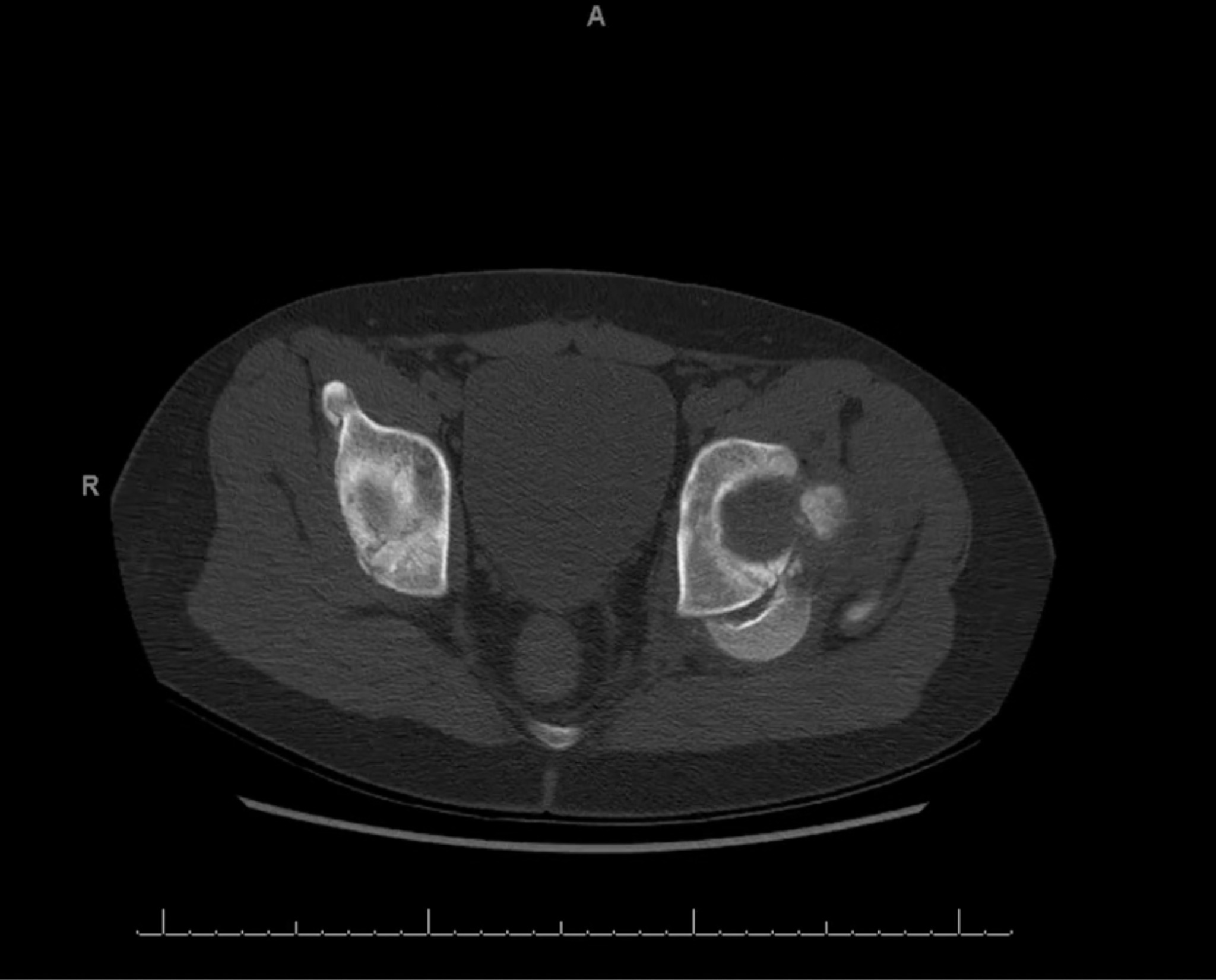
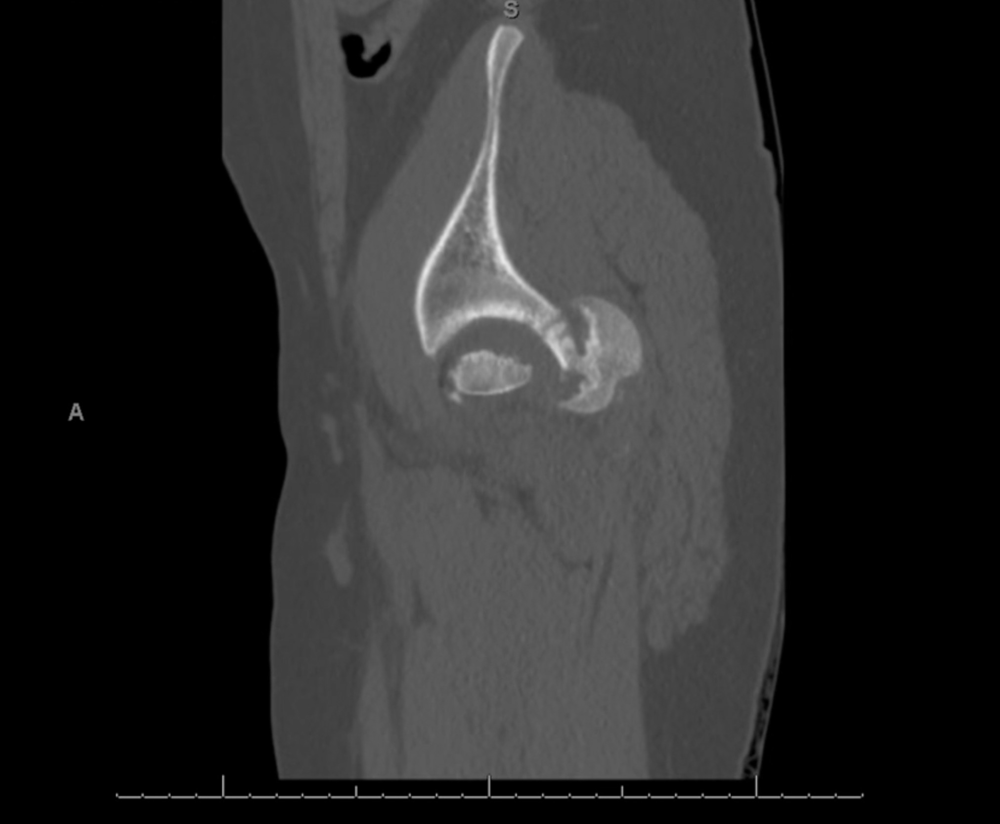
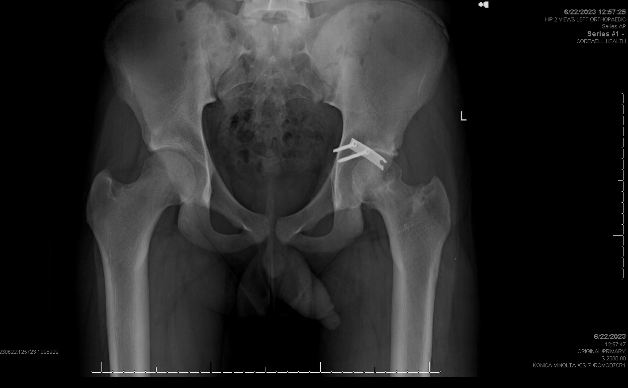
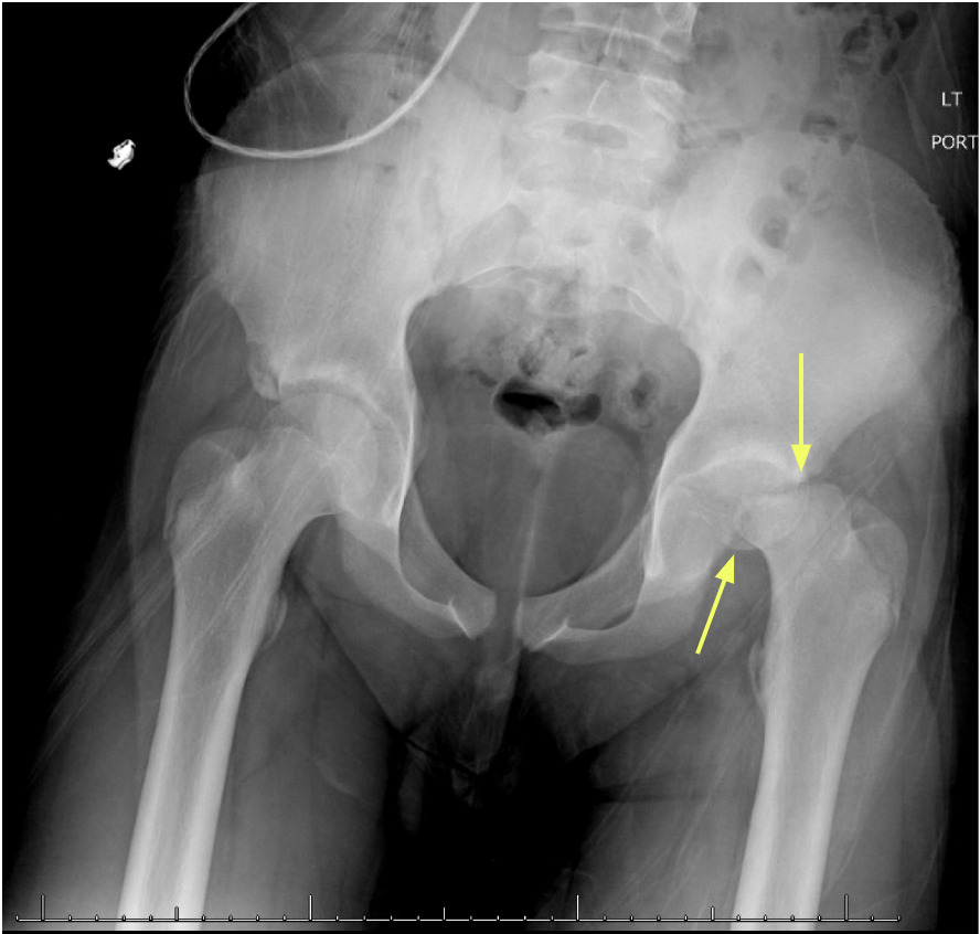
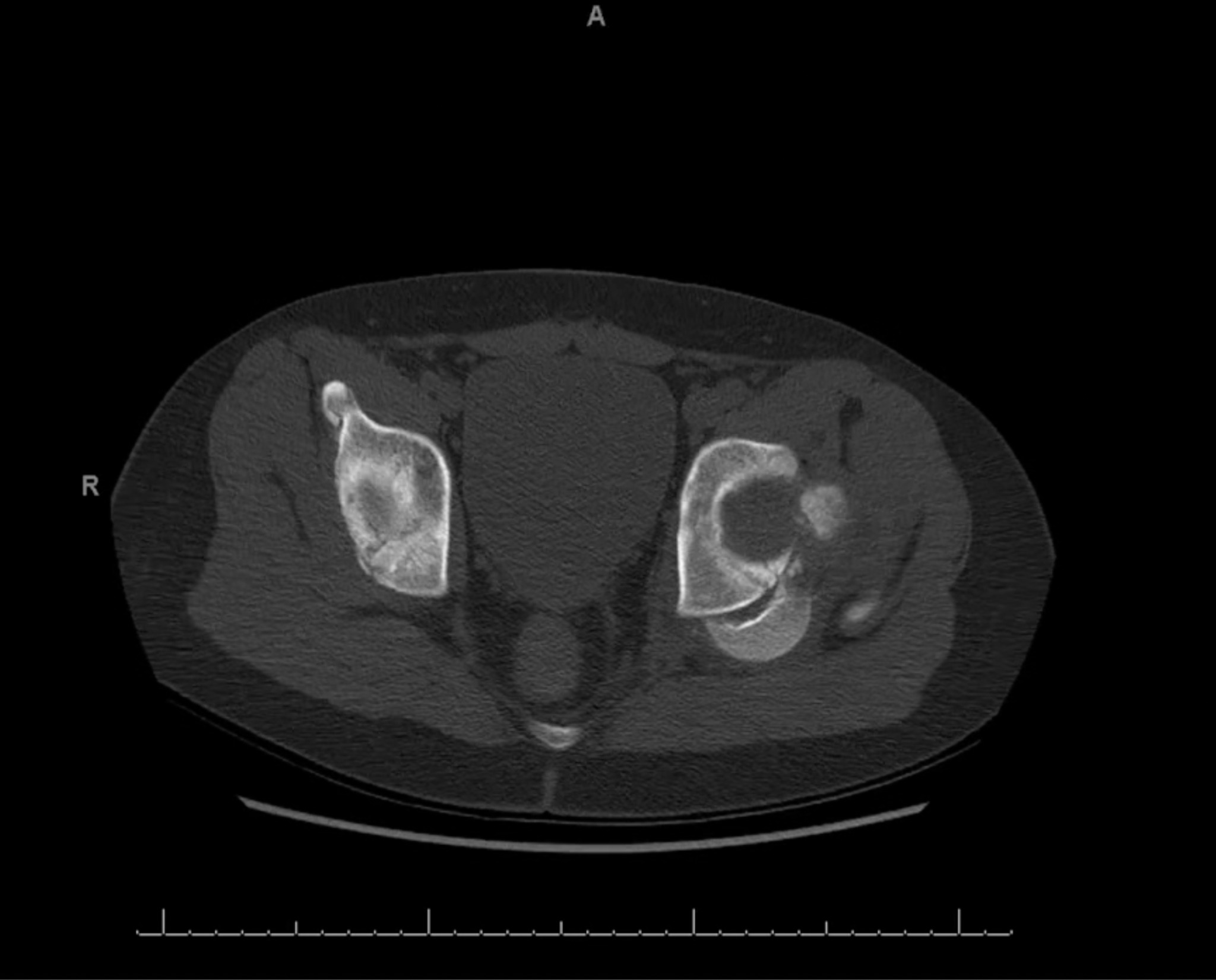
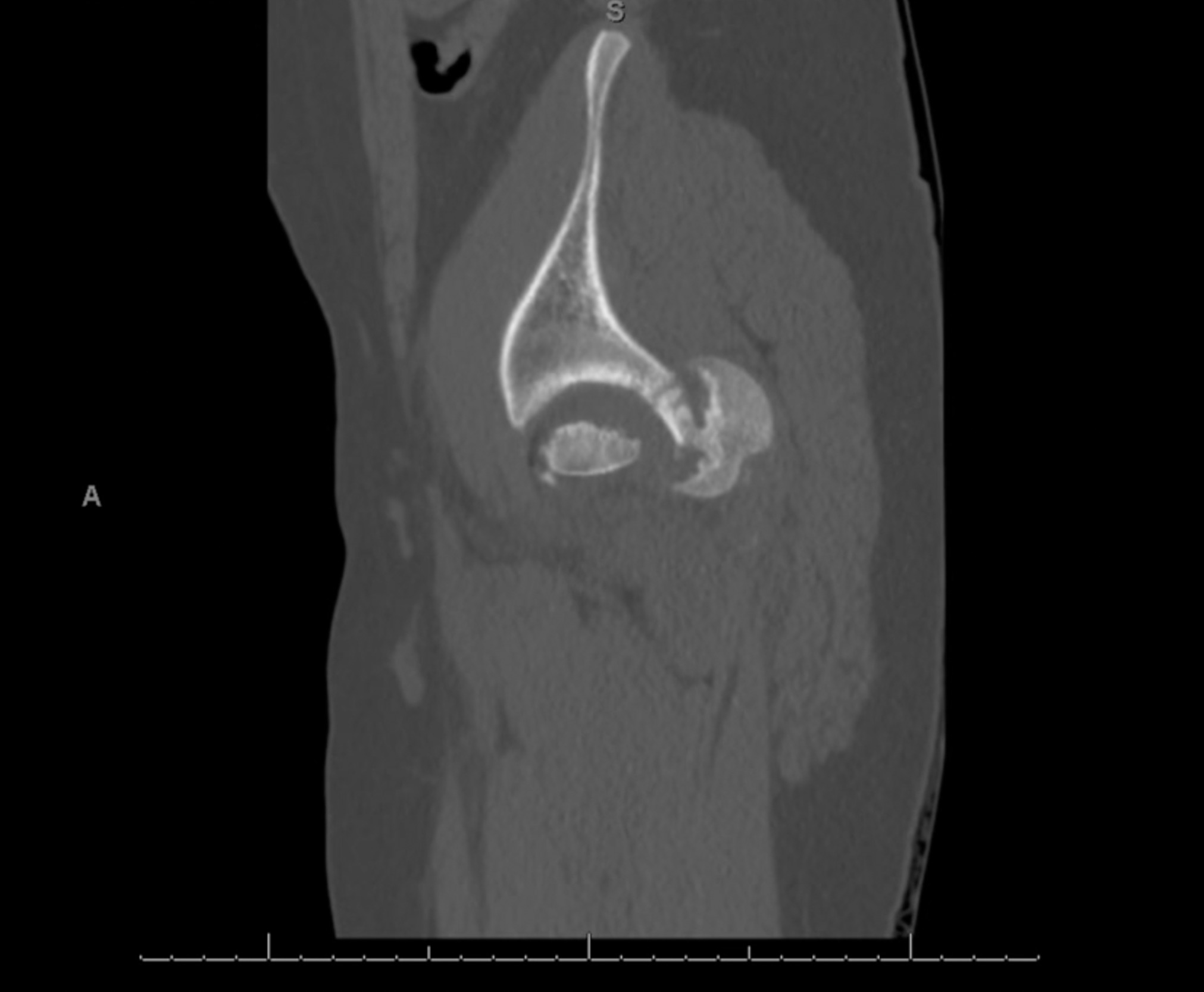
A 13-year-old male was brought into the emergency room following a scooter vs. car accident, complaining of severe pain in his left leg. An initial A/P radiograph (Figure 1) and axial CT (Figure 2) findings revealed an oblique fracture pattern of the femoral epiphysis consistent with a Delbet Type 1b fracture-dislocation of his left femoralR neck and a posterior wall fracture of the left acetabulum. The patient was transported to a Midwest level-1 trauma center for surgical intervention.
After resuscitation and stabilization, the patient was taken to the operating room. Despite the complexity of the injury, closed reduction was not attempted due to the severity of the dislocation and associated fractures. The patient was positioned in the lateral decubitus position and stabilized with Montreal pegs, with a roll beneath the chest and padding under the non operative leg. A longitudinal incision was made over the hip in line with the fascia lata. An anterolateral approach to the hip joint was utilized according to Ganz’s description, which preserves the deep branch of the medial circumflex artery. The subcutaneous tissues were divided using electrocautery to the fascia lata and gluteus fascia. The fascia lata was divided in line with the incision and the gluteal fascia was divided between the tensor fascia lata and the gluteus maximus muscles. The trochanteric bursa was then incised. The posterior border of the gluteus medius was identified. The vastus lateralis fascia was incised and the muscle was dissected from the intermuscular septum and extra periosteally from the lateral femur. A step-cut osteotomy of the greater trochanter was then performed, taking care to preserve the posterior superior tendinous attachment of the gluteus medius to the stable trochanter. The femoral epiphysis, intracapsular and deep to the external rotator muscles, was palpable along the posterior aspect of the ischium.
A Z-shaped capsulotomy was performed dividing the capsule along the anterolateral femoral neck towards the base of the trochanter, inferiorly from the base of the trochanter towards the base of the acetabulum diverging slightly from the intertrochanteric line, and posteriorly parallel to the acetabular labrum continuing along the ischium anterior to the piriformis muscle.
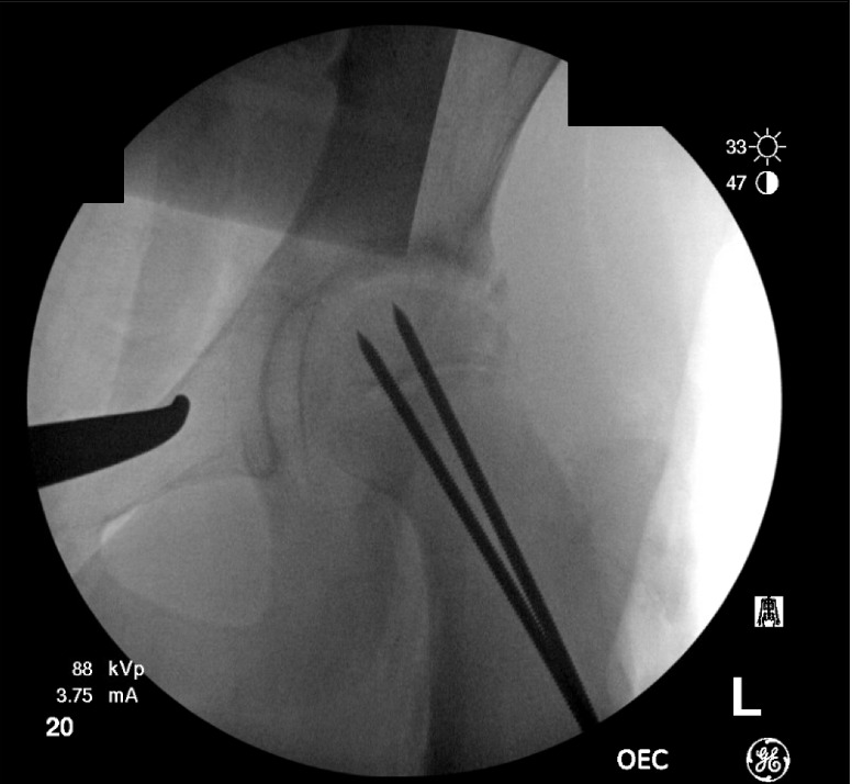
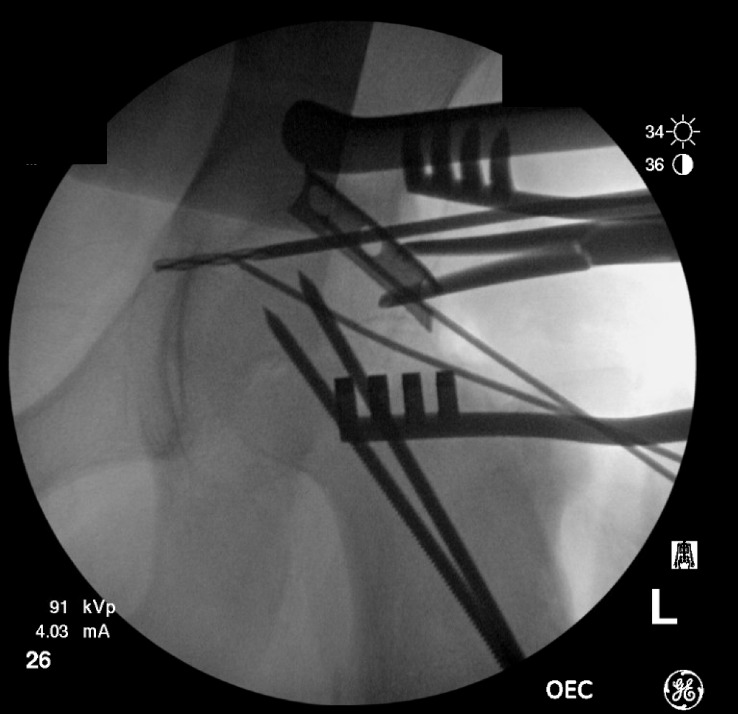
A retinacular flap was then developed. The periosteum of the anterior femoral neck was dissected longitudinally along the anterolateral femoral neck and dissected medially. A portion of the proximal greater trochanter was resected and the posterior retinaculum was developed all the way to the lesser trochanter. At this point, the epiphysis was mobilized and partially reduced onto the metaphysis using threaded Kirschner wires (Figure 3).
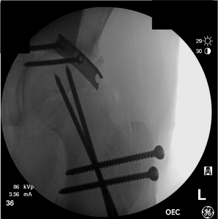
The acetabulum was then inspected. There was a large avulsed fragment of the epiphysis attached to the fovea which was then resected. The head split fragment had no repairable bone attached to the medial epiphyseal cartilage to facilitate repair of the cartilage of the medial epiphysis and therefore it was removed. The posterior acetabular wall was repaired using a spring plate (Figure 4). Finally, Two 4.5mm fully threaded cannulated screws were placed to achieve stable fixation (Figure 5) and the femoral head was reduced back into the acetabulum (Figure 6).
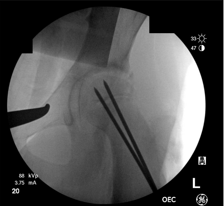
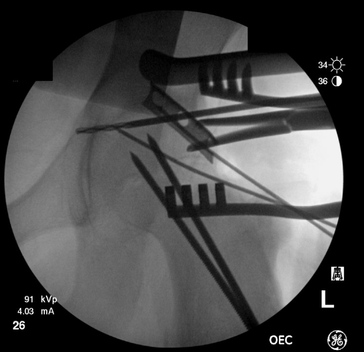
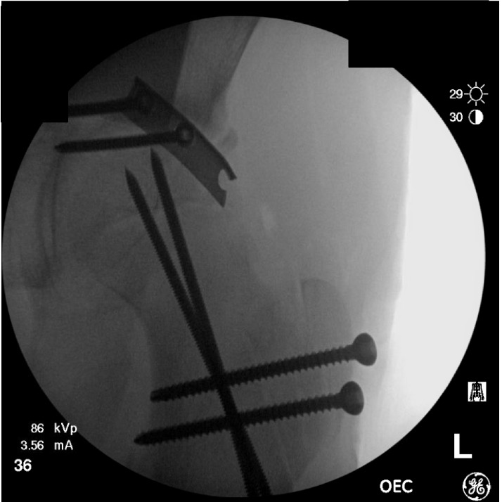
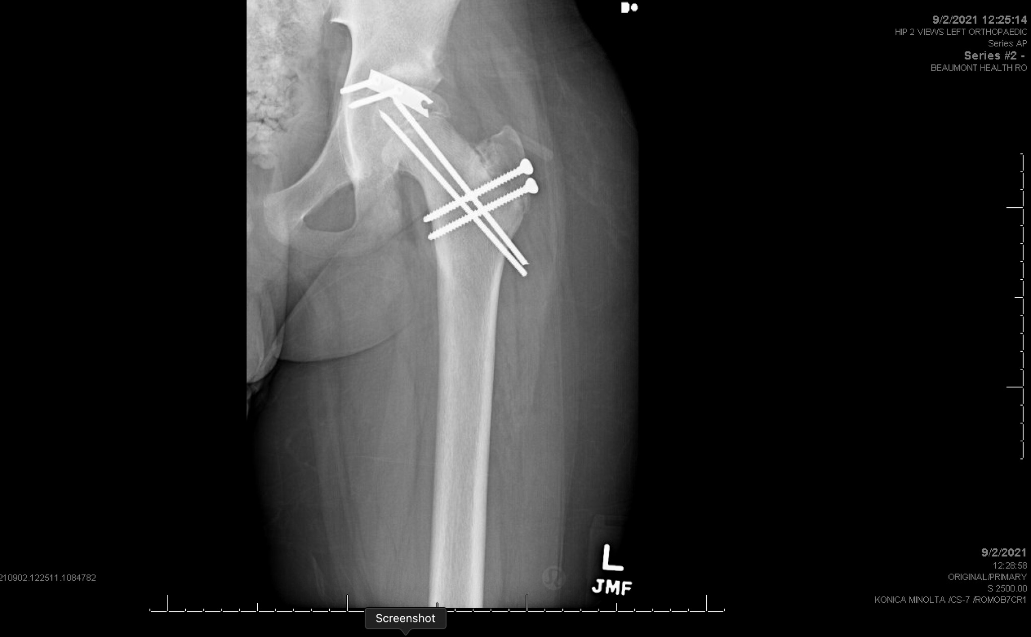
Seven months following the procedure, the patient was brought back to the operating room to remove the hardware. A proximal incision was made to remove the trochanteric screws and then a second longer incision was made for the pins. The pins required exposure and were both removed in their entirety. A final x-ray was taken to ensure proper hardware removal (Figure 7).
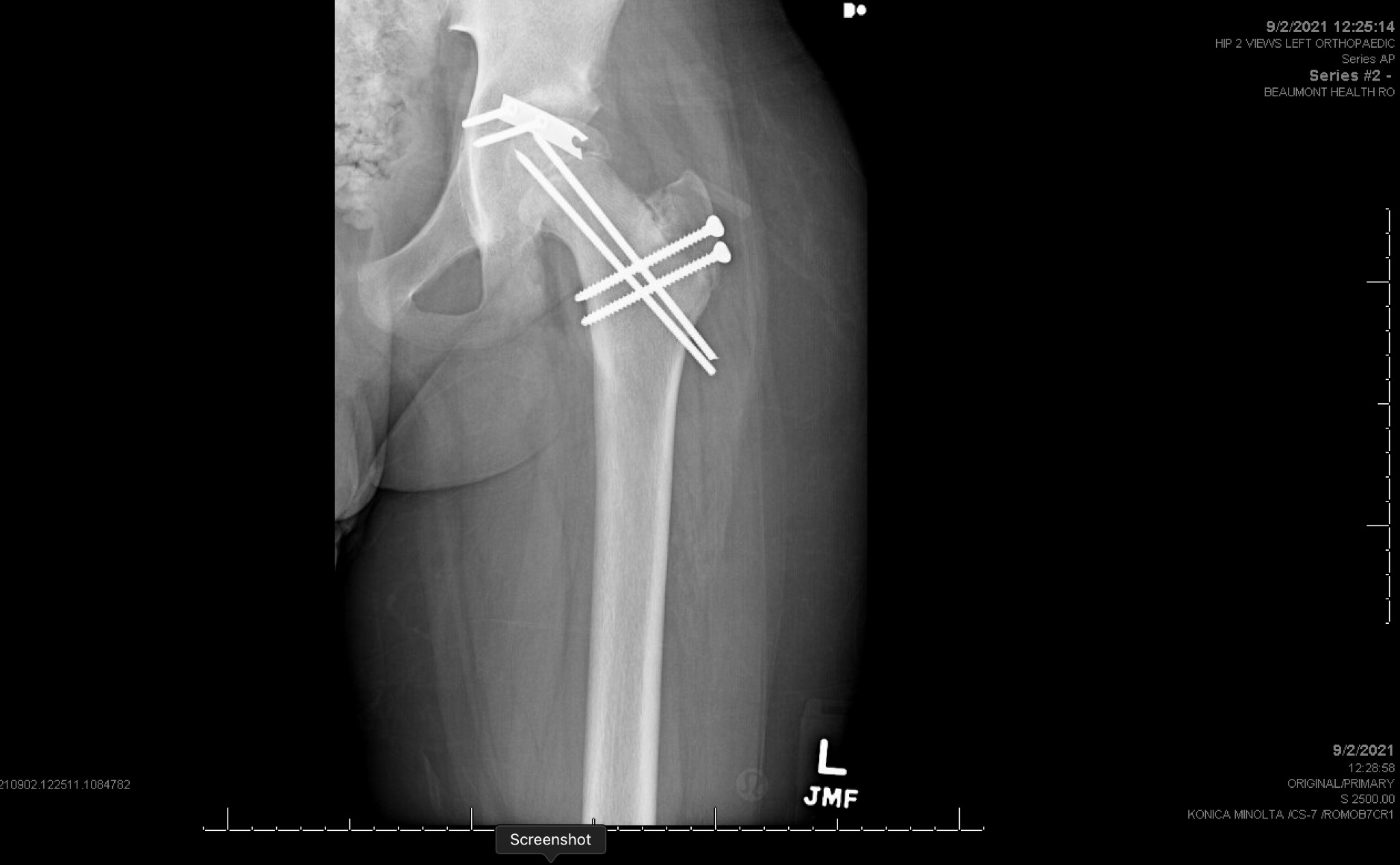
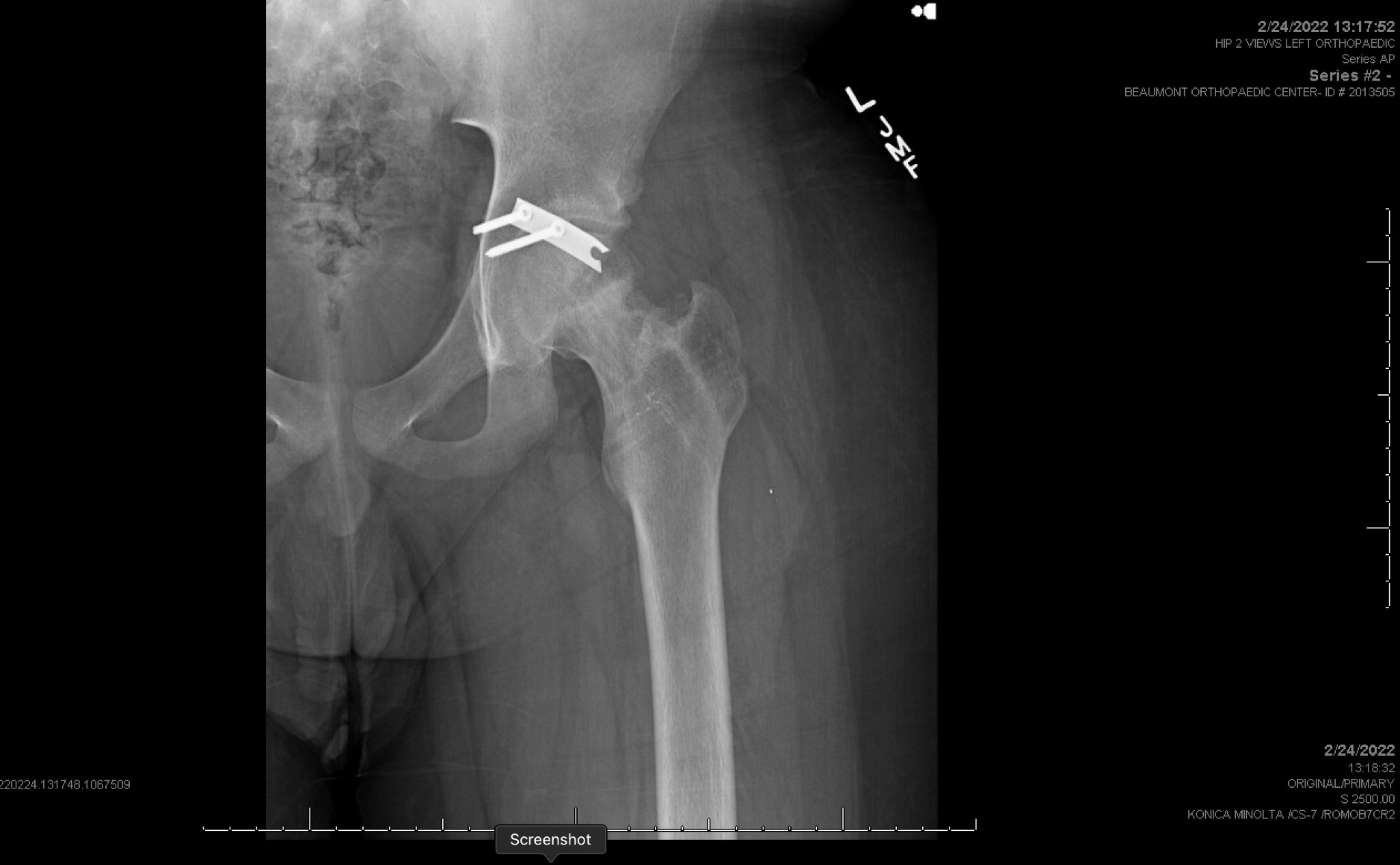
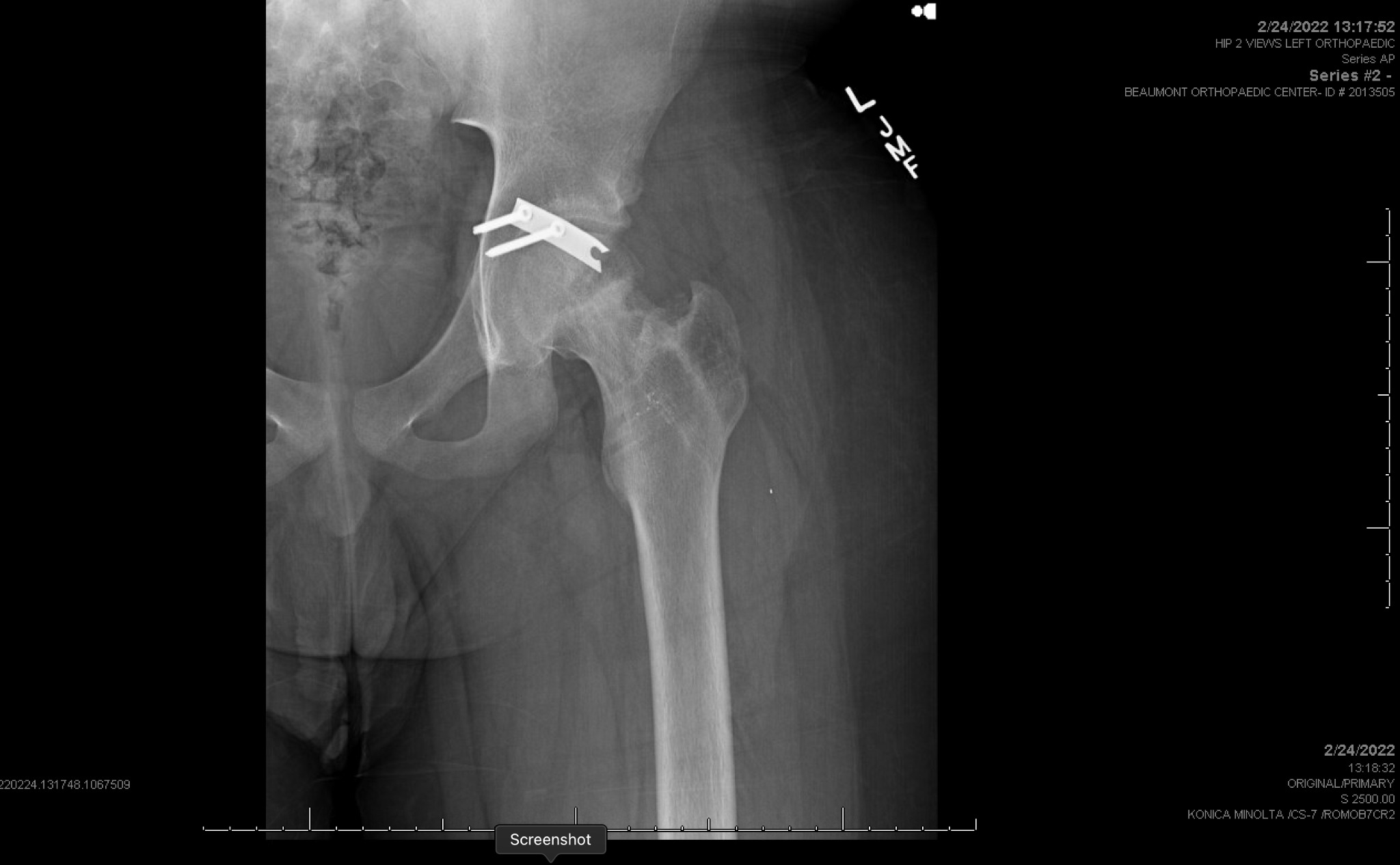
Two years following the incident, the male now 15 years old, presents to the clinic with his father for follow-up. He is doing well and is playing golf and soccer without any issues. X-ray imaging in the office showed that the left acetabular spring plate is still intact with maintained alignment (Figure 8). The femoral head is concentrically reduced within the acetabulum with no obvious femoral head lucency or collapse.
Discussion
Transphyseal femoral neck fractures, particularly of the Delbet Type 1 classification, are rare in the pediatric demographic.5,6 A comprehensive meta-analysis showed that transphyseal fractures only occurred in 8% of femoral neck fractures.7 Pediatric fractures of the femoral neck are usually a result of high-energy trauma and are often associated with simultaneous musculoskeletal injuries such as hip dislocation, ipsilateral fracture of the femur, or hip dislocation as seen in this patient.3
One of the most devastating complications associated with these injuries is avascular necrosis (AVN). Risk factors mitigating the incidence of AVN after fracture reduction include time between trauma and surgery, type of fracture, and patient age.8,9 Notably, in a case series done on 53 pediatric femur fractures with an average follow up rate of 10 years, only one patient with a transepiphyseal fracture was reported to have no signs of AVN.10 The unique anatomy and vascular supply of the pediatric hip is what makes AVN risk in these cases difficult to manage.5 Simultaneous hip dislocation with epiphyseal fractures are associated with higher rates of AVN.11,12 Remarkably, our patient exhibited no signs or indications of AVN two years postoperatively.
Using existing information from Sondur et. al. and a search through Pubmed Embase, and Google Scholar an extensive table of all previously reported pediatric Delbet type 1b fractures with concurrent acetabular fractures is provided in Table 1. This table can be used to compare the factors such as variations in surgical timing, approaches, and their effects on outcomes, particularly concerning avascular necrosis (AVN). The age of patients ranged from 10 to 15 years, with the time from injury to surgery extending up to 192 hours, demonstrating a wide spectrum of clinical scenarios. Despite differing approaches, including the Kocher Langenbeck and anterolateral approaches, outcomes such as AVN, restricted mobility, and varus malunion were inconsistently reported, suggesting that risk of AVN is determined by a multitude of factors rather than a sole determinant. Notably, our case is the first documented instance of employing the Ganz surgical hip dislocation with a digastric slide osteotomy of the trochanter for treating such injuries, resulting in no AVN at the 24-month follow-up. This outcome emphasizes the potential of specific surgical strategies, beyond just the timing of intervention, to influence recovery and long-term joint health in pediatric patients with complex hip injuries. The Ganz digastric osteotomy is a vital component of surgical hip dislocation, offering both safe access to the hip joint and preservation of critical vascular structures. This approach preserves the origin of the vastus lateralis and the insertion of the adductors, maintaining mechanical stability and minimizing postoperative risk of AVN. Unlike the trochanteric flip osteotomy, which involves detaching all abductors and external rotators in a posterior approach, the Ganz technique provides a safer, anatomically preserving alternative. Although there have been many reports of acetabular fractures associated with femoral neck fractures, this patient’s traumatic hip dislocation with a transphyseal femoral neck fracture which led to a posterior displacement of the femoral head relative to the acetabulum, has been rarely reported in literature.8,10,13
Current literature would suggest that each hour delaying surgical reduction of traumatic hip dislocation increases risk of AVN and non-union15,20 meaning this patient’s swift operation within approximately 9 hours of injury has surely contributed to his favorable outcome of no AVN. However, there is a need for continuous statistical analysis in order to determine a statistically significant association between early surgical reduction and favorable outcomes.9,21 Two particularly noteworthy cases from our review, reported by Sethurman et al. and Sondur et al., underscore the complexity of managing pediatric Delbet Type 1b fractures.12,13 Both cases resulted in the favorable outcome of no AVN after follow-up periods comparable to our case, despite markedly different intervals from injury to surgical intervention. Specifically, the patient in the study by Sethurman et al. received surgical treatment within 48 hours of injury, whereas the patient in the report by Sondur et al. underwent surgery more than a week post-injury. This variance in the timing of surgical intervention between cases that both achieved absence of AVN postoperatively prompts a reevaluation of the critical role that the urgency of surgical reduction may play in the prevention of AVN.
An open reduction was used in this case as the literature suggests a high risk for failure in attempts at closed reductions due to the inherent instability of these fractures.22,23 Coxa Vara is the second most common complication of femoral neck fractures after AVN, however, the increased age of this patient and open reduction intervention substantially lowers the risk of occurrence.24
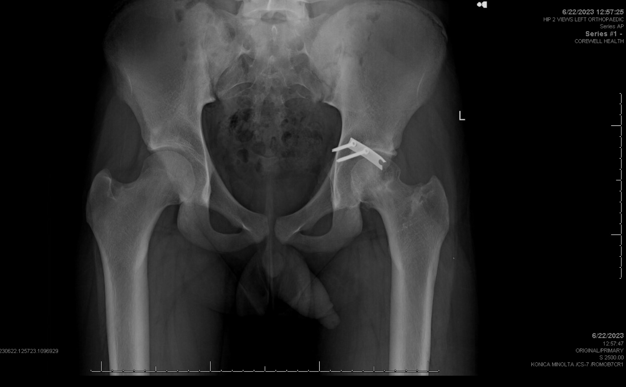
While this case report highlights an notable and rare outcome, it is not without its limitations. Advanced imaging with MRI to assess for avascular necrosis was not pursued, given the patient’s asymptomatic presentation and the lack of radiographic indications of early AVN. Nonetheless, the inclusion of MRI could have provided a more definitive evaluation of femoral head vascularity. Employing advanced imaging in similar cases may contribute to a deeper understanding of vascular integrity and long-term outcomes. Future studies should consider incorporating MRI assessments alongside radiographic evaluations to refine management strategies for such complex injuries. An additional consideration is the role of creeping substitution in explaining the normal radiographs observed. This process of bone resorption and replacement with viable bone may have contributed to the favorable plain radiographic appearance at 24 months. However, there is no definitive way to determine whether the observed outcome was directly due to creeping substitution due to the limited understanding of the process. It is also worth noting that a close review of the two-year follow-up radiographs (Figure 9) raises the possibility of early signs of sclerosis, cystic changes, or subchondral flattening, which could suggest early avascular necrosis (AVN). However, at this time, the lack of clinical symptoms and the minimal radiographic changes overall represent a better-than-expected outcome for a Delbet Type 1b fracture-dislocation. This interpretation highlights the need for continued follow-up to monitor for potential late-onset complications.
In summary, Delbet type 1b fractures present a significant challenge for pediatric patients, timely surgical intervention along with appropriate fixation methods can lead to more favorable outcomes. Further exploration of rates of the different complications in pediatric hip fractures at each stage of bone maturity would allow for improved preventative measures.
Ethical Statement
The authors take responsibility for every aspect of the work, ensuring that any concerns about its accuracy or integrity are thoroughly examined and addressed.
Declaration of Conflicting Interests
The author(s) declared no potential conflicts of interest.
Funding
The author(s) received no financial support for this article’s research, authorship, and/or publication.