
Introduction
Intertrochanteric fractures (ITFs) are extracapsular injuries of the proximal femur between the greater and the lesser trochanter. These fractures commonly affect older adults; however, they can occur in younger individuals as a result of significant trauma or in cases of pathological fracture. ITFs are almost always treated surgically, with traction tables used to achieve and maintain reduction before fixation. However, in the case of patients with ipsilateral lower-limb amputations, especially above-knee (AK), significant challenges are encountered in reduction and fixation strategies.1–3 Commonly used methods include inverted boot positioning, adhesive skin traction, and skeletal traction (pin-based methods).4–7 In this report, we describe a non-invasive traction technique using reinforced adhesive wraps for traction and successful proximal femoral nail (PFN) fixation in an above-knee amputee with an intertrochanteric fracture.
Case Report: A 40-year-old male presented to the Emergency Department complaining of acute right hip pain and inability to bear weight on his prosthesis following a fall from a ladder approximately three meters high. The patient was wearing his prosthesis at the time of the fall. The patient had a medical history of a traumatic transfemoral amputation for refractory chronic osteomyelitis of the distal femur five years before presentation. The amputation was well compensated by a sub-ischial prosthesis.
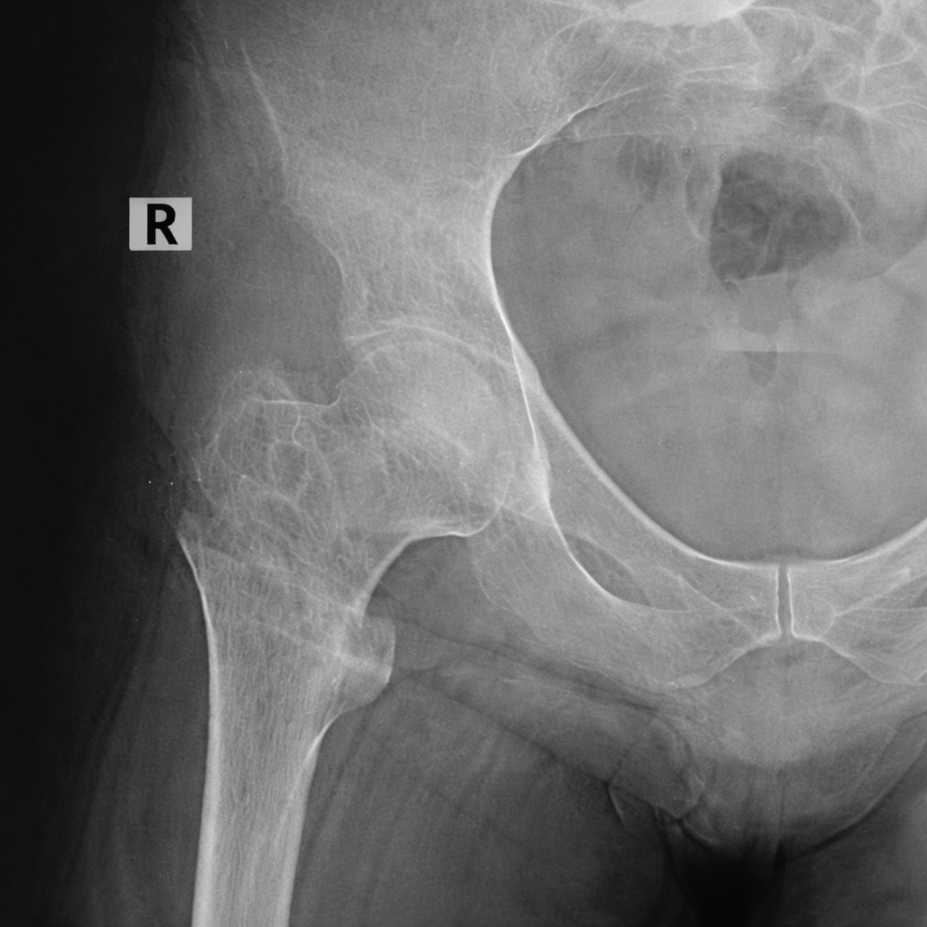
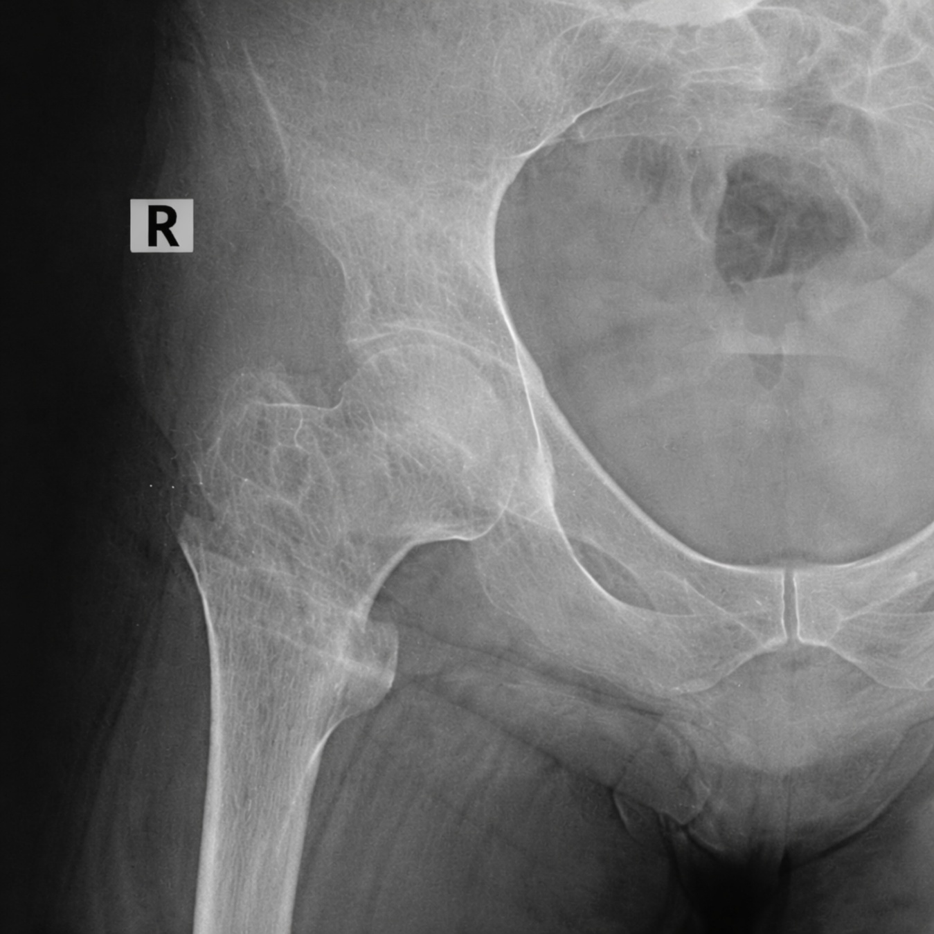
On examination, there was severe tenderness localized to the right hip and a palpable femoral pulse. Minimal movement of the distal stump elicited severe pain. Radiographs confirmed a displaced intertrochanteric fracture of the right femur (Fig. 1). Taking into consideration the instability of the fracture and the patient’s activity level, intramedullary fixation using a short proximal femoral nail (PFN) was planned. PFN was preferred over the dynamic hip screw (DHS) to avoid interference with the prosthetic socket and potential metal prominence. However, intraoperative positioning was challenging because the absence of a distal limb prevented attachment to a standard traction table.
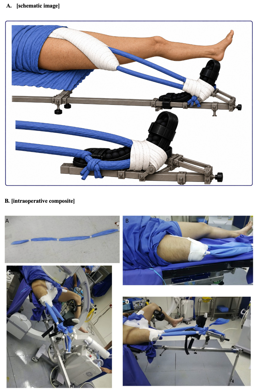
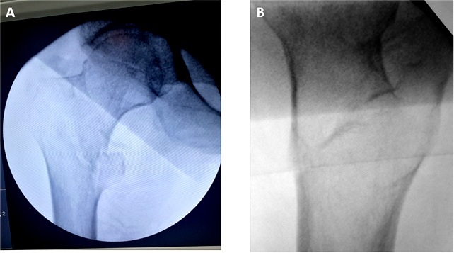
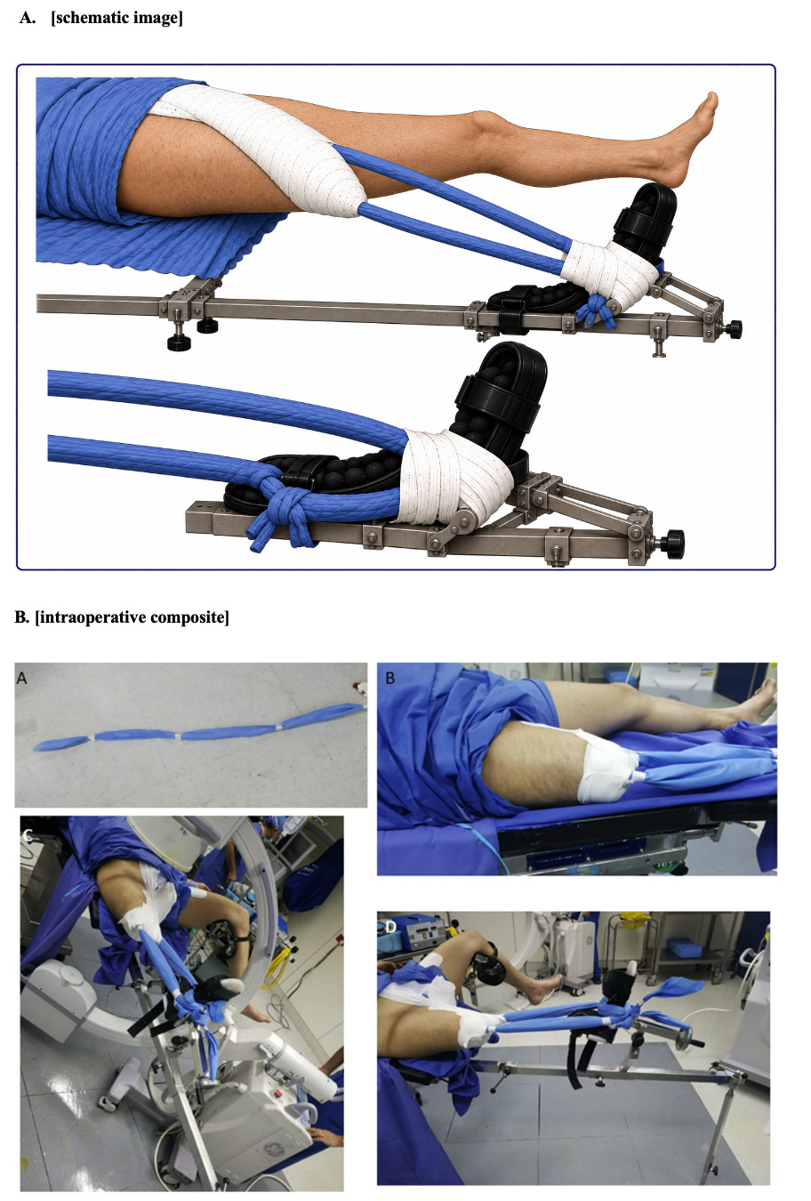
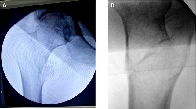
To achieve traction and fracture reduction, we devised a traction construct using two disposable sterilization wraps. Before folding, both wraps were approximately 30 cm wide; the lateral wrap measured approximately 130 cm in length, whereas the medial wrap measured approximately 160 cm. Each wrap was folded repeatedly into a rope-like construct to increase tensile strength. The construct was then secured to the stump using adhesive tape. The shorter wrap was applied longitudinally along the inferolateral aspect of the stump, leaving the proximal lateral thigh exposed. The longer wrap was applied longitudinally along the medial aspect of the thigh and secured with adhesive tape, extending proximally onto the suprapubic region above the pubic symphysis. The construct was further reinforced with an oblique circumferential layer of Tensoplast and Mefix tape extending from the proximal medial thigh to the distal lateral aspect of the stump (Fig. 2). The stability of the construct was tested manually before attachment to the traction table. Distally, both wraps were secured circumferentially around the traction boot before attachment to the traction table. This provided effective fracture reduction (Fig. 3), after which intramedullary fixation was performed using a standard proximal femoral nail (PFN).
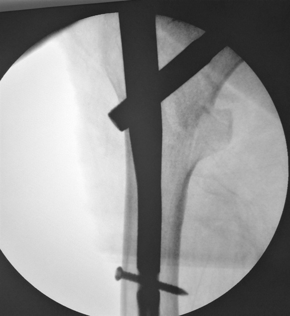
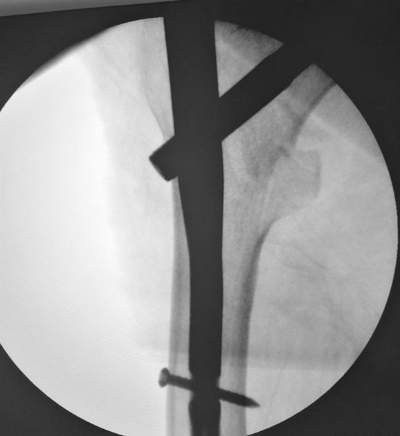
Fluoroscopy confirmed stable fixation and proper alignment (Fig. 4). Intravenous cefazolin was administered at induction and continued for 5 days because of the patient’s history of osteomyelitis.
Postoperative swelling delayed immediate prosthesis fitting. However, after postoperative swelling improved with cryotherapy and nonsteroidal anti-inflammatory medications, the patient was able to use his previous prosthesis and commenced full weight-bearing as tolerated with walking aids on postoperative day 3. By day five, he was comfortably ambulating with his previously used prosthesis. He was discharged on postoperative day six.
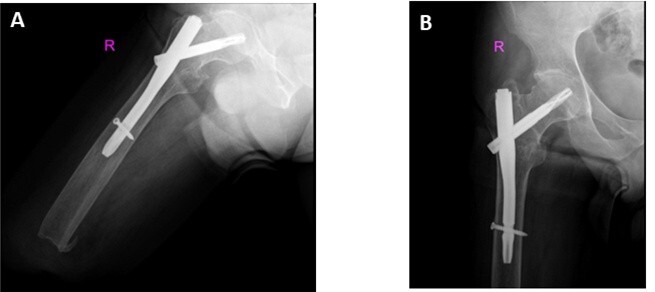
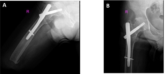
At six weeks, radiographs indicated maintenance of alignment and early signs of healing. At the 4-month follow-up, the patient reported feeling well and inquired about returning to manual work. Clinical examination and radiographs confirmed complete fracture union, and he was therefore cleared to resume work activities. At latest follow-up (3 years postoperatively), the patient reported no difficulties with ambulation, pain, or work activities (Fig. 5). He did not report changes in his lifestyle following fixation of the right hip. No modification or refitting of his prosthesis was required.
Discussion
Hip and proximal femur fractures in patients with lower-limb amputations are not common and are technically challenging. The absence of a distal limb presents challenges for axial traction and even more for rotational control on a fracture table, especially if the intertrochanteric patterns are unstable and accurate reduction becomes very challenging. Moreover, our patient’s short transfemoral stump and prior osteomyelitis limited our options substantially.
Recent case reports and mini reviews do not demonstrate the superiority of any single technique; the choice of technique essentially depends on the stump length/skin integrity, fracture displacement, equipment, and infection risk.5,6 Recent studies describe modified fracture table setups for above-knee amputees to obtain reliable traction without pins6,7; recent reports have also provided updated guidance on the reverse boot technique for below-knee amputees, including stepwise pearls and pitfalls8–10; and customized non-invasive constructs to assist in positioning when the boot is not feasible.2
In below-knee amputees, non-invasive boot-based methods (inverted “reverse boot”) are effective for many intertrochanteric fractures.8–10 Adhesive skin traction bands and tape constructs can also transmit traction to the table boot or support.2,3 However, these strategies become unreliable in short above-knee stumps, as bulky hardware may interfere with the operative field. Skeletal traction via Denham or Steinmann pins provides powerful traction and rotation in transfemoral amputees but carries risks of pin site infection and soft tissue injury. Moreover, pin cutout and interference with prosthetic interfaces are magnified in short stumps or those with prior infection.6,7
The described traction construct (Fig. 2) provides a simple, readily available, low-cost, radiolucent, and reproducible solution when the reverse boot was impractical, and pin traction was undesirable. The construct provided controlled axial traction and rotation without violating stump tissues, enabling fluoroscopic reduction (Fig. 3) and fixation with standard proximal femoral nail. In comparison with standard adhesive-only methods,8 our construct differs in both configuration and force transmission. Conventional adhesive traction usually uses wide adhesive strips applied symmetrically along the medial and lateral sides of the limb. However, the traction force is transmitted through relatively thin distal traction cords, which mainly provide axial traction and offer limited rotational control, particularly in short transfemoral stumps. In our technique, the sterilization wraps were folded repeatedly into thick, rope-like constructs to create a stronger traction unit. The longer medial wrap originated from the upper medial thigh and was reinforced circumferentially with Tensoplast and Mefix tape, while the shorter lateral wrap mainly acted as a stabilizer. We found that the thicker rope-like construct provided better control of rotation while maintaining effective axial traction and keeping most of the construct away from the operative field. In addition, unlike skeletal traction pins, this method avoids violation of soft tissues and bone. In patients with a history of refractory osteomyelitis, residual bone quality and soft tissue condition may be compromised, increasing concern for pin cutout, skin problems, or recurrence of infection, particularly in short stumps with limited soft tissue coverage.
Skin integrity ultimately determines the feasibility of the chosen technique; meticulous padding and avoidance of focal shear are essential. The construct should be manually tested before applying traction. No skin complications, pressure-related injuries, or construct failures were observed in this case. In cases with a very short stump or fragile skin, skeletal pin traction may be necessary. As current evidence is largely limited to case reports, multicenter series, or registry data would be valuable for comparing the safety and outcomes of different positioning and traction methods.
Conclusion
Intertrochanteric fractures in patients with ipsilateral above-knee amputation are uncommon and present unique surgical challenges. This case highlights that satisfactory reduction and fixation can be achieved with careful intraoperative planning using a non-invasive traction technique in patients with short transfemoral stumps and a history of osteomyelitis.
Acknowledgments
The authors would like to thank the patient for his cooperation and for providing written informed consent for publication.
Author Contributions (CRediT)
Mohammad Abu Hilal: Conceptualization, methodology, supervision, writing – original draft, and writing – review & editing.
Mohammed Tayyem: Investigation, data curation, and writing – review & editing.
Ethical Compliance
This case report was approved by the Ethics Committee of the Faculty of Medicine, Mutah University. All procedures were performed in accordance with the ethical standards of the institutional research committee and with the 1964 Declaration of Helsinki and its later amendments or comparable ethical standards. Written informed consent for publication was obtained from the patient. The patient’s identity has been anonymized, and all identifying information has been removed to protect confidentiality.
Conflicts of Interest
The authors declare no conflicts of interest.
Funding
None.